. 2021 Jan;41(1):302-316.
doi: 10.1161/ATVBAHA.120.315164. Epub 2020 Oct 8.

Cholesterol-Induced Phenotypic Modulation of Smooth Muscle Cells to Macrophage/Fibroblast-like Cells Is Driven by an Unfolded Protein Response

Affiliations

Cholesterol-Induced Phenotypic Modulation of Smooth Muscle Cells to Macrophage/Fibroblast-like Cells Is Driven by an Unfolded Protein Response

Abhijnan Chattopadhyay et al. Arterioscler Thromb Vasc Biol. 2021 Jan.

Abstract

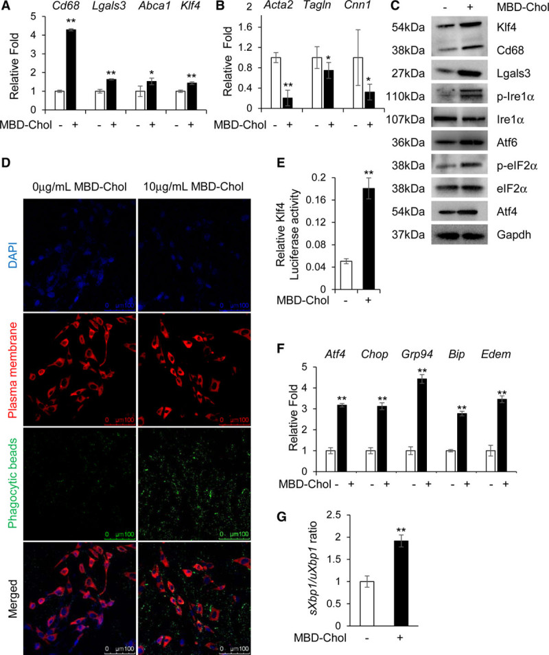
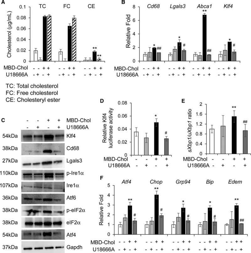
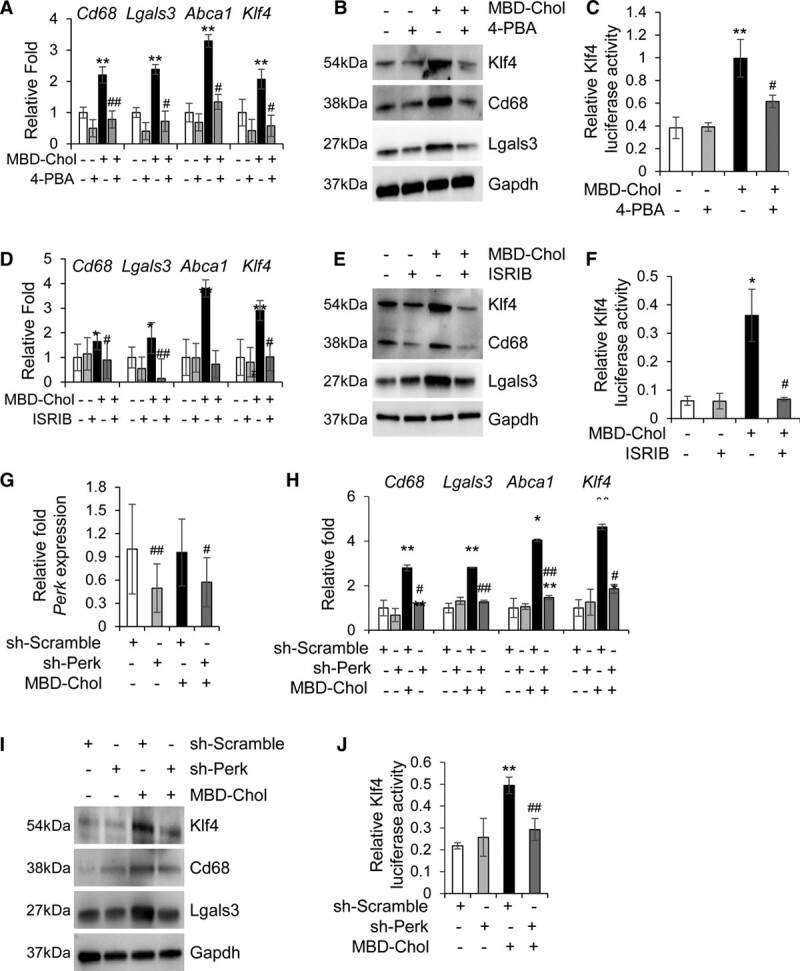
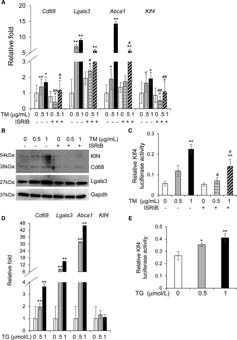
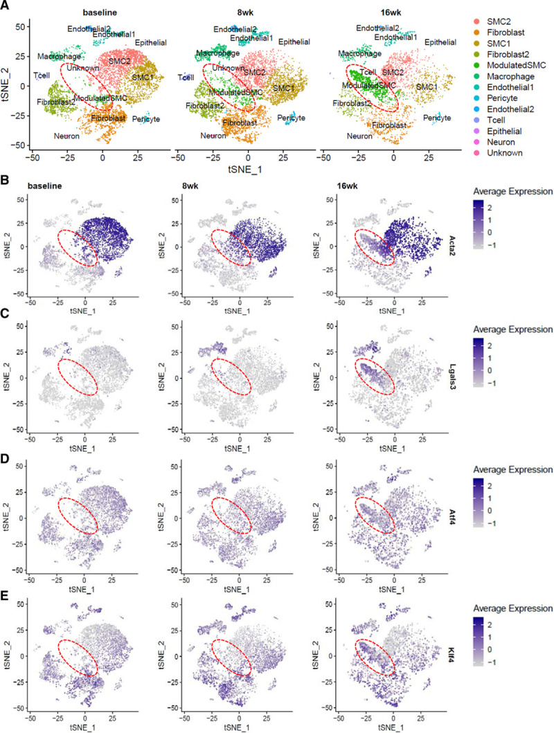
Objective: Vascular smooth muscle cells (SMCs) dedifferentiate and initiate expression of macrophage markers with cholesterol exposure. This phenotypic switching is dependent on the transcription factor Klf4 (Krüppel-like factor 4). We investigated the molecular pathway by which cholesterol induces SMC phenotypic switching. Approach and Results: With exposure to free cholesterol, SMCs decrease expression of contractile markers, activate Klf4, and upregulate a subset of macrophage and fibroblast markers characteristic of modulated SMCs that appear with atherosclerotic plaque formation. These phenotypic changes are associated with activation of all 3 pathways of the endoplasmic reticulum unfolded protein response (UPR), Perk (protein kinase RNA-like endoplasmic reticulum kinase), Ire (inositol-requiring enzyme) 1α, and Atf (activating transcription factor) 6. Blocking the movement of cholesterol from the plasma membrane to the endoplasmic reticulum prevents free cholesterol-induced UPR, Klf4 activation, and upregulation of the majority of macrophage and fibroblast markers. Cholesterol-induced phenotypic switching is also prevented by global UPR inhibition or specific inhibition of Perk signaling. Exposure to chemical UPR inducers, tunicamycin and thapsigargin, is sufficient to induce these same phenotypic transitions. Finally, analysis of published single-cell RNA sequencing data during atherosclerotic plaque formation in hyperlipidemic mice provides preliminary in vivo evidence of a role of UPR activation in modulated SMCs.

Conclusions: Our data demonstrate that UPR is necessary and sufficient to drive phenotypic switching of SMCs to cells that resemble modulated SMCs found in atherosclerotic plaques. Preventing a UPR in hyperlipidemic mice diminishes atherosclerotic burden, and our data suggest that preventing SMC transition to dedifferentiated cells expressing macrophage and fibroblast markers contributes to this decreased plaque burden.

Keywords: atherosclerosis; cholesterol; fibroblast; macrophage; phenotype; smooth muscle cell.


Conflict of interest statement

None.

Figures

Figure 1.
Figure 1.
Cholesterol induces phenotypic switching and endoplasmic reticulum (ER) stress in immortalized vascular smooth muscle cells (SMCs). A and B, With exposure to 10 µg/mL methyl-β-cyclodextrin cholesterol (MBD-Chol) for 72 h, expression of macrophage markers Cd68 and Lgals3 along with Abca1 and Klf4 (Krüppel-like factor 4) are upregulated (A), while contractile marker genes Acta2, Tagln and Cnn1 are downregulated (B). C, Immunoblots confirm increased protein levels of Cd68 (cluster of differentiation 68), Lgals3 (lectin, galactoside-binding, soluble, 3), and Klf4 along with induction of ER stress including phosphorylation of Ire (inositol-requiring enzyme) 1α and eIF2α (α-subunit of the eukaryotic elongation factor 2), induction of Atf (activating transcription factor) 4 and increased cleavage of Atf6. D, Exposure to 10 µg/mL MBD-Chol for 72 h increases phagocytic activity of SMCs as evident from the increased uptake of green fluorescent phagocytic beads. E, Klf4 transcriptional activity is increased when SMCs are exposed to cholesterol. F, Cholesterol exposure upregulates ER stress effector genes like Atf4 and Chop as well as the chaperones Grp94, Bip, and Edem. G, Cholesterol treatment increases splicing of Xbp1, indicating activation of the Ire1α pathway. Each result displayed here is representative of at least 3 independent biological replicates. P values were calculated using unpaired 2-tailed Student t test. *P<0.05 or **P<0.01, vs no cholesterol treatment. DAPI indicates 4′,6-diamidino-2-phenylindole; p-eIF2α, phosphorylated eIF2α; and p-Ire1α, phosphorylated Ire1α.
Figure 2.
Figure 2.
Blocking the movement of cholesterol from the plasma membrane to the endoplasmic reticulum (ER) blocks phenotypic switching and ER stress in immortalized smooth muscle cells. A, Cholesterol esterification is abrogated by 70 nmol/L U18666A, which prevents the trafficking of free cholesterol from the plasma membrane to the ER. B, Cholesterol-induced upregulation of macrophage markers and Krüppel-like factor 4 (Klf4) at the mRNA level is reduced to baseline upon concurrent exposure to 10 µg/mL methyl-β-cyclodextrin cholesterol (MBD-Chol) and 70 nmol/L U18666A for 72 h. C, Treatment with U18666A prevents cholesterol-induced increases in Klf4, macrophage marker proteins, and unfolded protein response effector proteins and signaling. D, Transcriptional activity of Klf4 is enhanced by cholesterol but reduced to baseline when U18666A blocks the movement of cholesterol from the plasma membrane to the ER. E, U18666A reduces cholesterol-induced splicing of Xbp1, a marker for Ire (inositol-requiring enzyme) 1α activation. F, Treatment with U18666A also reduces cholesterol-induced ER stress, as evidenced by decreased expression of activating transcription factor (Atf) 4, Chop, and the ER chaperones Grp94, Bip, and Edem. Each result displayed here is representative of at least 3 independent biological replicates. P values were calculated using 2-way ANOVA followed by Tukey Honest Significant Difference post hoc t test. *P<0.05 or **P<0.01 vs no cholesterol treatment. For the U18666A-treated samples, #P<0.05 or ##P<0.01 for U18666A treatment vs no treatment (DMSO). CE indicates cholesteryl esters; eIF2α, α-subunit of the eukaryotic elongation factor 2; FC, free cholesterol; Lgals3, lectin, galactoside-binding, soluble, 3; p-eIF2α, phosphorylated eIF2α; p-Ire1α, phosphorylated Ire1α; and TC, total cholesterol.
Figure 3.
Figure 3.
Inhibition of endoplasmic reticulum (ER) stress abrogates phenotypic switching of smooth muscle cells. A and B, Cholesterol-induced upregulation of macrophage markers is reduced to baseline upon concurrent exposure to 10 µg/mL methyl-β-cyclodextrin cholesterol (MBD-Chol) and 5 mmol/L 4-phenylbutyric acid (4-PBA) for 72 h at both the mRNA (A) and protein (B) levels. C, Transcriptional activity of Klf4 (Krüppel-like factor 4) that is enhanced by cholesterol is reduced to baseline by the ER stress inhibitor 4-PBA. D and E, Treatment with 200 nmol/L ISRIB (integrated stress response inhibitor) reverses cholesterol-induced upregulation of macrophage marker genes (D) and proteins (E). F, Exposure to ISRIB prevents the cholesterol-induced increase in Klf4 transcriptional activity. G, Quantitative polymerase chain reaction analysis demonstrates successful downregulation of protein kinase RNA-like ER kinase (Perk) mRNA by shRNA (short hairpin RNA) treatment. H, Perk knockdown reverses cholesterol-induced upregulation of macrophage marker genes. I, Immunoblots confirm downregulation of Perk abrogates cholesterol-induced increases in macrophage marker proteins. J, Downregulation of Perk also reduces cholesterol-induced increase in the transcriptional activation of Klf4. Each result displayed here is representative of at least 3 independent biological replicates. P values were calculated using 2-way ANOVA followed by Tukey Honest Significant Difference post hoc test. *P<0.05 and **P<0.01 vs no cholesterol treatment. For the 4-PBA–treated or ISRIB-treated samples, #P<0.05 or ##P<0.01 for drug treatment vs no treatment (DMSO). For the shRNA-treated samples, #P<0.05 or ##P<0.01 for comparison between scramble shRNA (sh-Scramble) and sh-Perk. Lgals3 indicates lectin, galactoside-binding, soluble, 3; sh-Perk, short hairpin RNA against Perk; and shRNA, short hairpin RNA.
Figure 4.
Figure 4.
Tunicamycin or thapsigargin treatment induces phenotypic switching in vascular smooth muscle cells (SMCs) independent of cholesterol. A and B, Exposure to 0.5 or 1 µg/mL tunicamycin (TM) for 5 h upregulates macrophage markers and Krüppel-like factor 4 (Klf4), and these increases are successfully reversed by ISRIB (integrated stress response inhibitor) treatment both at the mRNA (A) and protein (B) levels. C, TM treatment increases Klf4 transcriptional activity in a dose-dependent manner and this increase is reversed by ISRIB treatment. D, Treatment with 0.5 or 1µmol/L thapsigargin (TG) for 8 h induces phenotypic switching in SMCs but does not affect Klf4 expression. E, TG treatment causes a dose-dependent increase in the transcriptional activity of Klf4. Each result displayed here is representative of at least 3 independent biological replicates. P values were calculated using 2-way ANOVA followed by Tukey post hoc test. *P<0.05 or **P<0.01 vs no drug treatment (DMSO). #P<0.05 or ##P<0.01 for TM+ISRIB treatment vs TM treatment only. Lgals3 indicates lectin, galactoside-binding, soluble, 3.
Figure 5.
Figure 5.
Transcriptomic profiling of mouse aortic root atherosclerotic plaque shows increased unfolded protein response (UPR) in modulated vascular smooth muscle cells (SMCs). A, t-stochastic neighbor embedding (t-SNE) representation of various cell clusters detected in mouse aortic roots at baseline, and after 8 and 16 wk of high-fat diet (HFD) feeding. The disease-specific modulated SMC cluster, as identified by Wirka et al, is highlighted by the dotted red circle. B–E, t-SNE visualization of cell clusters at the 3 time points overlaid with expression of various genes shows decreased expression of the contractile marker Acta2 (B) and increased expression of the macrophage marker Lgals3 (C), the UPR effector Atf4 (D), and Klf4 (E), which is known to be responsible for phenotypic switching, in the modulated SMC cluster that appears over time on HFD. The scale on the right of each gene indicates level of expression. Analysis was performed on published single-cell RNA sequencing data (GSE131780) from Wirka et al where n=3 mice were used at each time point. Lgals3 indicates lectin, galactoside-binding, soluble, 3.
Figure 6.
Figure 6.
Cholesterol upregulates a subset of fibroblast markers which are driven by unfolded protein response (UPR). A, With exposure to 10 µg/mL methyl-β-cyclodextrin cholesterol (MBD-Chol) for 72 h, expression of a subset of fibroblast marker genes identified in modulated vascular smooth muscle cells (SMCs) is upregulated, and this upregulation is abrogated for all but one of these markers when UPR is inhibited by cotreatment with 5 mmol/L 4-phenylbutyric acid (4-PBA). B, Upregulation of fibroblast markers indicative of modulated SMCs induced by 10 µg/mL MBD-Chol is also prevented by specific inhibition of the Perk (protein kinase RNA-like endoplasmic reticulum kinase) pathway with 200 nmol/L ISRIB (integrated stress response inhibitor). Each result displayed here is representative of at least 3 independent biological replicates. P values were calculated using 2-way ANOVA followed by Tukey Honest Significant Difference post hoc test. *P<0.05 or **P<0.01 vs no cholesterol treatment. For the 4-PBA- or ISRIB-treated samples, #P<0.05 or ##P<0.01 for cholesterol+drug treatment vs no treatment (DMSO).

References

    1. Fatigati V, Murphy RA. Actin and tropomyosin variants in smooth muscles. Dependence on tissue type. J Biol Chem. 1984;259:14383–14388 - PubMed
    1. Nakashima Y, Fujii H, Sumiyoshi S, Wight TN, Sueishi K. Early human atherosclerosis: accumulation of lipid and proteoglycans in intimal thickenings followed by macrophage infiltration. Arterioscler Thromb Vasc Biol. 2007;27:1159–1165. doi: 10.1161/ATVBAHA.106.134080 - PubMed
    1. Nakashima Y, Wight TN, Sueishi K. Early atherosclerosis in humans: role of diffuse intimal thickening and extracellular matrix proteoglycans. Cardiovasc Res. 2008;79:14–23. doi: 10.1093/cvr/cvn099 - PubMed
    1. Dubland JA, Francis GA. So Much Cholesterol: the unrecognized importance of smooth muscle cells in atherosclerotic foam cell formation. Curr Opin Lipidol. 2016;27:155–161. doi: 10.1097/MOL.0000000000000279 - PubMed
    1. Ball RY, Stowers EC, Burton JH, Cary NR, Skepper JN, Mitchinson MJ. Evidence that the death of macrophage foam cells contributes to the lipid core of atheroma. Atherosclerosis. 1995;114:45–54. doi: 10.1016/0021-9150(94)05463-s - PubMed

Publication types

MeSH terms