. 2021 Jan;41(1):250-265.
doi: 10.1161/ATVBAHA.120.315187. Epub 2020 Oct 8.

Oxidative Stress Product, 4-Hydroxy-2-Nonenal, Induces the Release of Tissue Factor-Positive Microvesicles From Perivascular Cells Into Circulation

Affiliations

Oxidative Stress Product, 4-Hydroxy-2-Nonenal, Induces the Release of Tissue Factor-Positive Microvesicles From Perivascular Cells Into Circulation

Shabbir A Ansari et al. Arterioscler Thromb Vasc Biol. 2021 Jan.

Abstract

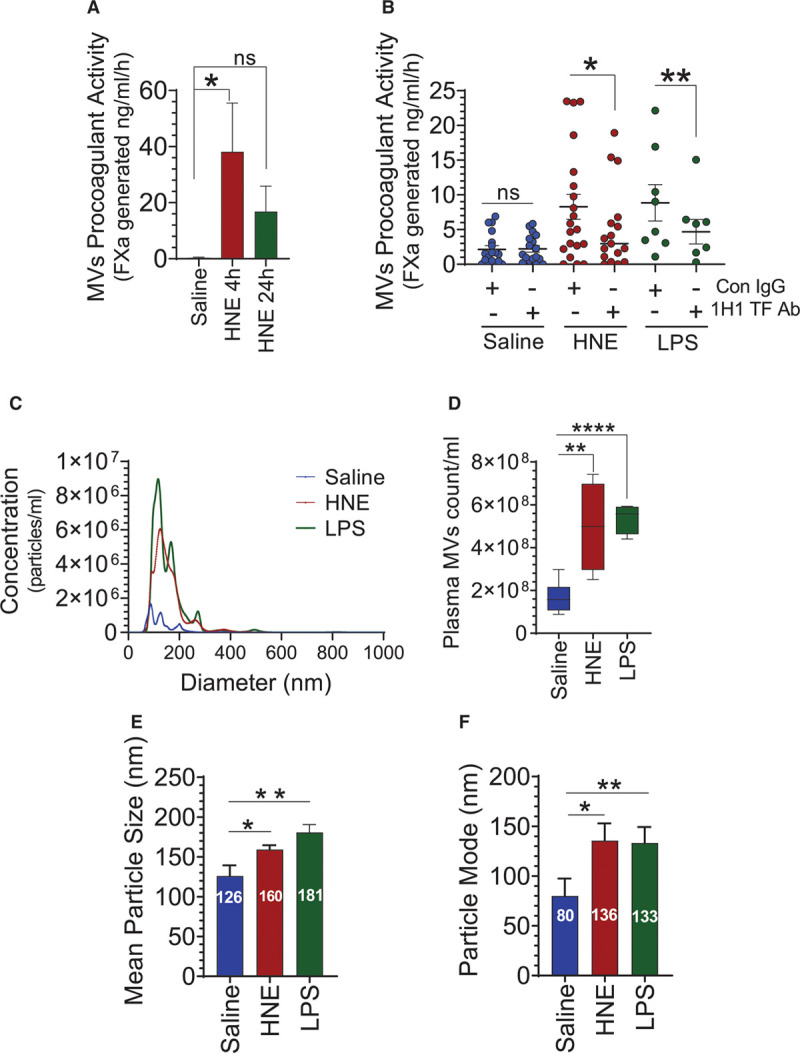
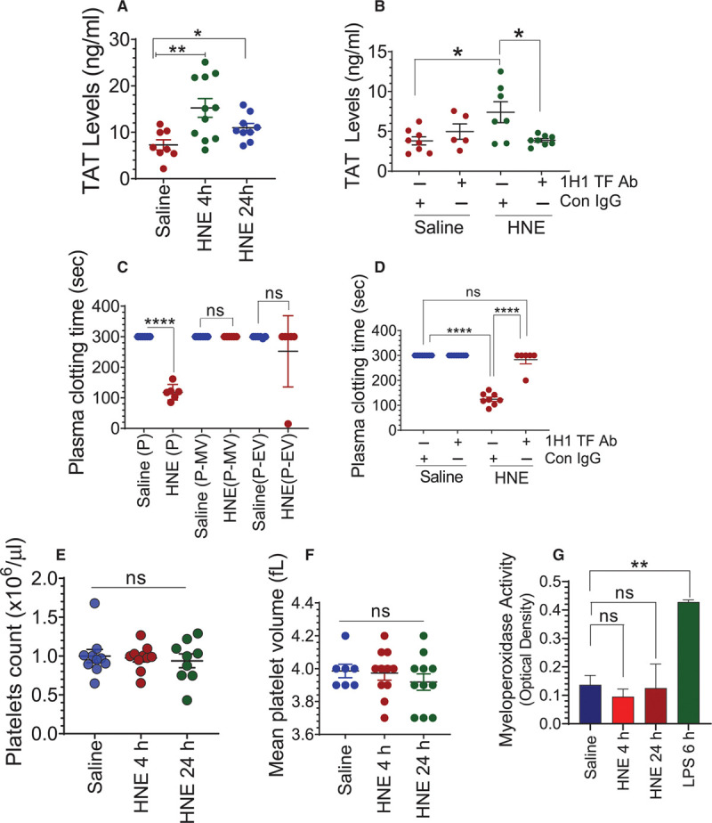
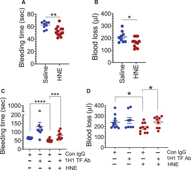
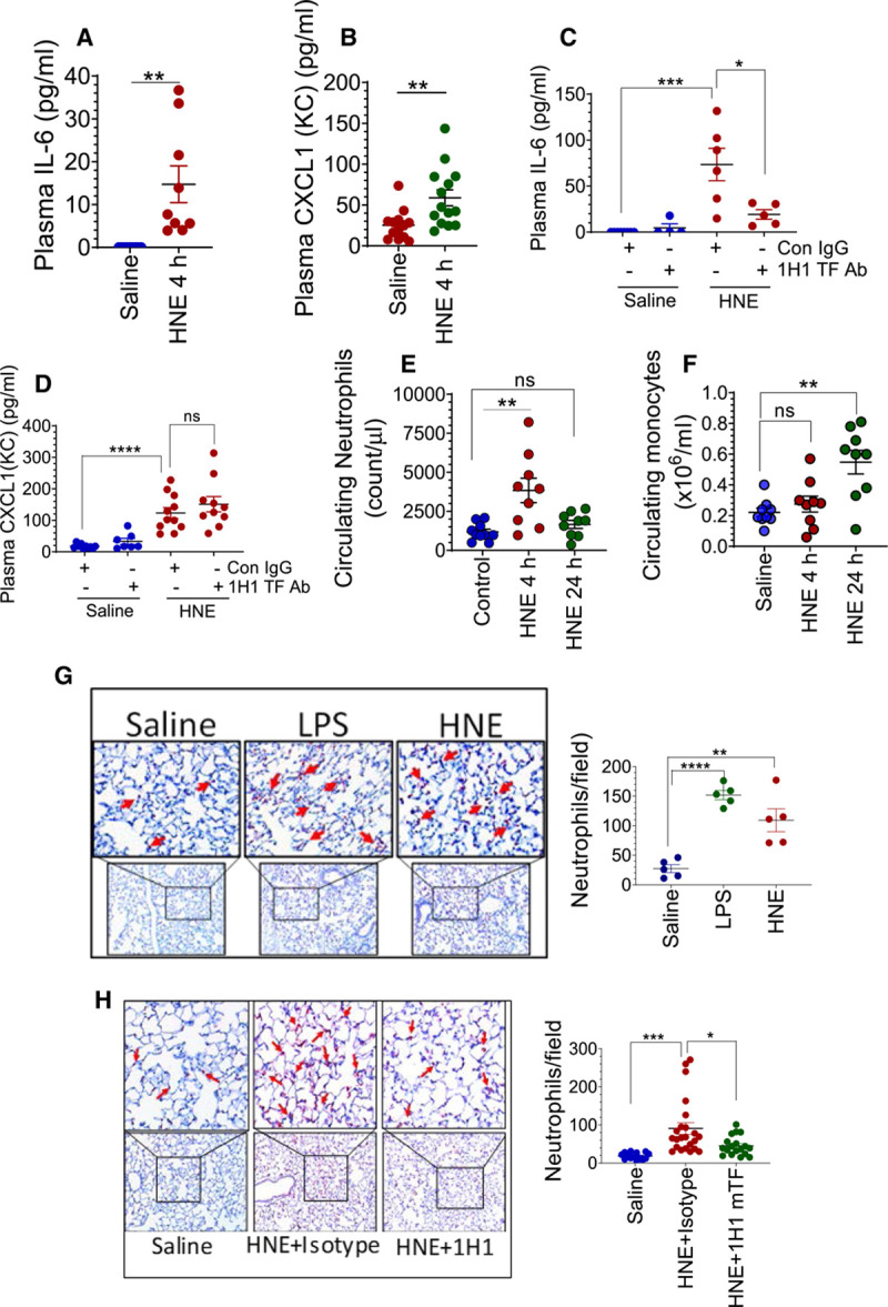
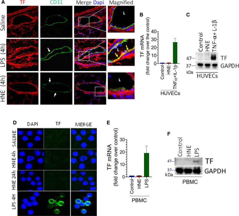
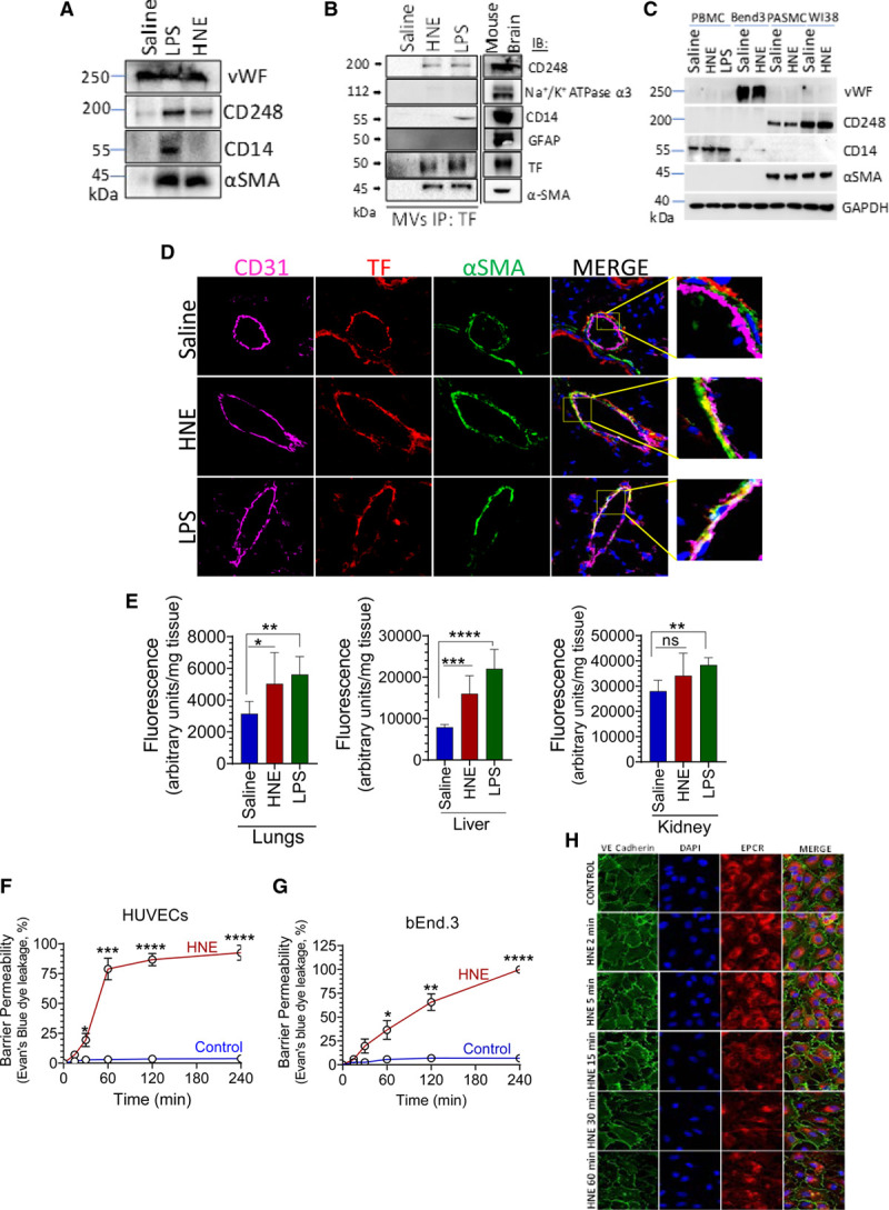
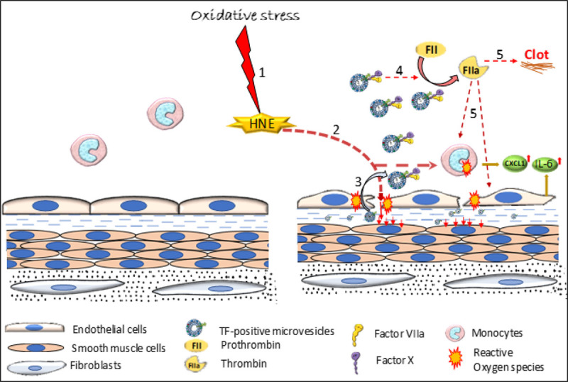
Objective: TF (Tissue factor) plays a key role in hemostasis, but an aberrant expression of TF leads to thrombosis. The objective of the present study is to investigate the effect of 4-hydroxy-2-nonenal (HNE), the most stable and major oxidant produced in various disease conditions, on the release of TF+ microvesicles into the circulation, identify the source of TF+ microvesicles origin, and assess their effect on intravascular coagulation and inflammation. Approach and Results: C57BL/6J mice were administered with HNE intraperitoneally, and the release of TF+ microvesicles into circulation was evaluated using coagulation assays and nanoparticle tracking analysis. Various cell-specific markers were used to identify the cellular source of TF+ microvesicles. Vascular permeability was analyzed by the extravasation of Evans blue dye or fluorescein dextran. HNE administration to mice markedly increased the levels of TF+ microvesicles and thrombin generation in the circulation. HNE administration also increased the number of neutrophils in the lungs and elevated the levels of inflammatory cytokines in plasma. Administration of an anti-TF antibody blocked not only HNE-induced thrombin generation but also HNE-induced inflammation. Confocal microscopy and immunoblotting studies showed that HNE does not induce TF expression either in vascular endothelium or circulating monocytes. Microvesicles harvested from HNE-administered mice stained positively with CD248 and α-smooth muscle actin, the markers that are specific to perivascular cells. HNE was found to destabilize endothelial cell barrier integrity.

Conclusions: HNE promotes the release of TF+ microvesicles from perivascular cells into the circulation. HNE-induced increased TF activity contributes to intravascular coagulation and inflammation.

Keywords: HNE; hemostasis; inflammation; neutrophils; oxidative stress; thrombosis; tissue factor.


Conflict of interest statement

None.

Figures

Figure 1.
Figure 1.
4-hydroxy-2-nonenal (HNE) promotes the release of TF+ (tissue factor) microvesicles. A, C57BL/6J mice were administered with HNE (10 mg/kg, IP) and 24 h following HNE administration, blood was collected via the submandibular vein into citrate anticoagulant. Microvesicles (MVs) were isolated from plasma and their procoagulant activity was measured in FVIIa (factor VIIa) activation of FX. B, MVs isolated from the plasma of saline-, HNE (10 mg/kg)-, or lipopolysaccharide (LPS; 10 mg/kg)-challenged mice (for 4 h) were incubated with control isotype IgG (Con IgG) or 1H1 murine TF mAb (1H1 TF Ab; 10 µg/mL) for 30 min before their procoagulant activity was measured in FX activation assay. C through F, MVs isolated from the plasma of saline-, HNE-, or LPS-administered mice were characterized by nanoparticle tracking analysis using NanoSight NS300. MVs number (C and D) and diameter (C, E, and F) were determined. *P<0.05; **P<0.01; and ****P<0.0001; ns, no statistically significant difference.
Figure 2.
Figure 2.
4-hydroxy-2-nonenal (HNE) activates intravascular blood coagulation in a TF (tissue factor)-dependent manner. A, C57BL/6J wild-type mice were administered with saline or HNE (10 mg/kg, IP). Four and 24 h following HNE administration, blood was collected into citrate anticoagulant via submandibular vein puncture. Levels of thrombin-antithrombin (TAT) complexes in the plasma were determined using ELISA. B, Mice were administered with isotype control IgG (Con IgG) or 1H1 murine TF mAb (1H1 TF Ab) immediately before HNE administration and 2 h following HNE administration (2 mg/kg, IP). Four hours following HNE administration, blood was collected from mice, and TAT levels in plasma were measured. C, Plasma (P), plasma depleted of microvesicle (MVs; P-MV) or plasma depleted of all extracellular vesicles (P-EV) of saline- or HNE-treated mice (for 4 h) were recalcified and the clotting times were measured using a semi-automated coagulizer (the maximum assay duration was 300 s). D, Plasma from saline- or HNE-treated mice (for 4 h) were incubated with either control IgG (Con IgG) or 1H1 murine TF mAb (1H1 TF Ab; 10 µg/mL) for 30 min, and then plasma was recalcified to measure the clotting time. E and F, Platelet count and mean platelet volumes were analyzed in an aliquot of blood collected from saline- or HNE-treated mice using HEMAVET. G, Myeloperoxidase activity levels in the plasma obtained from saline-, HNE-, or lipopolysaccharide (LPS)-administered mice. *P<0.05; **P<0.01; and ****P<0.0001; ns, no statistically significant difference.
Figure 3.
Figure 3.
4-hydroxy-2-nonenal (HNE) accelerates coagulation. A and B, C57BL/6J wild-type mice were administered with saline or HNE (10 mg/kg, IP). Four hours after HNE administration, bleeding was initiated by the saphenous vein incision. The average bleeding times were calculated from the number of hemostatic plugs formed in a 30-min bleeding period (A). The volume of blood leaked from the wound site was adsorbed onto Kimwipes for the entire duration of 30 min, and the blood loss was determined by extracting the hemoglobin from wipes and measuring it against known standards (B). C and D, Wild-type mice were administered with control IgG or 1H1 anti-murine TF (tissue factor) mAb (2 mg/kg) just before HNE administration and 2 h after HNE administration. Four hours after HNE administration, mice were subjected to the saphenous vein injury and the bleeding time (C) and the blood loss (D) were determined as described above. *P<0.05; **P<0.01; ***P<0.001; and ****P<0.0001.
Figure 4.
Figure 4.
4-hydroxy-2-nonenal (HNE) induces TF (tissue factor)-dependent proinflammatory responses in mice. Cytokines IL (interleukin)-6 (A) and CXCL1 (B) levels in the plasma of saline- or HNE-challenged (for 4 h) wild-type mice. C and D, Effect of murine TF antibody administration on HNE-induced increase in IL-6 (C) and CXCL1 (D) levels in plasma. Mice were administered with control isotype IgG or 1H1 murine TF mAb as described in Figure 2. E and F, HNE induces an increase in neutrophil and monocyte number in circulating blood. Wild-type mice were challenged with saline or HNE. Four and 24 h following HNE administration, the number of neutrophils (E) and monocytes (F) in blood were counted using HEMAVET. G and H, HNE induces neutrophil infiltration into the lungs, and the administration of murine TF antibody attenuates the HNE-induced response. G, Lung tissue sections from saline-, lipopolysaccharide (LPS)-, or HNE-treated mice (for 4 h) were immunostained for neutrophil marker Ly6G to detect neutrophil infiltration (left) and the number neutrophils in each field were counted (right). H, Mice were treated with isotype control IgG or 1H1 anti-murine TF antibody before HNE administration, as described in Figure 2. Lung tissue sections from saline- or HNE-administered mice (for 4 h) were stained for Ly6G (left), and the number of neutrophils in a field were counted (right). *P<0.05; **P<0.01; ***P<0.001; and ****P<0.0001; ns, no statistically significant difference.
Figure 5.
Figure 5.
4-hydroxy-2-nonenal (HNE) does not induce TF (tissue factor) expression in vascular cells. A, C57BL/6J mice were injected with saline, HNE (10 mg/kg), or lipopolysaccharide (LPS; 10 mg/kg). After 4 h, the lung tissues were harvested and immunostained with endothelial cell marker CD31 and TF. Images were focused on the immunostaining of blood vessels. Green fluorescence represents CD 31 staining, whereas red fluorescence indicates TF staining. White arrow marks point out endothelial denudation. Yellow arrows on merged images indicate TF staining on the endothelium. L, a lumen of the blood vessel. B and C, Monolayers of human umbilical vein endothelial cells (HUVECs) were treated with saline (control), HNE (40 µmol/L), or TNF (tumor necrosis factor)-α+IL (interleukin)-1β (10 ng/mL, each) for 2 or 4 h to analyze TF mRNA levels by quantitative real-time polymerase chain reaction (RT-PCR; B) or TF protein by western blot analysis (C), respectively. D through F, C57BL/6J mice were treated with saline, HNE (10 mg/kg), or LPS (10 mg/kg). Four hours following HNE or LPS administration, blood was collected, and PBMCs were isolated. TF expression was analyzed by confocal microscopy (D), measuring TF mRNA by quantitative RT-PCR (E), or TF protein by western blot analysis (F). In D, cell nuclei were stained with DAPI. Cells shown are monocytes (other mononuclear cells were much smaller in size than monocytes and were stained weakly with DAPI and negative for TF, and thus not readily visible).
Figure 6.
Figure 6.
4-hydroxy-2-nonenal (HNE) releases TF+ (tissue factor) microvesicles from perivascular cells and induces vascular barrier disruption. A, C57BL/6J mice were injected with saline, HNE (10 mg/kg), or lipopolysaccharide (LPS; 10 mg/kg). Microvesicle (MVs) harvested from the plasma of saline-, HNE-, or LPS-administered mice were lysed in an equal volume of 1 X SDS lysis buffer and subjected to immunoblot analysis for VWF (Von Willebrand factor), CD248, CD14, or α-SMA. B, MVs harvested from the plasma of saline-, HNE-, or LPS-administered mice were immunoprecipitated with anti-murine TF antibodies, and the immunoprecipitates were analyzed for the presence cell-specific markers by immunoblot analysis. Mouse whole brain lysate was used as a positive control. C, Cell extracts of PBMCs from saline-, HNE-, or LPS-treated mice, bEnd.3 cells, pulmonary artery smooth muscle cells (PASMC), and WI38 fibroblasts treated with or without HNE (20 µmol/L) for 4 h were subjected to immunoblot analysis and probed for VWF, CD248, CD14, or α-SMA. D, Lung tissue sections prepared from the saline-, HNE-, or LPS-treated mice were immunostained for CD31 (Magenta), TF (Red), and α-SMA (Green). A small portion of the merged image was enlarged to show colocalization. E, Three hours following saline-, HNE-, or LPS administration, mice were administered with fluorescein dextran (10 mg/kg) via the tail vein. One hour following fluorescein dextran administration, mice were euthanized, perfused with saline, and tissues were harvested. The extent of fluorescein dextran entered in tissues was determined by measuring the fluorescence intensity of tissue extracts. F and G, Human umbilical vein endothelial cell (HUVEC) or bEnd.3 endothelial cells were cultured in 24-transwell plates for 4 d to form tight confluent monolayers. Cells were treated with HNE (20 µmol/L) for varying periods. In controls, cells were treated for 4 h with a control vehicle. At the end of treatment, Evan blue dye was added to the apical chamber, and the amount of dye leaked into the lower chamber at 10 min was read in a spectrophotometer. H, HUVECs were cultured on coverslips and treated with HNE (20 µmol/L) for varying periods (2–60 min). Following HNE treatment, the cells were washed and fixed with 2% paraformaldehyde. The distribution of VE-cadherin was analyzed by immunostaining the cells with anti-VE-cadherin antibodies, followed by confocal microscopy. EPCR antibodies and DAPI were used to stain the cell surface and nucleus, respectively. *P<0.05; **P<0.01; ***P<0.001; and ****P<0.0001; ns, no statistically significant difference.
Figure 7.
Figure 7.
Schematic representation of 4-hydroxy-2-nonenal (HNE)-mediated intravascular coagulation and inflammation. HNE, a lipid peroxidation product that is generated in cardiovascular diseases, sepsis, and other disease conditions, destabilizes endothelial barrier integrity and induces the release of TF+ (tissue factor) microvesicles (MVs) from perivascular cells. Due to the barrier disruption, TF+ MVs from perivascular cells could enter the bloodstream and induce intravascular coagulation and thrombosis. Signaling induced by downstream proteases, such as thrombin, or TF-FVIIa (factor VIIa)-mediated direct signaling in perivascular cells could promote inflammation by inducing inflammatory cytokines and infiltration of neutrophils into tissues. Step 1: Oxidative stress generates HNE; Step 2: HNE induces ROS generation in monocytes and endothelium, and increases vascular permeability; Step 3: Breach in the vasculature leads to leakage of perivascular cells-derived TF+ MVs into the circulation; Step 4: TF+ MVs promote thrombin generation; Step 5: Thrombin induces clot formation and possibly upregulates cytokines expression.

Comment in

References

    1. Rapaport SI, Rao LV. The tissue factor pathway: how it has become a “prima ballerina”. Thromb Haemost. 1995;74:7–17 - PubMed
    1. Grover SP, Mackman N. Tissue factor: an essential mediator of hemostasis and trigger of thrombosis. Arterioscler Thromb Vasc Biol. 2018;38:709–725. doi: 10.1161/ATVBAHA.117.309846 - PubMed
    1. Drake TA, Morrissey JH, Edgington TS. Selective cellular expression of tissue factor in human tissues. Implications for disorders of hemostasis and thrombosis. Am J Pathol. 1989;134:1087–1097 - PMC - PubMed
    1. Fleck RA, Rao LV, Rapaport SI, Varki N. Localization of human tissue factor antigen by immunostaining with monospecific, polyclonal anti-human tissue factor antibody. Thromb Res. 1990;59:421–437. doi: 10.1016/0049-3848(90)90148-6 - PubMed
    1. Contrino J, Hair G, Kreutzer DL, Rickles FR. In situ detection of tissue factor in vascular endothelial cells: correlation with the malignant phenotype of human breast disease. Nat Med. 1996;2:209–215. doi: 10.1038/nm0296-209 - PubMed

Publication types

MeSH terms