. 2020 Jan-Dec:29:963689720963948.
doi: 10.1177/0963689720963948.

circRNA hsa_circ_104566 Sponged miR-338-3p to Promote Hepatocellular Carcinoma Progression

Affiliations

circRNA hsa_circ_104566 Sponged miR-338-3p to Promote Hepatocellular Carcinoma Progression

Guangming Liu et al. Cell Transplant. 2020 Jan-Dec.

Retraction in

  • Retraction Notice.
    [No authors listed] [No authors listed] Cell Transplant. 2024 Jan-Dec;33:9636897241298459. doi: 10.1177/09636897241298459. Cell Transplant. 2024. PMID: 39601276 Free PMC article. No abstract available.

Abstract

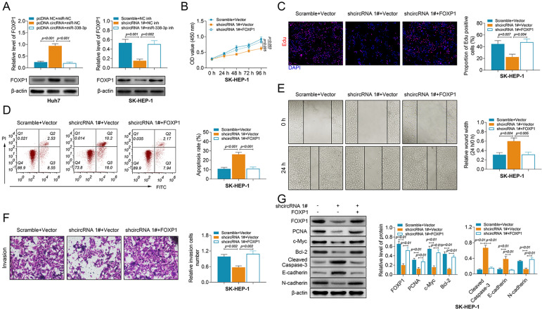
Circular RNAs (circRNAs) could sponge micro-RNAs (miRNAs) to regulate tumor progression of hepatocellular carcinoma (HCC). Hsa_circ_104566 contributes to papillary thyroid carcinoma progression. However, the tumorigenic mechanism of hsa_circ_104566 on HCC remains enigmatic. The role of hsa_circ_104566 on HCC was therefore evaluated in this study. First, the high expression of hsa_circ_104566 was found in HCC tissues, which was significantly associated with poor prognosis in HCC patients. Second, Hsa_circ_104566 promoted HCC progression by decreasing apoptosis and E-cadherin, while increasing cell viability, proliferation, migration, invasion, and N-cadherin. On the other hand, HCC progression was suppressed by knockdown of hsa_circ_104566. Hsa_circ_104566 could target miR-338-3p, and its expression was negatively correlated with miR-338-3p in HCC patients. Moreover, miR-338-3p suppressed protein expression of Forkhead box protein 1 (FOXP1) and had a negative correlation with FOXP1 in HCC patients. Functional assay further indicated that the promotion of HCC progression by hsa_circ_104566 was reversed by miR-338-3p, and miR-338-3p inhibitor could counteract the effect of hsa_circ_104566 knockdown on the suppression of HCC progression. In vivo assay indicated that hsa_circ_104566 knockdown suppressed HCC tumor growth and metastasis. In conclusion, hsa_circ_104566 sponged miR-338-3p to promote HCC progression, providing a potential therapeutic target for cancer intervention.

Keywords: FOXP1; HCC; hsa_circ_104566; miR-338-3p; progression.


Conflict of interest statement

Declaration of Conflicting Interests: The author(s) declared no potential conflicts of interest with respect to the research, authorship, and/or publication of this article.

Figures

Fig. 1.
Fig. 1.
Expression of hsa_circ_104566 was upregulated in HCC. (A) The expression levels of circRNA hsa_circ_104566 in HCC tissues, including high pathological grade (TNM III and IV) and low grade (TNM I and II), and adjacent noncancer tissues measured by qRT-PCR (N = 87). (B) Overall survival analysis of HCC patients with high and low levels of hsa_circ_104566 expression. (C) The expression levels of hsa_circ_104566 in HCC cell lines (SK-HEP-1, HLE, SNU449, Hep-3B, and Huh7) and MIHA measured by qRT-PCR. (D) Hsa_circ_104566 was resistant to RNase R digestion compared with linear PSD3 in both Huh7 and SK-HEP-1 cells. (E) Expression levels of hsa_circ_104566, GAPDH, and U6 in cytoplasm or nucleus of Huh7 and SK-HEP-1 cells measured by qRT-PCR, suggesting the mainly cytoplasm subcellular localization of hsa_circ_104566. circRNA: circular RNA; GAPDH: glyceraldehyde 3-phosphate dehydrogenase; HCC: hepatocellular carcinoma; qRT-PCR: quantitative real-time polymerase chain reaction; TNM: tumor node metastasis.
Fig. 2.
Fig. 2.
Hsa_circ_104566 promoted cell proliferation of hepatocellular carcinoma. (A) Efficiency of pcDNA-hsa_circ_104566 in Huh7 cells and sh-hsa_circ_104566 1# or 2# in SK-HEP-1 cells was measured by quantitative real-time polymerase chain reaction. (B) The influence of hsa_circ_104566 on cell viability of Huh7 and SK-HEP-1 cells measured by the cell counting kit-8. (C) The influence of hsa_circ_104566 on proliferation of Huh7 and SK-HEP-1 cells detected by the EdU staining assay. (D) The influence of hsa_circ_104566 on apoptosis of Huh7 and SK-HEP-1 cells detected by flow cytometry.
Fig. 3.
Fig. 3.
Hsa_circ_104566 promoted cell migration and invasion of hepatocellular carcinoma (HCC). (A) The influence of hsa_circ_104566 on cell migration of Huh7 and SK-HEP-1 cells measured by wound healing assay. (B) The influence of hsa_circ_104566 on cell invasion of Huh7 and SK-HEP-1 cells measured by Transwell assay. (C) The influence of hsa_circ_104566 on protein expression of E-cadherin and N-cadherin in Huh7 and SK-HEP-1 cells measured by immunofluorescence.
Fig. 4.
Fig. 4.
Hsa_circ_104566 bound miR-338-3p in HCC cells. (A) miR-338-3p was predicted as the potential binding target of hsa_circ_104566 via circular RNA Interactome (https://circinteractome.nia.nih.gov/). (B) The influence of miR-338-3p mimics on luciferase activities of pmirGLO-wt-hsa_circ_104566 or pmirGLO-mut-hsa_circ_104566 in Huh7 and SK-HEP-1 cells. (C) Subcellular localization of hsa_circ_104566 and miR-338-3p in Huh7 and SK-HEP-1 cells examined via RNA-fluorescence in situ hybridization. (D) The influence of hsa_circ_104566 on miR-338-3p expression in Huh7 and SK-HEP-1 cells. (E) The expression of miR-338-3p in HCC tissues and adjacent noncancer tissues measured by qRT-PCR (N = 87). (F) Negative correlation between miR-338-3p and hsa_circ_104566 in HCC patients. HCC: hepatocellular carcinoma; qRT-PCR: quantitative real-time polymerase chain reaction.
Fig. 5.
Fig. 5.
miR-338-3p bound FOXP1 in HCC cells. (A) FOXP1 was predicted as a potential miR-338-3p binding targets via Targetscan (http://www.targetscan.org/vert_71/). (B) The influence of miR-338-3p mimics on luciferase activities of pmirGLO-wt-FOXP1 or pmirGLO-mut-FOXP1 in Huh7 and SK-HEP-1 cells. (C) The influence of hsa_circ_104566 on protein expression of FOXP1 in Huh7 and SK-HEP-1 cells measured by western blot. (D) Immunohistochemical analysis of FOXP1 in HCC tissues and adjacent noncancer tissues. (E) The mRNA expression levels of FOXP1 in HCC tissues and adjacent noncancer tissues measured by qRT-PCR (N = 87). (F) Negative correlation between miR-338-3p and FOXP1 in HCC patients. Positive correlation between hsa_circ_104566 and FOXP1 in HCC patients. (G) Protein expression levels of SOX12 in clear cell renal cell carcinoma tissues and adjacent noncancer tissues measured by western blot. FOXP1: forkhead box protein 1; HCC: hepatocellular carcinoma; qRT-PCR: quantitative real-time polymerase chain reaction.
Fig. 6.
Fig. 6.
Hsa_circ_104566 promoted malignant behaviors of HCC through miR-338-3p/FOXP1. (A) The influence of hsa_circ_104566 and miR-338-3p on protein expression of FOXP1 in Huh7 and SK-HEP-1 cells measured by western blot. (B) The influence of hsa_circ_104566 and FOXP1 on cell viability of Huh7 and SK-HEP-1 cells measured by cell counting kit-8. (C) The influence of hsa_circ_104566 and FOXP1 on cell proliferation of Huh7 and SK-HEP-1 cells measured by the EdU staining assay. (D) The influence of hsa_circ_104566 and FOXP1 on cell apoptosis of Huh7 and SK-HEP-1 cells measured by flow cytometry. (E) The influence of hsa_circ_104566 and FOXP1 on cell migration of Huh7 and SK-HEP-1 cells measured by wound healing assay. (F) The influence of hsa_circ_104566 and FOXP1 on cell invasion of Huh7 and SK-HEP-1 cells measured by Transwell assay. (G) The influence of hsa_circ_104566 and FOXP1 on protein expression levels of FOXP1, PCNA, c-Myc, Bcl-2, cleaved caspase-3, E-cadherin, and N-cadherin in Huh7 and SK-HEP-1 cells measured by western blot. FOXP1: forkhead box protein 1; HCC: hepatocellular carcinoma; PCNA: proliferating cell nuclear antigen.
Fig. 7.
Fig. 7.
Knockdown of hsa_circ_104566 attenuated tumor growth. (A) Expression levels of hsa_circ_104566 or miR-338-3p in tissues from mice in shcircRNA 1# and Scramble groups. (B) The effect of sh-hsa_circ_104566 on hepatocellular carcinoma tumor growth in xenograft tumor mice. The tumor volume and weight were calculated. (C) Representative images of lung tissues from mice in shcircRNA 1# and Scramble groups. (D) Hematoxylin and eosin staining revealed morphologic features of lung tissues from mice in shcircRNA 1# and Scramble groups. The lung metastases have been shown by the arrows. (E) Immunohistochemistry staining was used to determine the expression of FOXP1, Ki-67, E-cadherin, and N-cadherin in tissues from mice in shcircRNA 1# and Scramble groups. Scale bar: 200 μm.

References

    1. Ziogas IA, Tsoulfas G. Advances and challenges in laparoscopic surgery in the management of hepatocellular carcinoma. World J Gastrointest Surg. 2017;9(12):233–245. - PMC - PubMed
    1. Ruzic M, Pellicano R, Fabri M, Luzza F, Boccuto L, Brkic S, Abenavoli L. Hepatitis C virus-induced hepatocellular carcinoma: a narrative review. Panminerva Med. 2018;60(4):185–191. - PubMed
    1. Petruzziello A. Epidemiology of Hepatitis B Virus (HBV) and Hepatitis C Virus (HCV) related hepatocellular carcinoma. Open Virol J. 2018;12:26–32. - PMC - PubMed
    1. Cao X, Quanlin A, Assaraf YG, Xiangdong W. Retinoids offer new and promising cancer therapeutic avenues. J Mol Clin Med. 2019;2(2):23–28.
    1. Jeck WR, Sharpless NE. Detecting and characterizing circular RNAs. Nat Biotechnol. 2014;32(5):453–461. - PMC - PubMed

Publication types

MeSH terms