. 2020 Dec;9(1):2322-2332.
doi: 10.1080/22221751.2020.1833687.

SARS-CoV-2 infection, disease and transmission in domestic cats

Affiliations

SARS-CoV-2 infection, disease and transmission in domestic cats

Natasha N Gaudreault et al. Emerg Microbes Infect. 2020 Dec.

Abstract

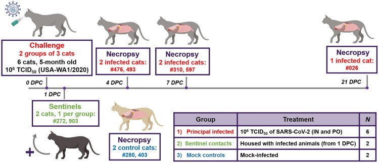
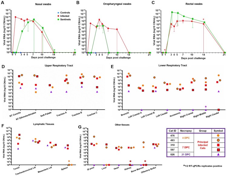
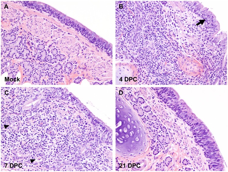
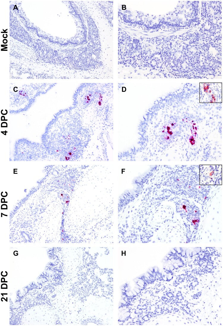
Severe Acute Respiratory Syndrome Coronavirus 2 (SARS-CoV-2) is the cause of Coronavirus Disease 2019 (COVID-19) and responsible for the current pandemic. Recent SARS-CoV-2 susceptibility studies in cats show that the virus can replicate in these companion animals and transmit to other cats. Here, we present an in-depth study of SARS-CoV-2 infection, disease and transmission in domestic cats. Cats were challenged with SARS-CoV-2 via intranasal and oral routes. One day post challenge (DPC), two sentinel cats were introduced. Animals were monitored for clinical signs, clinicopathological abnormalities and viral shedding. Postmortem examinations were performed at 4, 7 and 21 DPC. Viral RNA was not detected in blood but transiently in nasal, oropharyngeal and rectal swabs and bronchoalveolar lavage fluid as well as various tissues. Tracheobronchoadenitis of submucosal glands with the presence of viral RNA and antigen was observed in airways of the infected cats. Serology showed that both, principals and sentinels, developed antibodies to SARS-CoV-2. All animals were clinically asymptomatic during the course of the study and capable of transmitting SARS-CoV-2 to sentinels. The results of this study are critical for understanding the clinical course of SARS-CoV-2 in a naturally susceptible host species, and for risk assessment.

Keywords: COVID-19; SARS-CoV-2; cats; felines; susceptibility; transmission.


Conflict of interest statement

No potential conflict of interest was reported by the author(s).

Figures

Figure 1.
Figure 1.
Study design. Ten cats were placed into three groups. Group 1 (principal infected animals) consisted of six cats (three cats/housing unit) and was inoculated via intranasal (IN) and oral (PO) routes simultaneously with a total dose of 1 × 106 TCID50 of SARS-CoV-2 in 2 ml DMEM. The cats in Group 2 (n = 2; sentinel contact animals) and Group 3 (n = 2; mock control animals) were housed in a separate room. At 1-day post challenge (DPC), the two cats in Group 2 were co-mingled with the principal infected animals in Group 1 (one cat per cage) and served as sentinel contact controls. The remaining two cats in Group 3 were housed in a separate room and served as mock-infected negative controls. Principal infected animals were euthanized and necropsied at 4 (n = 2), 7 (n = 2) and 21 (n = 1) DPC to evaluate the course of disease. The two negative control animals in Group 3 were euthanized and necropsied at 3 DPC. The remaining three animals from Group 1 (one principal infected animal) and Group 2 (two sentinel contact animals) were maintained for future re-infection studies.
Figure 2.
Figure 2.
Shedding and presence of SARS-CoV-2 RNA in tissues. RT-qPCR was performed on nasal (A), oropharyngeal (B) and rectal swabs (C) collected from principal infected and sentinel cats over the course of the 21-day study, as well as, on the upper respiratory tract (D), lower respiratory tract (E), lymphatic (F) and other various tissues (G) from principal infected cats necropsied at 4, 7 and 21 days post challenge (DPC). Average viral copy number (CN) per mL or per mg tissues are shown. Astrisks (*) indicate 1 out of 2 of the RT-qPCR reactions were below the limit of detection. LN = lymph node; GI = gastrointestinal.
Figure 3.
Figure 3.
Histopathology of bronchi. Histological findings in the main bronchi of mock (A) and SARS-CoV-2 experimentally infected (B-D) cats. Histologic changes and their progression are similar to those observed in the trachea, with multifocal, widespread, mild to moderate lymphocytic and neutrophilic adenitis noted at 4 DPC (B) and 7 DPC (C). Necrotic debris within distorted submucosal glands are indicated with arrowheads (C), and few transmigrating lymphocytes are indicated with an arrow (B). No histologic changes are noted at 21 DPC (D). H&E. Total magnification: 200X.
Figure 4.
Figure 4.
SARS-CoV-2 RNA and antigen detection in bronchi. SARS-CoV-2 tropism in bronchi of mock (A and B) experimentally (C-H) infected cats determined by S-specific RNAscope® in situ hybridization (Fast Red) and anti-N-specific immunohistochemistry (IHC; Fast Red). The viral tropism is limited to glandular and ductular epithelial cells of multifocal, scattered submucosal glands. Viral RNA is detected within infected cells at 4 days post-challenge (DPC; C and D) and, to a lower degree at 7 DPC (E and F). Few scattered glandular epithelial cells are positive for SARS-CoV-2 N antigen by IHC (D and F, insets). No viral RNA or antigen is detected at 21 DPC (G and H). Total magnification: 100X (A, C, E and G) and 200X (B, D, F, H).

References

    1. Fehr AR, Perlman S.. Coronaviruses: an overview of their replication and pathogenesis. Methods Mol Biol. 2015;1282:1–23. - PMC - PubMed
    1. Gorbalenya AE, Baker SC, Baric RS, et al. The species severe acute respiratory syndrome-related coronavirus: classifying 2019-nCoV and naming it SARS-CoV-2. Nature Microbiology. 2020;5(4):536–544. - PMC - PubMed
    1. Fung TS, Liu DX.. Human Coronavirus: host-pathogen Interaction. Annu Rev Microbiol. 2019;73:529–557. - PubMed
    1. Saif LJ. Animal coronaviruses: what can they teach us about the severe acute respiratory syndrome? Rev Sci Tech. 2004;23(2):643–660. - PubMed
    1. Woo PC, Lau SK, Li KS, et al. Genetic relatedness of the novel human group C betacoronavirus to Tylonycteris bat coronavirus HKU4 and Pipistrellus bat coronavirus HKU5. Emerg Microbes Infect. 2012;1(11):e35. - PMC - PubMed

MeSH terms