

Short-term changes in the anterior segment and retina after small incision lenticule extraction
- PMID: 33028265
- PMCID: PMC7539406
- DOI: 10.1186/s12886-020-01668-7
Short-term changes in the anterior segment and retina after small incision lenticule extraction
Abstract
Background: To analyse short-term changes in the anterior segment and retina after small incision lenticule extraction (SMILE).
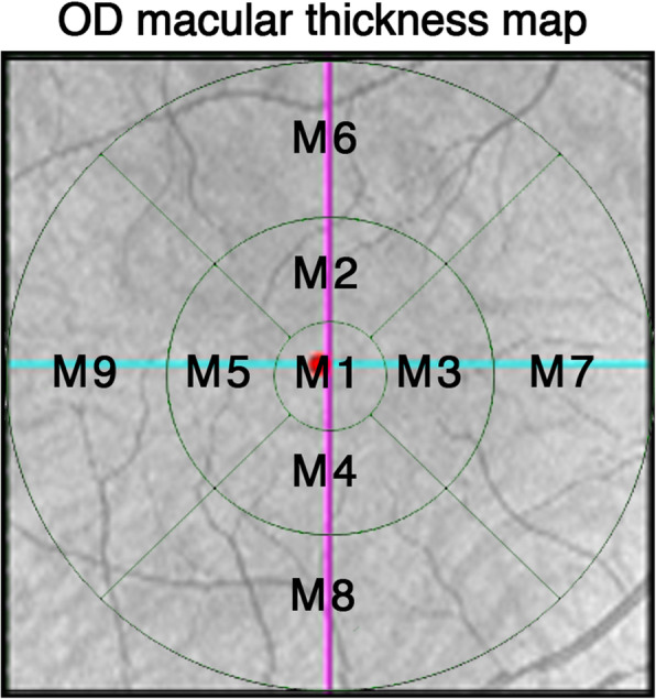
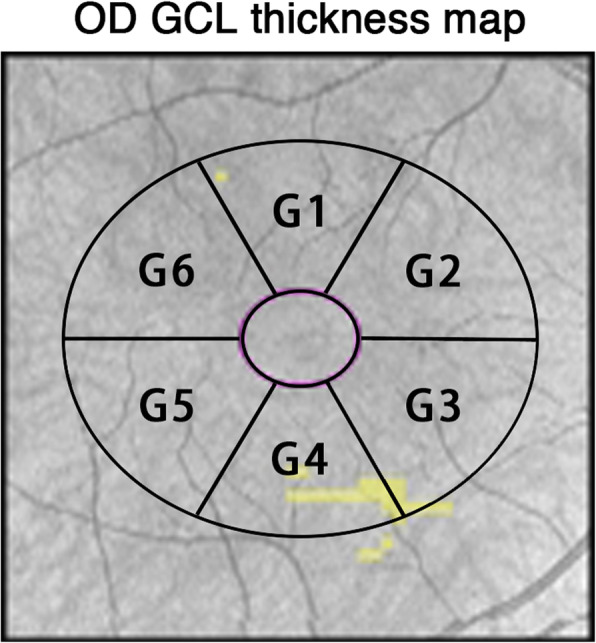
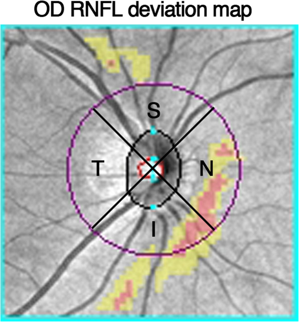
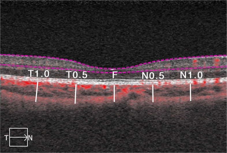
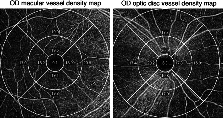
Methods: Patients with myopia scheduled for SMILE were recruited from Ruijin Hospital, Shanghai, China. Basic patient information such as age, sex, and refractive errors was recorded. Ocular measurements were taken before surgery, and 1 day and 1 week after surgery; they included axial length (AL), central corneal thickness (CCT), anterior chamber depth (ACD), lens thickness (LT), white to white (WTW), pupil diameter (PD), macular thickness (MT), ganglion cell layer thickness (GCL), retinal nerve fiber layer thickness (RNFL), choroidal thickness (CT), macular vessel density, and optic disc vessel density.
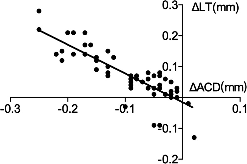
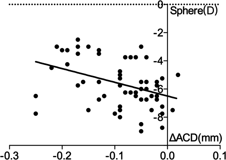
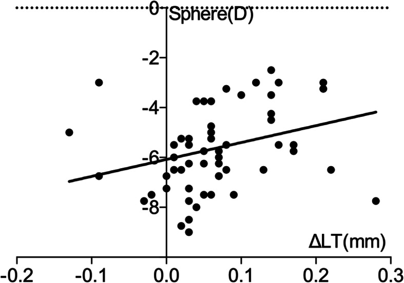
Results: Sixty-one eyes of 31 patients were selected for this study. AL, CCT, ACD, and postoperative PD were significantly reduced (p < 0.05), while LT was thickened after surgery (p < 0.05). MT at the fovea decreased 1 day and 1 week after surgery (p < 0.05). GCL showed no significant changes after surgery. RNFL was unchanged 1 day after surgery, but the inferior sector was thickened 1 week after surgery. CT was thicker at the fovea 1 day after surgery and 1.0 mm from the fovea in the nasal sector 1 week after surgery. Macular vessel density was significantly decreased 1 day after surgery and most recovered in 1 week. Optic disc vessel density decreased at the peripapillary part 1 day after surgery and recovered after 1 week. ΔACD and ΔLT showed no significant correlation 1 day after surgery. ΔACD was negatively correlated with ΔLT and sphere 1 week after surgery (r = - 0.847, p < 0.000; r = - 0.398, p = 0.002). ΔLT was positively correlated with the sphere 1 week after surgery (r = 0.256, p = 0.048).
Conclusion: The anterior segment was the most affected, while the retina also underwent changes with regard to MT, RNFL, CT, macular vessel density, and peripapillary vessel density.
Keywords: Anterior segment; Optical coherence tomography angiography (OCTA); Retina; Small incision lenticule extraction.
Conflict of interest statement
The authors declare that they have no competing interests.
Figures
Similar articles
-
Reliability of Vessel Density Measurements in the Peripapillary Retina and Correlation with Retinal Nerve Fiber Layer Thickness in Healthy Subjects Using Optical Coherence Tomography Angiography.Ophthalmologica. 2018;240(4):183-190. doi: 10.1159/000485957. Epub 2018 Apr 25. Ophthalmologica. 2018. PMID: 29694957
-
[Peripapillary and macular vessel density in eyes with different phases of thyroid-associated ophthalmopathy].Zhonghua Yan Ke Za Zhi. 2020 Nov 11;56(11):824-831. doi: 10.3760/cma.j.cn112142-20191115-00574. Zhonghua Yan Ke Za Zhi. 2020. PMID: 33152840 Chinese.
-
Compensatory Changes in the Anterior Segment and Vascular System of the Eye in Myopic Children After Orthokeratology.Front Pediatr. 2021 Sep 9;9:663644. doi: 10.3389/fped.2021.663644. eCollection 2021. Front Pediatr. 2021. PMID: 34568237 Free PMC article.
-
[Optic nerve morphology and vessel density in eyes with different phases of non-arteritic anterior ischemic optic neuropathy].Zhonghua Yan Ke Za Zhi. 2019 Sep 11;55(9):677-686. doi: 10.3760/cma.j.issn.0412-4081.2019.09.010. Zhonghua Yan Ke Za Zhi. 2019. PMID: 31495153 Chinese.
-
Ganglion cell-inner plexiform layer and retinal nerve fiber layer thickness according to myopia and optic disc area: a quantitative and three-dimensional analysis.BMC Ophthalmol. 2017 Mar 11;17(1):22. doi: 10.1186/s12886-017-0419-1. BMC Ophthalmol. 2017. PMID: 28283025 Free PMC article.
Cited by
-
Change in choroid thickness and vascularity index associated with accommodation and aberration after small-incision lenticule extraction.Int J Ophthalmol. 2025 Apr 18;18(4):672-682. doi: 10.18240/ijo.2025.04.14. eCollection 2025. Int J Ophthalmol. 2025. PMID: 40256035 Free PMC article.
-
Analysis of risk and protective factors associated with retinal nerve fiber layer defect in a Chinese adult population.Int J Ophthalmol. 2023 Mar 18;16(3):427-433. doi: 10.18240/ijo.2023.03.14. eCollection 2023. Int J Ophthalmol. 2023. PMID: 36935788 Free PMC article.
-
Quantitative changes in iris and retinal blood flow after femtosecond laser-assisted in situ keratomileusis and small-incision lenticule extraction.Front Med (Lausanne). 2022 Aug 5;9:862195. doi: 10.3389/fmed.2022.862195. eCollection 2022. Front Med (Lausanne). 2022. PMID: 35991655 Free PMC article.
References
MeSH terms
LinkOut - more resources
Full Text Sources
Medical
