

Transurethral resection of a bladder trigone leiomyoma: a rare case report
- PMID: 33028269
- PMCID: PMC7542762
- DOI: 10.1186/s12894-020-00722-2
Transurethral resection of a bladder trigone leiomyoma: a rare case report
Abstract
Background: Bladder leiomyomas are rare and benign tumors of the bladder. They account for 0.43% of all bladder tumors, and only 250 cases have been reported in English literature. Based on the size and localization of the lesion, their symptoms vary considerably. Women seem to be more affected, and obstructive symptoms predominate. Surgical treatment is almost always highly effective, leaving a low recurrence rate.
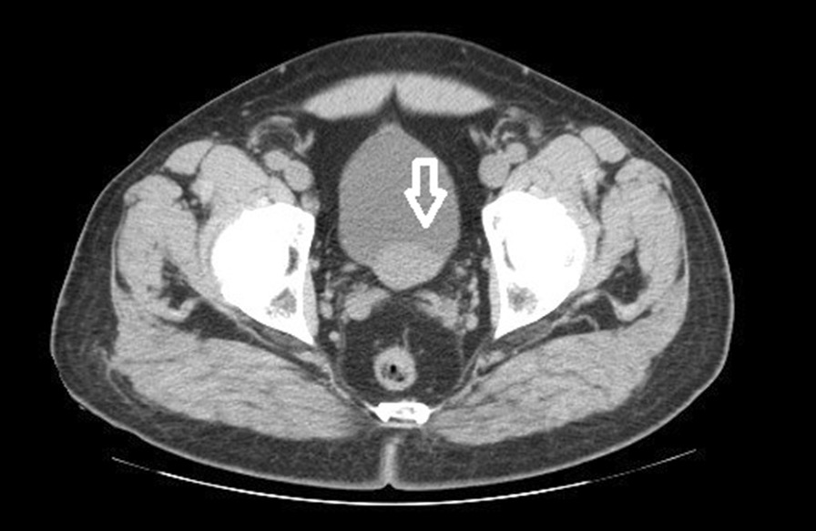
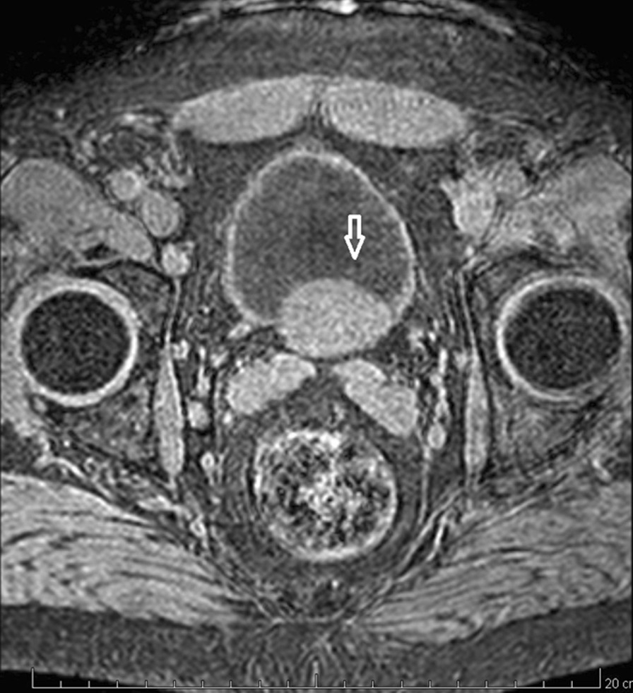
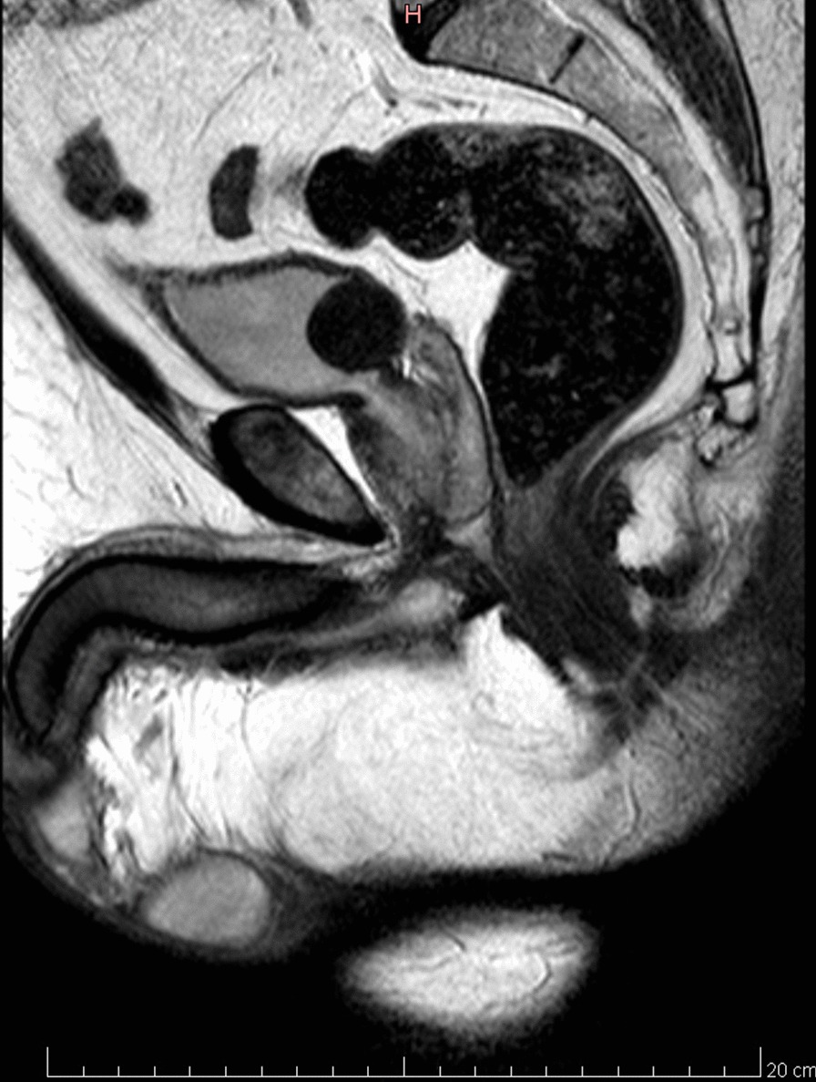
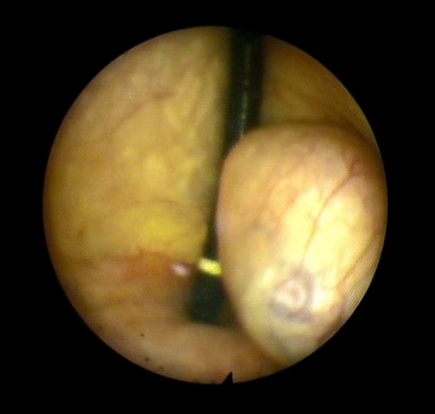
Case presentation: We present a clinical case of a 52-year old man with macroscopic hematuria and obstructive lower urinary tract symptoms due to a large bladder trigone leiomyoma. CT and MRI showed a well-defined large bladder leiomyoma and cystoscopy established the initial findings. The patient underwent successful transurethral resection of the lesion, and pathology findings confirmed the diagnosis.
Conclusions: This case report demonstrates that transurethral resection of a large bladder trigone leiomyoma is a feasible and successful procedure. Long term follow-up proves that there is neither scarring distortion of the bladder trigone area nor damage in the ureteral orifices, even though there was a thorough removal of the trigone wall.
Keywords: Bladder; Leiomyoma; Obstruction; Transurethral resection; Trigone.
Conflict of interest statement
The authors declare that they have no competing interests.
Figures
Similar articles
-
Transurethral Resection of a Large Urinary Bladder Leiomyoma: A Rare Case Report.Urol J. 2017 Jul 2;14(4):4052-4054. Urol J. 2017. PMID: 28670675
-
Transurethral resection of a giant bladder leiomyoma.BMC Urol. 2025 Apr 21;25(1):97. doi: 10.1186/s12894-025-01774-y. BMC Urol. 2025. PMID: 40259295 Free PMC article.
-
Endoscopic partial cystectomy for bladder leiomyoma using retroperitoneoscopic and transurethral procedures.Int J Urol. 2002 Mar;9(3):190-2. doi: 10.1046/j.1442-2042.2002.00447.x. Int J Urol. 2002. PMID: 12010334
-
[Leiomyoma of the urinary bladder: report of three cases].Hinyokika Kiyo. 2003 Nov;49(11):671-4. Hinyokika Kiyo. 2003. PMID: 14719456 Review. Japanese.
-
Bladder leiomyoma presenting as dyspareunia: Case report and literature review.Medicine (Baltimore). 2016 Jul;95(28):e3971. doi: 10.1097/MD.0000000000003971. Medicine (Baltimore). 2016. PMID: 27428187 Free PMC article. Review.
Cited by
-
Characteristics and outcomes in bladder Leiomyoma management: a systematic review of case reports and case series from the past 20 years.BMC Urol. 2024 Nov 13;24(1):252. doi: 10.1186/s12894-024-01624-3. BMC Urol. 2024. PMID: 39538239 Free PMC article.
-
Imaging findings of atypical leiomyoma of urinary bladder simulating ureterocele.J Surg Case Rep. 2022 Jun 22;2022(6):rjac256. doi: 10.1093/jscr/rjac256. eCollection 2022 Jun. J Surg Case Rep. 2022. PMID: 35769310 Free PMC article.
-
Recurrent atypical leiomyoma in bladder trigone, confused with uterine fibroids: A case report.World J Clin Cases. 2022 Oct 16;10(29):10728-10734. doi: 10.12998/wjcc.v10.i29.10728. World J Clin Cases. 2022. PMID: 36312486 Free PMC article.
-
An Atypical Case of Bladder Lipoma Presenting as Gross Hematuria: A Case Report.Cureus. 2023 Jul 26;15(7):e42471. doi: 10.7759/cureus.42471. eCollection 2023 Jul. Cureus. 2023. PMID: 37521590 Free PMC article.
-
Robotic transvesical bladder leiomyoma excision.Urol Case Rep. 2022 Mar 17;43:102054. doi: 10.1016/j.eucr.2022.102054. eCollection 2022 Jul. Urol Case Rep. 2022. PMID: 35345668 Free PMC article.
References
-
- Kretschmer JL. Leiomyoma of the bladder with a report of a case and a review of the literature. J Urol. 1931;24:26–57.
Publication types
MeSH terms
LinkOut - more resources
Full Text Sources
Medical
