. 2020 Nov 15;147(22):dev189787.
doi: 10.1242/dev.189787.

The ETS-transcription factor Pointed is sufficient to regulate the posterior fate of the follicular epithelium

Affiliations

The ETS-transcription factor Pointed is sufficient to regulate the posterior fate of the follicular epithelium

Cody A Stevens et al. Development. .

Abstract

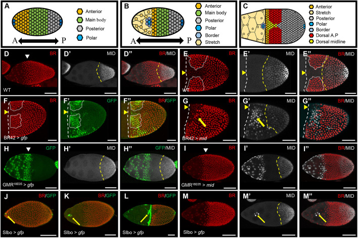
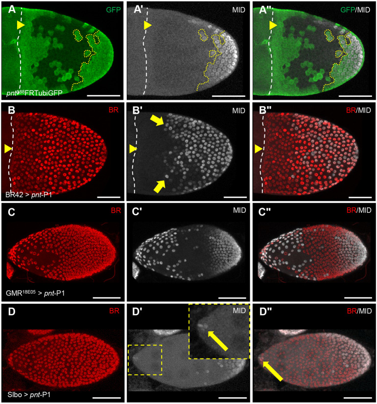
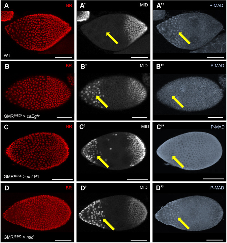
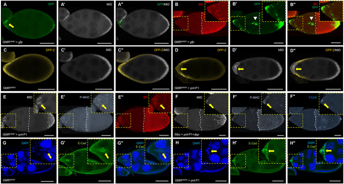
The Janus-kinase/signal transducer and activator of transcription (JAK/STAT) pathway regulates the anterior posterior axis of the Drosophila follicle cells. In the anterior, it activates the bone morphogenetic protein (BMP) signaling pathway through expression of the BMP ligand decapentaplegic (dpp). In the posterior, JAK/STAT works with the epidermal growth factor receptor (EGFR) pathway to express the T-box transcription factor midline (mid). Although MID is necessary for establishing the posterior fate of the egg chamber, we show that it is not sufficient to determine a posterior fate. The ETS-transcription factor pointed (pnt) is expressed in an overlapping domain to mid in the follicle cells. This study shows that pnt is upstream of mid and that it is sufficient to induce a posterior fate in the anterior end, which is characterized by the induction of mid, the prevention of the stretched cells formation and the abrogation of border cell migration. We demonstrate that the anterior BMP signaling is abolished by PNT through dpp repression. However, ectopic DPP cannot rescue the anterior fate formation, suggesting additional targets of PNT participate in the posterior fate determination.

Keywords: Anterior-posterior axis coordination; Cell morphogenesis; EGFR signaling; ETS-transcription factor.


Conflict of interest statement

Competing interestsThe authors declare no competing or financial interests.

Figures

Fig. 1.
Fig. 1.
The midline gene is necessary but not sufficient for posterior fate determination. (A-C) Cartoon representation of egg chambers at stage 8 (S8) (A), S9 (B) and S10 (C). The different domains are color coded: anterior domain, main body follicle cells, posterior domain, polar cells, border cells, stretch cells, dorsal A.P (appendage primordia) and dorsal midline. (D-E″) Wild-type expression of midline (MID, white) in early S9 (D-D″) (n=7) and later at S10B (E-E″) of oogenesis (n=10). The pattern of Broad (BR) is used as a spatial reference for the dorsal midline at S10B. (F-F″) The pattern of BR42-GAL4 driver expressing GFP (F′, green) (n=4). We focus on the BR domain, which is outlined with a dotted white line. (G-G″) Ectopic expression of MID by the BR42-GAL4 driver (yellow arrow, G′) disrupts BR patterning (G and G″) (n=5). (H-H″) The pattern of GMR18E05-GAL4 expressing GFP (H, green) in the anterior domain (n=6). (I-I″) Ectopic expression of MID by GMR18E05-GAL4 (n=8). (J-L) The pattern of Slbo-GAL4 driver expressing GFP (green) during S8 (J, n=6), S9 (K, n=5) and S10B (L, n=10) of oogenesis. Yellow arrows indicate the migrating border cells. (M-M″) Ectopic expression of MID by the Slbo-GAL4 (M′,M″, yellow arrows) (n=8). Yellow dashed line (D′,D″,E′,E″,G′,H′,H″,I′,M′) marks the anterior boundary of MID. Broad (BR, red) is expressed early uniformly in the follicular epithelium (D-D″), and later marks the dorsal appendage primordia (E-E″, white dotted outline). White arrowheads (D,H,I) mark posterior migration of follicular epithelium. (E-G″) White dashed lines mark the anterior boundary of follicular epithelium. Yellow arrowheads mark the dorsal midline. In all images, the anterior is to the left. Scale bars: 50 µm. n, the number of images with similar results.
Fig. 2.
Fig. 2.
Pointed is an upstream regulator of midline in the follicular epithelium. (A-A″) Null clones of pointed (pntΔ88) negatively marked by the loss of GFP (A). The clonal boundary is indicated by a yellow dotted line exhibiting cell-autonomous loss of MID (A′,A″) (n=46). (B-B″) Ectopic expression of pnt-P1 by the BR42-GAL4 driver exhibiting ectopic MID (B′) and loss of BR (B), and merge (B″) (n=4). Yellow arrows in B′ mark the ectopic MID. (C-C″) Ectopic expression of pnt-P1 in the anterior domain using the GMR18E05-GAL4 exhibiting ectopic MID (C′). BR is used as a spatial marker (C) and merge (C″) (n=11). (D-D″) Ectopic expression of pnt-P1 in the border cells exhibiting ectopic MID (D′). BR is used as a spatial marker (D) and merge (D″) (n=12). Yellow arrow (D′, inset) marks ectopic MID (D′,D″). Yellow arrowheads (A-B″) mark the dorsal midline. White dashed lines mark the anterior boundary of follicular epithelium. In all images, anterior is to the left. Scale bars: 50 µm. n, the number of images with similar results.
Fig. 3.
Fig. 3.
Ectopic pointed represses BMP signaling. (A-A″) Wild-type expression of BR (A), MID (A′) and P-MAD pattern (A″) at early stage 9 of oogenesis. Yellow arrows in A′ and A″ indicate the lack of anterior MID and presence of P-MAD, respectively (n=6). (B-B″) Using the GMR180E5-GAL4 driver to ectopically express a constitutively active EGFR (caEgfr) in the anterior epithelium at early stage 9 of oogenesis. BR (B), gain of MID (B′) and loss of P-MAD (B″) are observed, yellow arrows (n=5). (C-C″) Using the GMR180E5-GAL4 driver to ectopically express pnt-P1 in the anterior epithelium. BR (C), gain of MID expression (C′) and loss of P-MAD (C″) are observed, marked by a yellow arrow (n=11). (D-D″) Using the GMR18E05-GAL4 driver to ectopically express MID in the anterior epithelium. BR (D), gain of MID expression (D′) and intact P-MAD pattern are observed, marked by a yellow arrow (n=13). In all images, BR marks the follicular epithelium. In all images, the anterior of the egg chamber is to the left. Scale bars: 50 µm. n, number of images with similar results.
Fig. 4.
Fig. 4.
Pointed represses dpp and anterior cell morphogenesis independent of BMP signaling. (A-A″) GMR43H01-GAL4 driving expression of GFP at S9 egg chamber, yellow arrow (A) marks anterior GFP (n=6). (A′) The pattern of MID. (A″) Merged image. (B-B″) GMR43H01-GAL4 driving expression of GFP at S10 (n=10). (B) Broad, (B′) GFP, (B″) merge. Expression is observed in some of the anterior stretch follicle cells, migrating border cell cluster and posterior cells (B′,B″, white arrowheads). We focus on the anterior and border cells (insets). (C-C″) The pattern of a LacZ-DPP reporter (DPP-Z) in the GMR43H01-GAL4 driver background cross-section of early S9 egg chamber (C), MID pattern (C′) and merged image (C″) (n=6). (D-D″) GMR43H01-GAL4 driver expressing pnt-P1 in the anterior resulting in ectopic expression of MID (D′, dotted white lines, yellow arrows) and loss of DPP-Z (D,D″ merge, yellow arrows) (n=11). (E-E″) Ectopic expression of pnt-P1 using the GMR43H01-GAL4 driver in anterior stretch cells induced MID (E, yellow arrow, inset) and contained P-MAD (E′, yellow arrow, inset). These cells did not stretch and remained clustered in the anterior, marked by BR (E″, yellow arrow, inset) (n=5). (F-F″) Slbo-GAL4 driving expression of both pnt-P1 and dpp. (F) MID expression observed in anterior polar cells (yellow arrow, inset). (F′) P-MAD was present in these cells (yellow arrow, inset). (F″) FASIII marks polar cells failing to migrate posteriorly (yellow arrow, inset) (n=6). (G-G″) GMR43H01-GAL4 exhibiting cell nuclei marked by DAPI (G) and cell boundaries marked by E-cadherin (E-Cad) (G′), insets showcase migrating border cells (G-G″, yellow arrows) (n=6). (H-H″) Ectopic expression of pnt-P1 in anterior stretch cells. (H) DAPI marks cell nuclei, (H′) E-Cad marks cell boundaries. Insets showcase accumulation of E-Cad and loss of border cell migration (H-H″, yellow arrows) (n=7). Broad (BR) in B,E″ marks follicular epithelium. White dashed lines (E-F″) mark the anterior boundary of follicular epithelium. In all images, the anterior of the egg chamber is to the left. For A-D,G-H, cross-sections are shown. Scale bars: 50 µm. n, the number of images with similar results.

Similar articles

Cited by

References

    1. Bastock R. and St Johnston D. (2008). Drosophila oogenesis. Curr. Biol. 18, R1082-R1087. 10.1016/j.cub.2008.09.011 - DOI - PubMed
    1. Berg C. A. (2005). The Drosophila shell game: patterning genes and morphological change. Trends Genet. 21, 346-355. 10.1016/j.tig.2005.04.010 - DOI - PubMed
    1. Boisclair Lachance J.-F., Fregoso-Lomas M., Eleiche A., Bouchard Kerr P. and Nilson L. A. (2009). Graded Egfr activity patterns the Drosophila eggshell independently of autocrine feedback. Development 136, 2893-2902. 10.1242/dev.036103 - DOI - PubMed
    1. Brigaud I., Duteyrat J.-L., Chlasta J., Le Bail S., Couderc J.-L. and Grammont M. (2015). Transforming growth factor β/activin signalling induces epithelial cell flattening during Drosophila oogenesis. Biol. Open 4, 345-354. 10.1242/bio.201410785 - DOI - PMC - PubMed
    1. Chen Y. and Schüpbach T. (2006). The role of brinker in eggshell patterning. Mech. Dev. 123, 395-406. 10.1016/j.mod.2006.03.007 - DOI - PubMed

Publication types

MeSH terms