Review
. 2020 Oct 7;5(1):228.
doi: 10.1038/s41392-020-00313-5.

Emerging role of tumor cell plasticity in modifying therapeutic response

Affiliations
Review

Emerging role of tumor cell plasticity in modifying therapeutic response

Siyuan Qin et al. Signal Transduct Target Ther. .

Abstract

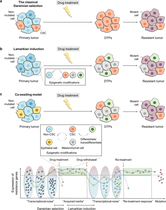
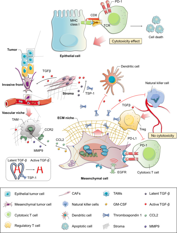
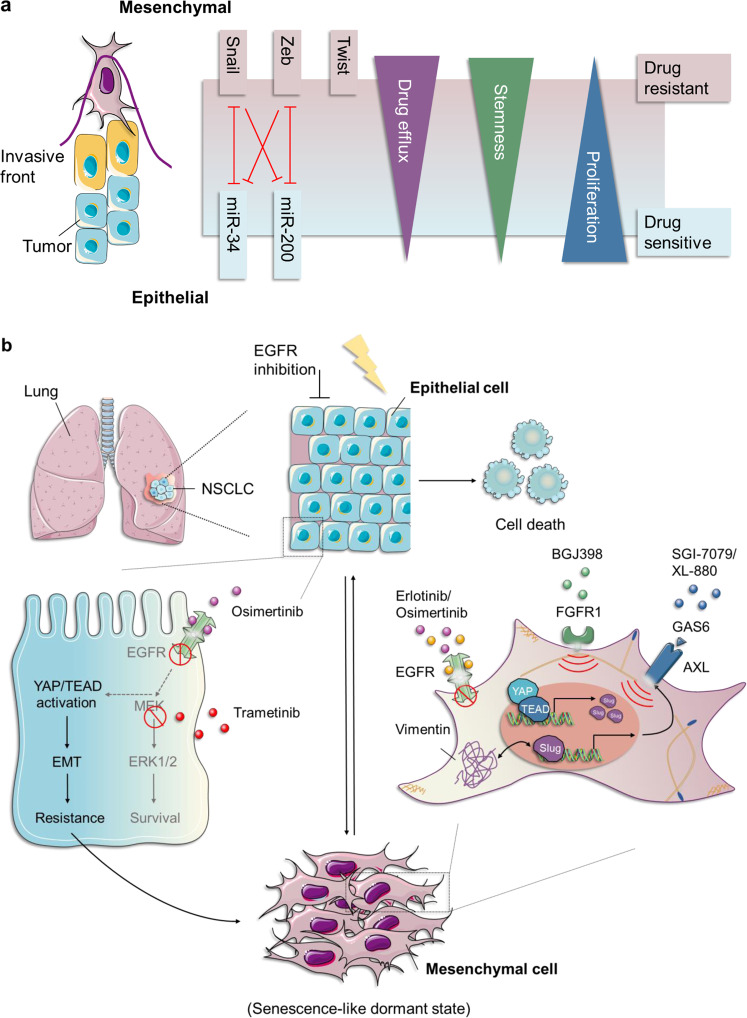
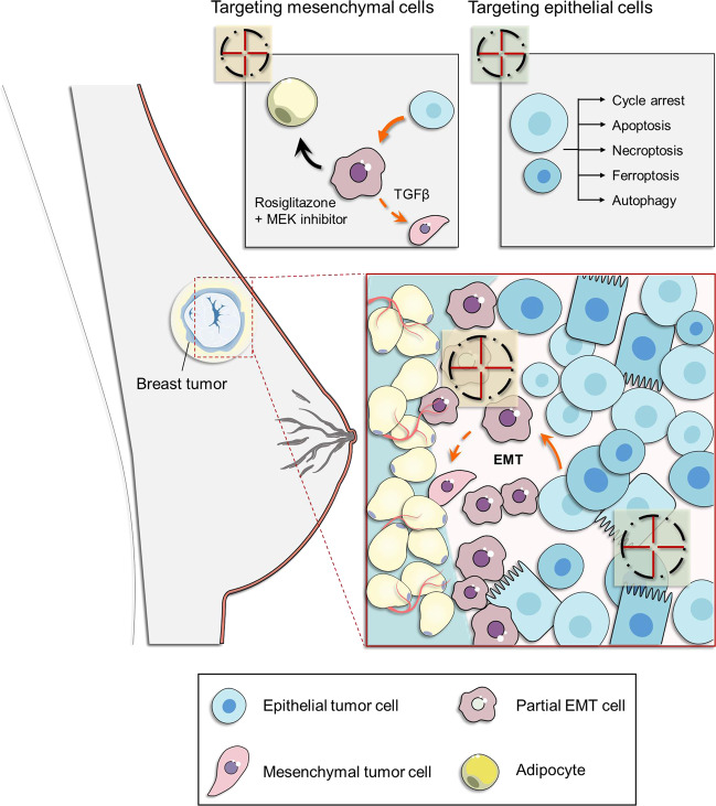
Resistance to cancer therapy is a major barrier to cancer management. Conventional views have proposed that acquisition of resistance may result from genetic mutations. However, accumulating evidence implicates a key role of non-mutational resistance mechanisms underlying drug tolerance, the latter of which is the focus that will be discussed here. Such non-mutational processes are largely driven by tumor cell plasticity, which renders tumor cells insusceptible to the drug-targeted pathway, thereby facilitating the tumor cell survival and growth. The concept of tumor cell plasticity highlights the significance of re-activation of developmental programs that are closely correlated with epithelial-mesenchymal transition, acquisition properties of cancer stem cells, and trans-differentiation potential during drug exposure. From observations in various cancers, this concept provides an opportunity for investigating the nature of anticancer drug resistance. Over the years, our understanding of the emerging role of phenotype switching in modifying therapeutic response has considerably increased. This expanded knowledge of tumor cell plasticity contributes to developing novel therapeutic strategies or combination therapy regimens using available anticancer drugs, which are likely to improve patient outcomes in clinical practice.


Conflict of interest statement

The authors declare that they have no conflict of interest.

Figures

Fig. 1
Fig. 1
The genesis of DTPs according to natural selection theory (classical Darwinian selection), the Lamarckian induction concept, and the coexisting model. a The natural selection theory shows that the preexisting DTPs, here represented by CSCs, can be selected and enriched upon drug exposure. b The concept of Lamarckian induction attaches importance to the natural aptitude of tumor cells in adapting to pharmacologic interventions through different levels of epigenetic modifications, giving rise to the emergence and coexistence of DTPs in varying tolerant states. c The coexisting model suggests the dynamic transcriptional fluctuation at a single-cell level of resistance-related markers (“transcriptional noise”). A small fraction of tumor cells, whose expression of these resistance-related genes exceeds a certain threshold at the moment of treatment, can survive and be selected (the blue and yellow dot), marking a return to classical Darwinian selection. However, with increasing duration of drug exposure, such a stochastic, transient, fluctuated “survival mode” arrives at drug-refractory state through epigenetic modifications, ultimately resulting in the establishment of a DTP pool. These alterations in the epigenome, which can be summed up as “acquired inertia,” are in agreement with the concept of Lamarckian induction. The solid line represents the changes of resistance-related markers expression with treatment, while the dotted line represents those without treatment (below). CSC cancer stem cell, DTPs drug-tolerant persisters
Fig. 2
Fig. 2
The tumor microenvironment (TME) contributes to resistance via EMT. Increased levels of TSP-1 (the activator of TGF-β1) secreted by mesenchymal tumor cells on the one hand facilitates the further activation of the TGF-β signaling pathway, contributing to a positive feedback effect on EMT; on the other hand, it promotes the generation of Foxp3+ Tregs from naive CD4+ CD25 T cells that antagonizes the activity of cytotoxic T cells, together with the induction of impaired DCs and inhibition of NK cells within the TME, thus ultimately resulting in immunotherapy, and even chemotherapy resistance. Tumors arising from the mesenchymal cells express a higher level of PD-L1 and lower level of MHC-I, together with more Tregs within TME in comparison with those formed by the epithelial cells, supporting the immunosuppressive role of EMT programs, which at least in part contributes to the resistance to cancer therapies. The TAMs, known as the most plentiful immune-related stromal components in TME, have been shown to infiltrate mainly at the invasive fronts of tumors. CCL2, synthesized by cancer cells, triggers the recruitment of circulating monocytes with the expression of CCR2 into tumors with the subsequent acquisition of a TAM phenotype. ZEB1-expressing macrophages promote their own polarization toward a stronger protumor phenotype; and meanwhile, upregulate the expression of CCL2 and CD74 in cancer cells through an increased release of MMP9, resulting in a mesenchymal/stem-like state. This forms a CCR2-MMP9-CCL2+ feedback loop between TAMs and the cancer cells. TSP-1 thrombospondin-1, PD-L1 programmed cell death-ligand 1, Tregs regulatory T cells, DCs dendritic cells, MHC-I the class-I major histocompatibility complex, TAMs tumor-associated macrophages, CAFs cancer-associated fibroblasts
Fig. 3
Fig. 3
The role of EMT in EGFR-TKI resistance. a Cancer cells undergoing EMT are characterized by morphotypic switching from “cobblestone-like” shapes to “fibroblast-like” forms. This process can be achieved via several EMT-TFs (Snail, Zeb, and Twist) and miRNAs in response to paracrine and autocrine stimuli, endowing cancer cell with a more aggressive phenotype, including enhanced invasive capacity, therapeutic resistance (enhanced drug efflux and slow cell proliferation), and stemness properties. b In EGFR-mutant NSCLC, upregulation of TEAD-mediated YAP promotes the transcription of Slug, which further induces the upregulation of AXL in NSCLC cells. AXL signaling, whose activation relies on interactions with its specific ligand GAS6, promotes EMT that drives Slug-overexpressing mesenchymal cells to acquire resistance with erlotinib. In addition, the mesenchymal cells display enhanced resistance to EGF816 accompanied by a significant activation of the FGFR1 pathway, implicating the potential of FGFR1 as a drug target for evading resistance to EGF816. A subpopulation of cancer cells can enter a senescence-like state to escape cell death upon administration of EGFRi (osimertinib) in combination with MEKi (tretinamib), resulting in resistance. This change is characterized by YAP/TEAD-mediated activation of EMT programs. The therapeutic strategy of pharmacologically cotargeting YAP/TEAD (by MYF-01-37) and EGFR/MEK leads to synthetic lethality. AXL anexelekto, GAS6 growth arrest-specific protein 6, SGI-7079/XL-880 AXL inhibitor, EGF816 the third-generation EGFR-TKIs, FGFR1 fibroblast growth factor receptor 1, BGJ398: FGFR inhibitor
Fig. 4
Fig. 4
EMT can be hijacked for therapeutic purposes by forcing trans-differentiation. EMT frequently occurs at the invasive front of the individual tumor, which also allows cancer cells to achieve a high plasticity level due to the mechanistic correlation and functional overlap between the EMT process and the CSC phenotype. The mesenchymal characteristics of those tumor cells are embodied in their potential for re-differentiation and possibly even trans-differentiation. Meanwhile, cancer cells also achieve resistance to a variety of conventional therapeutics during the EMT process, commonly resulting in tumor recurrence. However, the plasticity of those cancer cells can be utilized for therapeutic purposes by forcing their trans-differentiation process towards postmitotic and well-differentiated phenotypes rather than by direct killing. The treatment of an MEK inhibitor—trametinib—together with an adipogenesis inducer—rosiglitazone—can strongly promote the direct lineage conversion of those aggressive cancer cells to “peaceful” adipocytes. This provides the potential of preventing treatment failure by combining trans-differentiation therapy with multiple conventional therapies, efficiently killing the proliferative cancer cells that form the bulk of the tumor as well as eradicating invasive cells that escape conventional therapies by the development of an EMT/CSC phenotype. EMT epithelial–mesenchymal transition, CSC cancer stem cell
Fig. 5
Fig. 5
Trans-differentiation from castration-sensitive PCa to CRPC to NEPC: involvement of two generations of AR pathway-targeted agent. a ADTs mediate CRPC generation in an AR-independent manner, while ARPIs trigger NEPC formation dependent on AR signaling. ADTs blocking AR-related signaling, which exhibit remarkable activity causing tumor regression, lead to the emergence of aggressive CRPC with tumor recurrence. Likewise, the novel ARPIs by targeting specific ADT resistance contribute to tumor regression, whereas inducing a more aggressive NEPC phenotype in the process of neuroendocrine trans-differentiation promotes later acquisition of therapy resistance. b The characteristics of lineage switching from CRPC to NEPC in terms of clinical histology and molecular levels. Alteration of cellular identity from CRPC to NEPC is mainly characterized by the absence of AR and PSA. NEPC is also different from CRPC due to deletion of TP53 and RB1, enhancement of MYCN or AURKA, and upregulation of EZH2. c Aggressive behavior accompanying functional transformation from CRPC to NEPC. As lineage switching occurs, CRPC is converted to NEPC accompanied by increased invasiveness, intensive drug resistance, and elevated stem-like cell properties. CRPC castration-resistant prostate cancer, NEPC neuroendocrine prostate cancer, AR androgen receptor, ADTs androgen deprivation therapies, ARPIs AR pathway inhibitors, PSA serum prostate-specific antigen
Fig. 6
Fig. 6
Two models describing the mechanism of lineage switching from CRPC to NEPC. a Following ARPI treatment, NEPC can originate from a small subpopulation of mutated neuroendocrine cells surrounding the primary tumor in CRPC, or derive from overgrown CSCs in CRPC, which undergo a differentiation process to acquire an AR-independent basal-like phenotype. b AR-dependent luminal epithelial cells initially undergo developmental reprogramming to neurological PCSCs, followed by differentiation into an AR-independent basal-like NEPC by ARPI-induced neuroendocrine trans-differentiation. Due to dynamic reversible cancer cell plasticity, the newly acquired NEPC can be reverted to the luminal epithelial-like CRPC by restoring TP53 and RB1, re-exposure to androgen or inhibition of EZH2 and SOX2 implicated in pluripotency networks. PCSCs prostate cancer stem cells, SOX2 SRY-box transcription factor 2, EZH2 enhancer of zeste homolog 2
Fig. 7
Fig. 7
Overview of the molecular basis of re-activation of developmental programs contributing to cancer cell plasticity—major inhibitors of Hh, Wnt, and Notch signaling pathways for targeted therapy. The Hedgehog (Hh), wingless/integrated (Wnt), and Notch signaling pathways play a crucial part in acquisition and expansion of CSC phenotype after being stimulated by internal factors or extrinsic stimuli, for instance, HGF and docetaxel. Re-activation of these developmental programs promotes the corresponding transcription factors entering into the nucleus to regulate expression of downstream effectors that are closely related to CSCs regeneration and maintenance, as well as multiple biological functions. To prevent cancer cell plasticity, particularly phenotypic switching from non-CSC to CSC states induced by Hh, Wnt, and Notch signaling pathways, a range of inhibitors targeting these pathways have been approved for clinical use or are under development, as shown in blue, purple, and green. CAFs cancer-associated fibroblasts, Ihh/Dhh/Shh: Sonic hedgehog/Indian hedgehog/Desert hedgehog, PTCH1 2 patched1 and patched2, SMO smoothened, ATO arsenic trioxide, HDAC6i histone deacetylase 6 inhibitor, GLI glioma-associated oncogene homolog, LRP5/6 lipoprotein receptor-related protein 5/6, HGF hepatocyte growth factor, VA valproic acid, EMT epithelial–mesenchymal transition, NICD Notch intracellular region, ADAM a disintegrin and metalloprotease, DLL1, 3, 4 Delta-like ligand 1, 3, 4, Jagged 1, 2 Serrate-like ligand 1, 2, IR ionizing radiation, NT neoadjuvant therapy, Rova-T rovalpituzumab tesirine, DIM 3,3′-diindolylmethane, DiFiD 3,5-bis (2,4-difluorobenzylidene)-4-piperidone, EGCG epigallocatechin-3-gallate

References

    1. Bedard PL, Hyman DM, Davids MS, Siu LL. Small molecules, big impact: 20 years of targeted therapy in oncology. Lancet. 2020;395:1078–1088. doi: 10.1016/S0140-6736(20)30164-1. - DOI - PubMed
    1. Carneiro, B. A. & El-Deiry, W. S. Targeting apoptosis in cancer therapy. Nat. Rev. Clin. Oncol. 17, 395–417 (2020). - PMC - PubMed
    1. Vasan N, Baselga J, Hyman DM. A view on drug resistance in cancer. Nature. 2019;575:299–309. doi: 10.1038/s41586-019-1730-1. - DOI - PMC - PubMed
    1. Redmond KM, Wilson TR, Johnston PG, Longley DB. Resistance mechanisms to cancer chemotherapy. Front. Biosci. 2008;13:5138–5154. doi: 10.2741/3070. - DOI - PubMed
    1. Fojo T. Multiple paths to a drug resistance phenotype: mutations, translocations, deletions and amplification of coding genes or promoter regions, epigenetic changes and microRNAs. Drug Resist. Updat. 2007;10:59–67. doi: 10.1016/j.drup.2007.02.002. - DOI - PubMed

Publication types

MeSH terms

Substances