. 2020 Oct 7;10(1):16671.
doi: 10.1038/s41598-020-72978-5.

In situ-forming collagen hydrogel crosslinked via multi-functional PEG as a matrix therapy for corneal defects

Affiliations

In situ-forming collagen hydrogel crosslinked via multi-functional PEG as a matrix therapy for corneal defects

Gabriella Maria Fernandes-Cunha et al. Sci Rep. .

Abstract

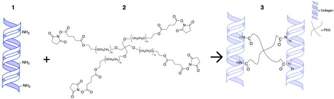
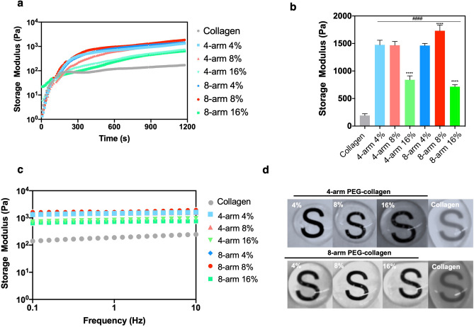
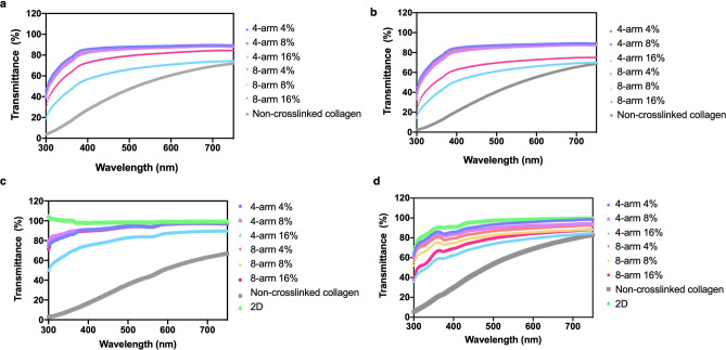
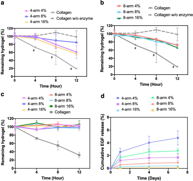
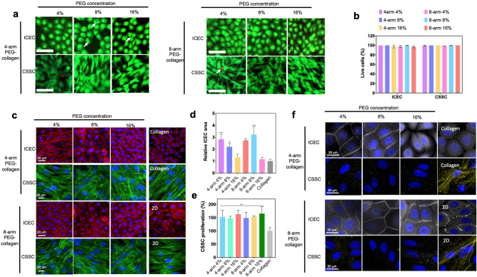
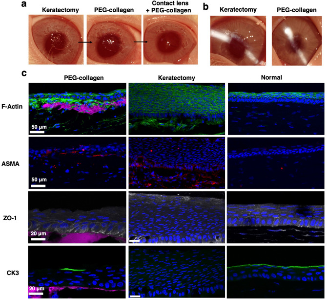
Visually significant corneal injuries and subsequent scarring collectively represent a major global human health challenge, affecting millions of people worldwide. Unfortunately, less than 2% of patients who could benefit from a sight-restoring corneal transplant have access to cadaveric donor corneal tissue. Thus, there is a critical need for new ways to repair corneal defects that drive proper epithelialization and stromal remodeling of the wounded area without the need for cadeveric donor corneas. Emerging therapies to replace the need for donor corneas include pre-formed biosynthetic buttons and in situ-forming matrices that strive to achieve the transparency, biocompatibility, patient comfort, and biointegration that is possible with native tissue. Herein, we report on the development of an in situ-forming hydrogel of collagen type I crosslinked via multi-functional polyethylene glycol (PEG)-N-hydroxysuccinimide (NHS) and characterize its biophysical properties and regenerative capacity both in vitro and in vivo. The hydrogels form under ambient conditions within minutes upon mixing without the need for an external catalyst or trigger such as light or heat, and their transparency, degradability, and stiffness are modulated as a function of number of PEG arms and concentration of PEG. In addition, in situ-forming PEG-collagen hydrogels support the migration and proliferation of corneal epithelial and stromal cells on their surface. In vivo studies in which the hydrogels were formed in situ over stromal keratectomy wounds without sutures showed that they supported multi-layered surface epithelialization. Overall, the in situ forming PEG-collagen hydrogels exhibited physical and biological properties desirable for a corneal stromal defect wound repair matrix that could be applied without the need for sutures or an external trigger such as a catalyst or light energy.


Conflict of interest statement

Author D.M. holds a patent on the portable ophthalmic camera system used to photograph the animal eyes, and authors D.M., GM.F-C, and H.L. have a patent application on the hydrogel technology used in the studies. All other authors declare no competing interests.

Figures

Figure 1
Figure 1
Schematic of the crosslinking between primary amines on collagen and NHS groups on multi-arm PEG polymers; (1) collagen, (2) 4-arm PEG-NHS (3) PEG-collagen hydrogel.
Figure 2
Figure 2
Physical properties of chemically crosslinked PEG-collagen hydrogels by NHS chemistry and non‐crosslinked (physical) collagen hydrogels. (a) Dynamic moduli of 4-arm PEG-collagen and 8-arm PEG-collagen hydrogels using different concentrations of PEG polymer as function of time during gelation. The hydrogels were mounted immediately after mixing. (b) Dynamic moduli of 4-arm PEG-collagen and 8-arm PEG-collagen hydrogels at different concentrations of PEG polymer. 4 and 8-arm PEG-collagen at 16% v/v PEG show lower storage modulus (**** p < 0.0001) compared to 4 and 8arm PEG-collagen at 4 and 8%. 8-arm PEG-collagen hydrogel at 8% showed higher storage modulus (**** p < 0.0001) compared to all other PEG concentrations and type. Collagen crosslinked with 4 and 8-arm PEG polymer at all concentrations showed significant higher storage modulus (#### p < 0.0001) compared to non-crosslinked collagen hydrogel; data is presented as mean ± SD, two-way ANOVA (p < 0.005) was used to detect statistical differences followed by Tukey’s multiple comparisons test. (c) Dynamic moduli of 4-arm PEG-collagen and 8-arm PEG-collagen hydrogels as a function of frequency. (d) Photographs of 4 and 8-arm PEG-collagen and non-crosslinked (physical) collagen hydrogels.
Figure 3
Figure 3
Transmittance spectra of collagen hydrogels from 350 to 800 nm (a) before swelling (b) after swelling for 24 h in PBS, (c) after 24 h in the presence of ICECs and (d) after 24 h in the presence CSSCs.
Figure 4
Figure 4
Degradation of (a) 4 and (b) 8-arm PEG-collagen hydrogels at different PEG concentrations (4%, 8%, and 16%) 24 h after swelling was evaluated in the presence of 1 mg/mL of collagenase for 12 h. Data is presented as mean ± SD, two-way ANOVA (p < 0.005) was used to detect statistical differences followed by Tukey’s multiple comparisons test. (c) Degradation profile of 4 and 8-arm PEG-collagen in the presence of CSSCs for 12 h. (d) EGF release from 4 and 8-arm PEG-collagen hydrogels during 7 days in PBS. For each condition n = 3 was used.
Figure 5
Figure 5
(a) Live/dead assays on ICEC and CSSC seeded on 4-arm PEG-collagen and 8-arm PEG-collagen hydrogels using different concentrations of PEG after 2 days in culture. Scale bar: 100 µm—live cells (green) and dead cells (red). (b) Viability of corneal cells seeded on PEG-collagen hydrogels. (c) F‐actin staining ICEC (red) and CSSC (green) showing cell morphology when seeded on 4-arm PEG-collagen and 8-arm PEG-collagen hydrogels using different concentrations of PEG after 2 days in culture. The nucleus (blue) was stained for both cells. Scale bar: 20 µm. (d) Relative ICEC cell area 2 days after seeding on PEG-collagen hydrogel. Areas of the cells seeded on 4 and 8-arm PEG-collagen with 4 and 8% PEG were statistically different compared to non-crosslinked collagen (****p < 0.0001, ***p = 0.0002, **p = 0.003). The data was normalized by cells seeded on non-crosslinked collagen hydrogels, with n  =  3 for each condition and ordinary one-way ANOVA applied, followed by Dunnett’s multiple comparison test. (e) Relative CSSC proliferation on 4-arm and 8-arm PEG-collagen hydrogels using different concentrations of PEG polymer after 2 days in culture. The data was normalized by cells seeded on non-crosslinked collagen hydrogels. A sample size of 3 and an ordinary one-way ANOVA was used followed by Dunnett’s multiple comparison test. (**p < 0.01 vs non crosslinked collagen). (f) ZO-1 (gray) staining of ICEC and ASMA staining of CSSC proliferated on PEG-collagen hydrogels for 2 days. Scale bar: 20 µm.
Figure 6
Figure 6
(a) Photographs of rabbit corneas right after keratectomy showing a rough surface and then a smooth surface after application of PEG-collagen hydrogel and contact lens. (b) Appearance of untreated and treated corneas on post-op day 7. (c) PEG-collagen hydrogel (stained magenta) underneath a multi-layered, migrated epithelial layer can be observed 7 days after treatment. For the keratectomy-only group, epithelial hyperplasia was observed. ASMA (red) was observed for the rabbits that received PEG-collagen as well as in the keratectomy group. Normal epithelial cell phenotype was detected by the presence of ZO-1 (gray) in the wing and superficial cells and CK3 (green). Minimal ZO-1 expression and no focal CK3 expression was observed for the keratectomy group.

References

    1. Channa R, et al. Epidemiology of eye-related emergency department visits. JAMA Ophthalmol. 2016;134:312–319. doi: 10.1001/jamaophthalmol.2015.5778. - DOI - PubMed
    1. Lagali N. Corneal stromal regeneration: current status and future therapeutic potential. Curr. Eye Res. 2019 doi: 10.1080/02713683.2019.1663874. - DOI - PubMed
    1. Samarawickrama C, et al. Collagen-based fillers as alternatives to cyanoacrylate glue for the sealing of large corneal perforations. Cornea. 2018;37:609–616. doi: 10.1097/ico.0000000000001459. - DOI - PubMed
    1. Trujillo-de Santiago G, et al. Ocular adhesives: design, chemistry, crosslinking mechanisms, and applications. Biomaterials. 2019;197:345–367. doi: 10.1016/j.biomaterials.2019.01.011. - DOI - PMC - PubMed
    1. Gain P, et al. Global survey of corneal transplantation and eye banking. JAMA Ophthalmol. 2016;134:167–173. doi: 10.1001/jamaophthalmol.2015.4776. - DOI - PubMed

Publication types