. 2020 Oct 7;10(1):16680.
doi: 10.1038/s41598-020-73497-z.

Fabrication of monodispersed copper oxide nanoparticles with potential application as antimicrobial agents

Affiliations

Fabrication of monodispersed copper oxide nanoparticles with potential application as antimicrobial agents

Fisseha A Bezza et al. Sci Rep. .

Abstract

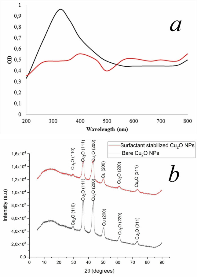
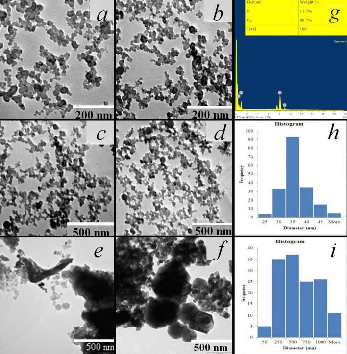
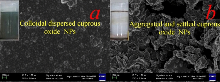
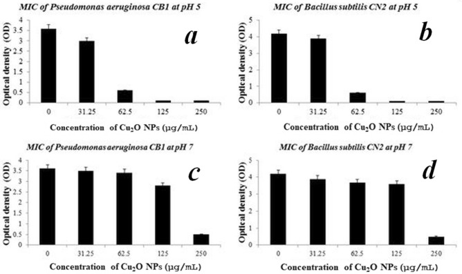
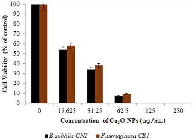
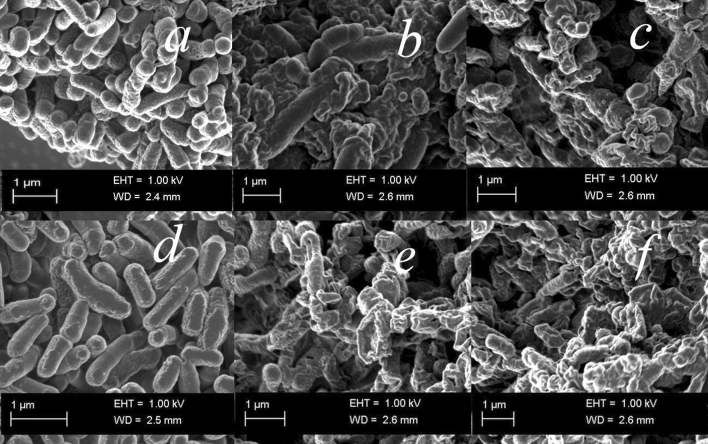
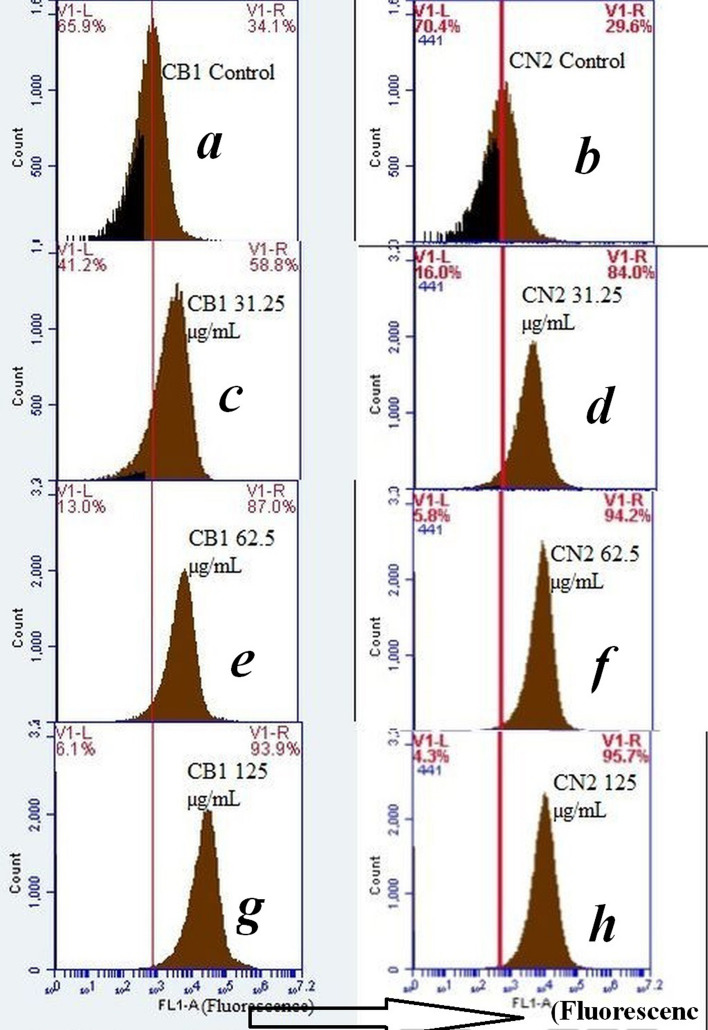
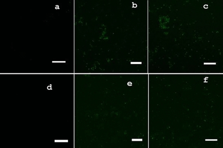
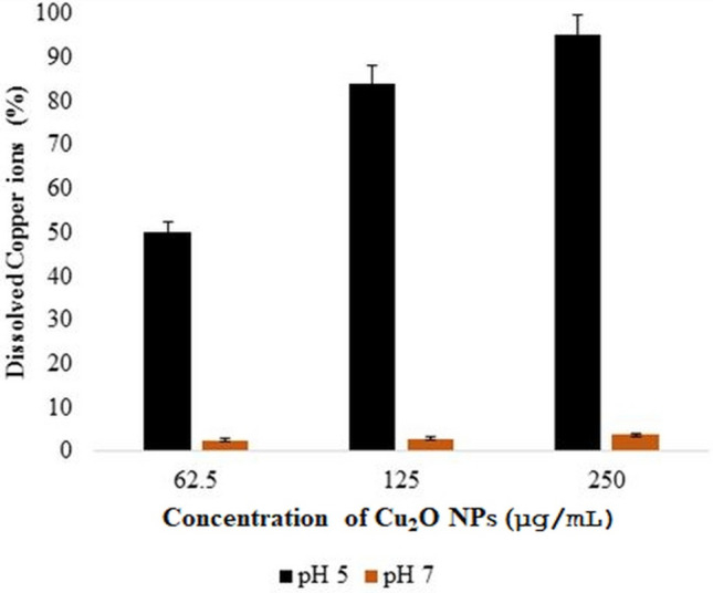
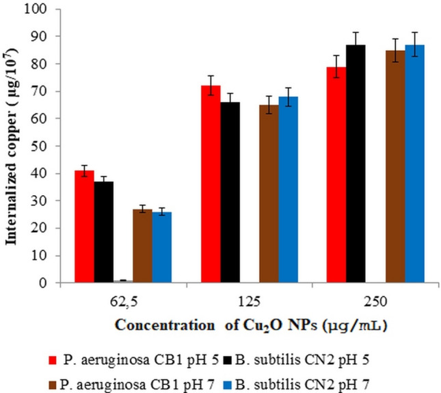
Cuprous oxide nanoparticles (Cu2O NPs) were fabricated in reverse micellar templates by using lipopeptidal biosurfactant as a stabilizing agent. Scanning electron microscopy (SEM), transmission electron microscopy (TEM), energy dispersive x-ray spectrum (EDX) and UV-Vis analysis were carried out to investigate the morphology, size, composition and stability of the nanoparticles synthesized. The antibacterial activity of the as-synthesized Cu2O NPs was evaluated against Gram-positive B. subtilis CN2 and Gram-negative P. aeruginosa CB1 strains, based on cell viability, zone of inhibition and minimal inhibitory concentration (MIC) indices. The lipopeptide stabilized Cu2O NPs with an ultra-small size of 30 ± 2 nm diameter exhibited potent antimicrobial activity against both Gram-positive and Gram-negative bacteria with a minimum inhibitory concentration of 62.5 µg/mL at pH5. MTT cell viability assay displayed a median inhibition concentration (IC50) of 21.21 μg/L and 18.65 μg/mL for P. aeruginosa and B. subtilis strains respectively. Flow cytometric quantification of intracellular reactive oxygen species (ROS) using 2,7-dichlorodihydrofluorescein diacetate staining revealed a significant ROS generation up to 2.6 to 3.2-fold increase in the cells treated with 62.5 µg/mL Cu2O NPs compared to the untreated controls, demonstrating robust antibacterial activity. The results suggest that lipopeptide biosurfactant stabilized Cu2O NPs could have promising potential for biocompatible bactericidal and therapeutic applications.


Conflict of interest statement

The authors declare no competing interests.

Figures

Figure 1
Figure 1
UV–Vis spectra of Cu2O NPs synthesised in the presence of lipopeptide microbial surfactant (a, black series) and in the absence of lipopeptide microbial surfactant (a, red series). Representative XRD patterns of the surfactant stabilized Cu2O NPs (b, red line) compared to the bare Cu2O NPs (b, black line), reflecting smaller size of the surfactant stabilized Cu2O NPs.
Figure 2
Figure 2
Representative TEM images of (a, c), Cu2O NPs at 1 g/L, (b, d), Cu2O NPs at 2 g/L lipopeptide biosurfactant additives at different magnifications; (e, f) Cu2O NPs with no lipopeptide biosurfactant additive, (g) EDX of the synthesised Cu2O NPs with an inset chart displaying percentage elemental composition of copper and oxygen. (h, i) Particle size distribution histograms of Cu2O NPs synthesised in the presence of 2 g/L lipopeptide biosurfactant additive and in its absence respectively.
Figure 3
Figure 3
Representative SEM images of Cu2O NPs prepared (a) in the presence of 2 g/L lipopeptide surfactant, (b) in the absence of the lipopeptide microbial surfactant, displaying larger aggregated particles. Scale bar: 200 nm. The vials in the insets show colloidal stability of the resultant products after standing undisturbed for two weeks.
Figure 4
Figure 4
MIC of Cu2O NPs against P. aeruginosa CB1 and B. subtilis CN2 at different pHs evaluated by measuring optical density at 600 nm (OD600) after incubation at 37 °C, 180 rpm for 24 h. Data are presented as mean ± SD of three independent experiments, each performed in triplicates. Variation between sample means are analysed by ANOVA.
Figure 5
Figure 5
Cell viability percentages of Gram-positive B. subtilis CN2 and Gram-negative P. aeruginosa CB1 cells against various concentrations of Cu2O NPs after 24 h of exposure; results are expressed as percentages of control cells. Values are mean ± SD of three independent experiments, each performed in triplicates and considered statistically significant when p < 0.05.
Figure 6
Figure 6
Representative TEM micrographs of untreated B. subtilis CN2 cells (a), showing intact and high electron density morphology, 100 µg/mL (b), 125 µg/mL (c) dosage of Cu2O NPs treated cells indicating cytoplasmic injury with disintegrated outer membrane (f, yellow arrow). P. aeruginosa CB1 cells treated with 100 µg/mL (e), 125 µg/mL (f) dosage of Cu2O NPs and untreated control (d). Considerable size of adhered nanoparticles was observed (b, red arrow) attached to the surface of the cells of the bacteria, and disrupted cell wall and membrane leakage was observed (e, green arrow). Scale bar is 1 µm.
Figure 7
Figure 7
Representative SEM micrographs of untreated B. subtilis CN2 cells (a), showing intact and high electron density morphology, 100 µg/mL (b), 125 µg/mL (c) dosage Cu2O NPs treated cells indicating cytoplasmic injury with disintegrated outer membrane. P. aeruginosa CB1 untreated cells (d), treated with 100 µg/mL (e) and 125 µg/mL (f) dosage of Cu2O NPs. Scale bar is 1 µm.
Figure 8
Figure 8
Oxidative stress response at various doses of Cu2O NPs on the Gram-negative P. aeruginosa CB1 (left column) and Gram-positive B. subtilis CN2 (right column) bacterial cells. (a, b) Control cells; (c, d) cells treated with 31.25 µg/mL; (e, f) cells treated with 62.5 µg/mL; (g, h) cells treated with 125 µg/mL for 24 h. After treatment of cells with designated concentrations of Cu2O NPs for 24 h, intracellular ROS generation was quantified by oxidation of cell permeable dye 2,7-dichlorodihydrofluorescein diacetate (DCFDA) staining using flow cytometer. The DCF fluorescence is proportional to the ROS generated. The FL1-A corresponds to the green emission of the DCF. The V1-L and V1-R markers correspond to ROS negative and positive cells respectively. Scale Bar: 5 µm.
Figure 9
Figure 9
Confocal microscopy of green fluorescence images of ROS in Cu2O NPs treated, and untreated control cells measured by 2,7-dichlorofluorescin diacetate (DCFH-DA) fluorescence-based assay. Cells were treated with 0, 62.5 and 125 μg/mL of surfactant stabilized Cu2O NPs for 24 h. (ac) green fluorescence images of 0, 62.5 and 125 μg/mL dose Cu2O NPs exposed Gram-positive B. subtilis CN2 cells. (df) green fluorescence images of 0, 62.5 and 125 μg/mL dose Cu2O NPs exposed Gram-negative P. aeruginosa CB1 cells. Untreated cells were used as a negative control (a, d). Scale Bar: 5 µm.
Figure 10
Figure 10
Percentage of dissolved copper ions released from various dosages of Cu2O NPs at different pH values after 24 h of exposure at 37 °C. Data are means ± standard deviations of three independent experiments each performed in triplicates. There was no statistically significant difference between the amount of Cu1+ dissolved at 62.5, 125, and 250 µg/mL treatments at the pHs administered.
Figure 11
Figure 11
Cellular uptake of Cu2O NPs after 24 h of exposure of bacterial strains to various dosages of Cu2O NPs at 37 °C. Data are means ± standard deviations of three independent experiments each performed in triplicates. There was no statistically significant difference between the amount of copper internalized at 62.5, 125, and 250 µg/mL treatments at pH 5 and pH 7 in each of the strains (p > 0.05).

References

    1. Hajipour MJ, Fromm KM, Ashkarran AA, de Aberasturi DJ, de Larramendi IR, Rojo T, Serpooshan V, Parak WJ, Mahmoudi M. Antibacterial properties of nanoparticles. Trends Biotechnol. 2012;30:499–511. doi: 10.1016/j.tibtech.2012.06.004. - DOI - PubMed
    1. Pelgrift RY, Friedman AJ. Nanotechnology as a therapeutic tool to combat microbial resistance. Adv. Drug Deliv. Rev. 2013;65:1803–1815. doi: 10.1016/j.addr.2013.07.011. - DOI - PubMed
    1. Palmer AC, Kishony R. Understanding, predicting and manipulating the genotypic evolution of antibiotic resistance. Nat. Rev. Genet. 2013;14:243–248. doi: 10.1038/nrg3351. - DOI - PMC - PubMed
    1. Ventola CL. The antibiotic resistance crisis: part 1: causes and threats. Pharm. Ther. 2015;40:277. - PMC - PubMed
    1. Huh AJ, Kwon YJ. “Nanoantibiotics”: a new paradigm for treating infectious diseases using nanomaterials in the antibiotics resistant era. J. Control. Release. 2011;156:128–145. doi: 10.1016/j.jconrel.2011.07.002. - DOI - PubMed

Publication types