. 2020 Oct 7;10(1):16706.
doi: 10.1038/s41598-020-73713-w.

Rosettes integrity protects Plasmodium vivax of being phagocytized

Affiliations

Rosettes integrity protects Plasmodium vivax of being phagocytized

Letusa Albrecht et al. Sci Rep. .

Abstract

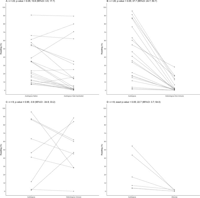
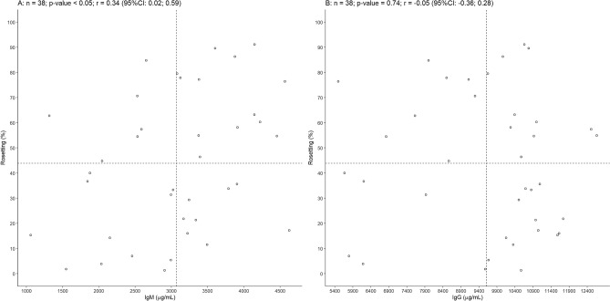
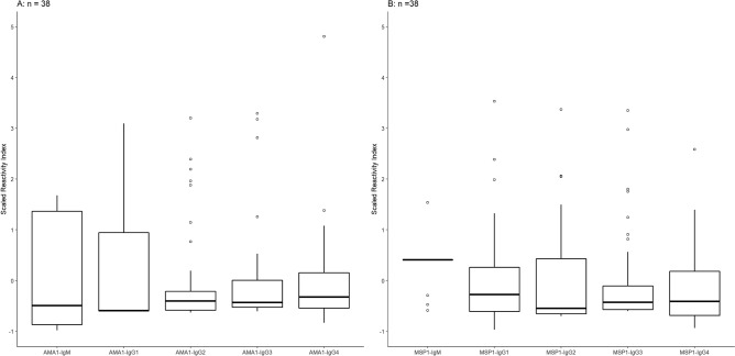
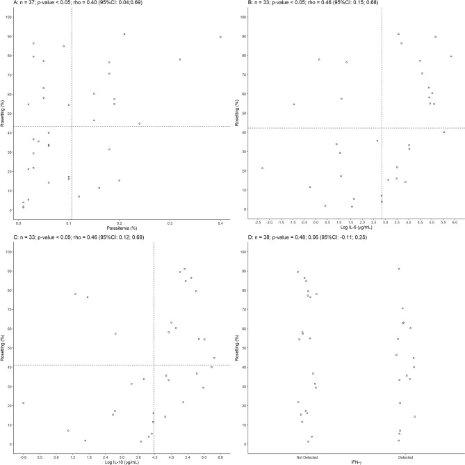
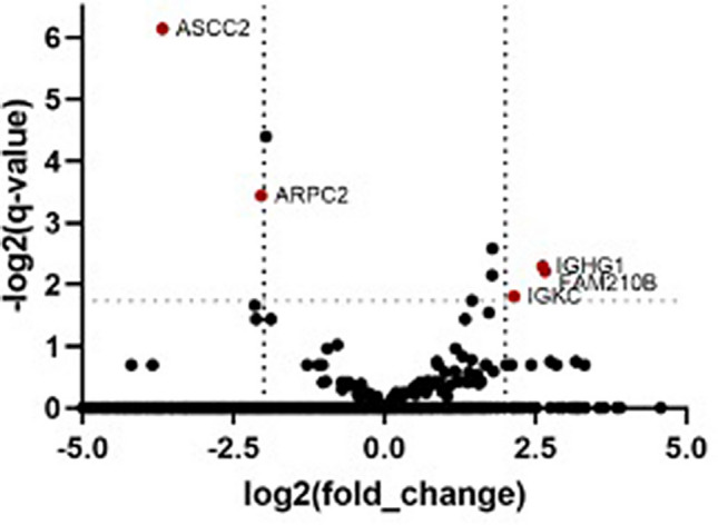
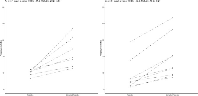
Plasmodium vivax is the most prevalent cause of malaria outside of Africa. P. vivax biology and pathogenesis are still poorly understood. The role of one highly occurring phenotype in particular where infected reticulocytes cytoadhere to noninfected normocytes, forming rosettes, remains unknown. Here, using a range of ex vivo approaches, we showed that P. vivax rosetting rates were enhanced by plasma of infected patients and that total immunoglobulin M levels correlated with rosetting frequency. Moreover, rosetting rates were also correlated with parasitemia, IL-6 and IL-10 levels in infected patients. Transcriptomic analysis of peripheral leukocytes from P. vivax-infected patients with low or moderated rosetting rates identified differentially expressed genes related to human host phagocytosis pathway. In addition, phagocytosis assay showed that rosetting parasites were less phagocyted. Collectively, these results showed that rosette formation plays a role in host immune response by hampering leukocyte phagocytosis. Thus, these findings suggest that rosetting could be an effective P. vivax immune evasion strategy.


Conflict of interest statement

The authors declare no competing interests.

Figures

Figure 1
Figure 1
Features of Plasmodium vivax rosettes. (A) Rosetting in the presence of autologous native or heat-inactivated plasma (Student’s t-test, p < 0.05, n = 23). (B) Rosetting of P. vivax in the presence of heterologous nonimmune or autologous plasma. Rosetting was eightfold higher in the presence of autologous plasma (Student’s t-test, p < 0.05, n = 20). (C) Rosetting of P. vivax in the presence of heterologous plasma from a high rosetting isolate (heterologous immune plasma). No difference in rosetting was observed between autologous and heterologous immune plasma (Student’s t-test, p = 0.95, n = 9) (D) Rosettes did not form on medium containing Albumax II (Wilcoxon test, p < 0.05, n = 6). CI: confidence interval.
Figure 2
Figure 2
Total IgM but not IgG correlated with rosetting in vivax malaria. (A) Correlation of total IgM (Pearson correlation coefficient r = 0.34, p < 0.05, n = 38) and (B) total IgG (Pearson correlation coefficient r = − 0.05, p = 0.74, n = 38) from malaria infected patients and rosetting rate of P. vivax isolates. CI: confidence interval.
Figure 3
Figure 3
Naturally acquired IgG and IgM antibodies to merozoite antigens and its relationship with rosetting. (A) Boxplot showing median and interquartile ranges of scaled reactivity index for IgG subclasses (IgG1, IgG2, IgG3 and IgG4) and IgM towards PvAMA-1 and (B) PvMSP-1 recombinant proteins. Open circles denote outliers observations.
Figure 4
Figure 4
Parasitemia and plasmatic levels of IL-6 and IL-10 correlated with rosetting in vivax malaria. (A) Positive correlation between peripheral parasitemia and rosetting (Spearman’s correlation coefficient rho = 0.40, p =  < 0.05, n = 37). (B) Correlation of P. vivax rosetting and log IL-6 (Spearman’s correlation coefficient rho = 0.46, p < 0.05, n = 33). (C) log IL-10 (Spearman’s correlation coefficient rho = 0. 46, p < 0.05 and n = 33) and (D) IFN-γ (Student’s t-test, p = 0.48 and n = 38). CI: confidence interval.
Figure 5
Figure 5
RNAseq analysis of PBMCs from patients infected with P. vivax. Volcano plot showing the range of the log2(fold change) relative to the − log2(q-values) of mapped genes from PBMCs of P. vivax infected patients between moderate and low rosetting isolates differential expression data analysis. Identified genes with a p-value < 0.05, q-value < 0.3 and log2 (fold change) > 2 cut-offs obtained from RNAseq differential gene expression analysis are put in evidence (dark red dots).
Figure 6
Figure 6
Non-rosetting parasites are more likely to be phagocyted. (A) Rosetting-disrupted P. vivax parasites were preferentially phagocyted in vitro by THP-1 cells (Wilcoxon test, p = 0.02, n = 7) as well as for (B) patients peripheral blood mononuclear cells (Wilcoxon test, p = 0.004, n = 9). CI: confidence interval.

References

    1. Costa FT, et al. On the pathogenesis of Plasmodium vivax malaria: Perspectives from the Brazilian field. Int. J. Parasitol. 2012;42:1099–1105. doi: 10.1016/j.ijpara.2012.08.007. - DOI - PubMed
    1. Baird KJ, Maguire JD, Price RN. Diagnosis and treatment of Plasmodium vivax malaria. Adv. Parasitol. 2012;80:203–270. doi: 10.1016/B978-0-12-397900-1.00004-9. - DOI - PubMed
    1. Russell BM, Cooke BM. The rheopathobiology of Plasmodium vivax and other important primate malaria parasites. Trends Parasitol. 2017;33:321–334. doi: 10.1016/j.pt.2016.11.009. - DOI - PubMed
    1. Suwanarusk R, et al. The deformability of red blood cells parasitized by Plasmodium falciparum and P. vivax. J. Infect. Dis. 2004;189:190–194. doi: 10.1086/380468. - DOI - PubMed
    1. Carvalho BO, et al. On the cytoadhesion of Plasmodium vivax-infected erythrocytes. J. Infect. Dis. 2010;202:638–647. doi: 10.1086/654815. - DOI - PubMed

Publication types