. 2020 Oct;586(7829):424-428.
doi: 10.1038/s41586-020-2812-9. Epub 2020 Oct 7.

Relatives of rubella virus in diverse mammals

Affiliations

Relatives of rubella virus in diverse mammals

Andrew J Bennett et al. Nature. 2020 Oct.

Erratum in

Abstract

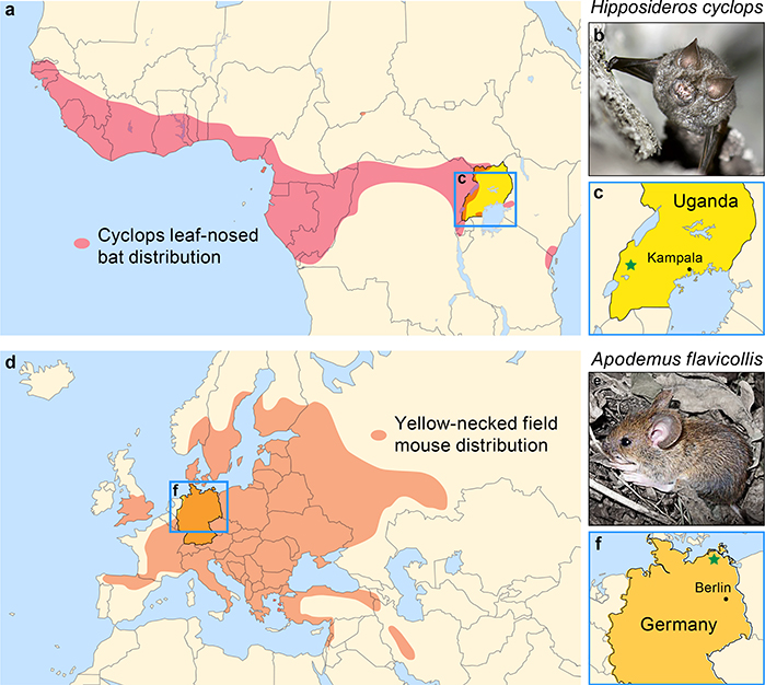
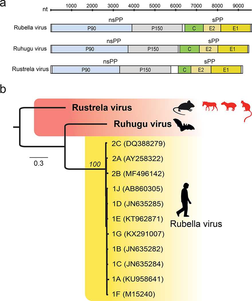
Since 1814, when rubella was first described, the origins of the disease and its causative agent, rubella virus (Matonaviridae: Rubivirus), have remained unclear1. Here we describe ruhugu virus and rustrela virus in Africa and Europe, respectively, which are, to our knowledge, the first known relatives of rubella virus. Ruhugu virus, which is the closest relative of rubella virus, was found in apparently healthy cyclops leaf-nosed bats (Hipposideros cyclops) in Uganda. Rustrela virus, which is an outgroup to the clade that comprises rubella and ruhugu viruses, was found in acutely encephalitic placental and marsupial animals at a zoo in Germany and in wild yellow-necked field mice (Apodemus flavicollis) at and near the zoo. Ruhugu and rustrela viruses share an identical genomic architecture with rubella virus2,3. The amino acid sequences of four putative B cell epitopes in the fusion (E1) protein of the rubella, ruhugu and rustrela viruses and two putative T cell epitopes in the capsid protein of the rubella and ruhugu viruses are moderately to highly conserved4-6. Modelling of E1 homotrimers in the post-fusion state predicts that ruhugu and rubella viruses have a similar capacity for fusion with the host-cell membrane5. Together, these findings show that some members of the family Matonaviridae can cross substantial barriers between host species and that rubella virus probably has a zoonotic origin. Our findings raise concerns about future zoonotic transmission of rubella-like viruses, but will facilitate comparative studies and animal models of rubella and congenital rubella syndrome.


Conflict of interest statement

Competing interest declaration. The authors declare no competing interests.

Figures

Extended Data Figure 1 |
RNA in situ hybridization of rustrela virus. a-e) Detection of rustrela virus RNA in brain tissues of a donkey (a), Bennett’s tree-kangaroo (b), capybara (c) and yellow-necked field mice (d, e). Chromogenic labelling (fast red) with probes to rustrela virus NSP-coding region are visible in neuronal cell bodies (arrow) but not in adjacent glial cells (arrow head). Mayer’s hematoxylin counter stain. Scale bar = 50 μm f). Negative control probe to bacterial gene dapB encoding dihydrodipicolinate reductase. Lack of chromogenic labelling (fast red). Mayer’s hematoxylin counter stain. Scale bar = 100 μm. RNAscope results were evaluated on at least 3 slides per animal, yielding comparable results in all cases. In situ hybridization was performed according to the manufacturer’s instructions, including a positive control probe, peptidylprolyl isomerase B (cyclophilin B, ppib), and a negative control probe, dihydrodipicolinate reductase (DapB). Evaluation and interpretation were performed by a board certified pathologist (DiplECVP) with more than 13 years experience.
Extended Data Figure 2 |
Extended Data Figure 2 |. Average substitution rates at non-synonymous (dN; dashed lines) and synonymous (dS; grey lines) sites, and the ratio of dN/dS (solid lines), for aligned, concatenated amino acid sequences comparing RuV and RuhV (a), RuV and RusV (b), and RuhV and RusV (c) using sliding window analysis (100 residue window length, 10 residue steps).
Protein domains are labeled on the X axes: MT=methyltransferase; Y, Q, and X=domains of unknown function; Pro=protease; Hel=helicase; RdRp=RNA-directed RNA polymerase; NT1=neutralizing epitope 1.
Extended Data Figure 3 |
Extended Data Figure 3 |. Phylogenetic analyses of the coding sequences of envelope glycoprotein E1 (a) and helicase and RNA-directed RNA polymerase p90 (b) of RusV and RuhV and RusV, including all sequences obtained in this study (GenBank accession numbers in parentheses).
Numbers above branches represent bootstrap values; scale bars indicate amino acid substitutions per site.
Fig. 1 |
Fig. 1 |
Geographic locations of viruses and their hosts. a) Summary map of estimated cyclops leaf-nosed bat distribution in Africa (red) and Uganda (blue box). b) Cyclops leaf-nosed bat in Kibale National Park, Uganda (photo credit: Caley Johnson). c) Location of bat sample collection and discovery of ruhugu virus (Kibale National Park, Uganda, green star). d) Summary map of estimated yellow-necked field mouse distribution in Eurasia (orange) and Germany (blue box). e) Yellow-necked field mouse in northeastern Germany (photo credit: Ulrike M. Rosenfeld). f) Location of zoo animals and discovery of rustrela virus in Germany (southern Baltic Sea region, green star).
Fig. 2 |
Fig. 2 |
Histopathology and immune reaction of rustrela virus in the brain of a capybara, Bennett’s tree-kangaroo and donkey. a–c) Non-suppurative meningoencephalitis with mononuclear, perivascular cuffing, brain, capybara (a), Bennett’s tree-kangaroo (b), and donkey (c). d) Mononuclear meningeal infiltrates, brain, donkey. e) Glial nodules, brain, donkey. f) Neuronal necrosis (arrow) and degeneration with satellitosis (arrow head), brain, donkey. HE stain; scale bar 20 μm (a–c, e–f), 50 μm (d). g–l) Immune reaction by immunohistochemistry, perivascular, brain, Bennett’s tree kangaroo; and in glial nodules, brain, donkey (j–l), numerous CD-3 labelled T- lymphocytes (g, j), Iba-1 positive microglial cells and macrophages (h, k), CD79a immunoreactive B- lymphocytes (i, l). Immunohistochemistry, AEC chromogen, Mayer’s haematoxylin counter stain, scale bar 20 μm. m–n) Apoptosis, few active Caspase-3 labelled cells (arrows), perivascular and scattered throughout the neuropil, brain, Bennett’s tree-kangaroo (m), brain, capybara (n). Immunohistochemistry, AEC chromogen, Mayer’s haematoxylin counter stain; scale bar 20 μm. o) Hemorrhage, Prussian Blue reaction demonstrates multiple iron deposits (arrows) within mononuclear cells found perivascularly, admixed with red blood cell accumulations, indicating intra-vital haemorrhage; scale bar 20μm. Immunohistochemistry was performed on at least 4 slides per animal, yielding comparable results in all cases. In each run, positive control slides and negative control primary antibodies were included. Evaluation and interpretation were performed by a board certified pathologist (DiplECVP) with more than 13 years experience
Fig. 3 |
Fig. 3 |
Evolutionary relationships among viruses. a) Comparative genome architecture of RuV, RuhV, and RusV, showing five open reading frames (colored), two untranslated regions at the 5′ and 3′ termini (white), and an intergenic region (white) between the ORFs encoding the non-structural (nsPP) and structural (sPP) polyproteins. b) Maximum likelihood phylogenetic tree of rustrela virus, ruhugu virus, and rubella virus genotypes 1A–1J and 2A–2C. Black silhouettes represent natural hosts of each virus, and red silhouettes represent spillover hosts in the case of RusV. Numbers beside nodes indicate bootstrap values (%; only values for major branches are shown); the scale bar indicates amino acid substitutions per site.
Fig. 4 |
Fig. 4 |
Comparisons of the rubella (RuV), ruhugu (RuhV), and rustrela virus (RusV) E1 envelope glycoproteins. a) Amino acid alignment and sequence logo of an immunoreactive region of E1 for RuhV, RusV, and 13 RuV genotypes (GenBank accession numbers in parentheses). Lines indicate locatons of putative linear neutralizing B-cell epitopes NT1-NT4. b) Homology-based model of RuhV E1 homotrimer structure in the post-fusion state showing receptor-binding site view (left) and profile view (right). Global model quality estimates (QMEAN) indicate a good model fit relative to the crystal structure of the RuV E1 in the post-fusion form (Protein Data Bank biological assembly 4adg.1). c) Homology-based model of the RusV E1 homotrimer structure in the post-fusion state, as described above for RuhV. Key differences are seen in the modeled neutralizing epitopes NT3 and NT4 and in Fusion Loops 1 and 2 (FL1 and FL2). RuhV FL1 and FL2 residues are highly similar to those of RuV, whereas RusV FL2 residues differ from those of RuV FL2 to a greater extent. The color scale indicates normalized QMEAN local score.

References

    1. Lambert N, Strebel P, Orenstein W, Icenogle J & Poland GA Rubella. Lancet 385, 2297–2307, doi:10.1016/S0140-6736(14)60539-0 (2015). - DOI - PMC - PubMed
    1. Zhou Y, Ushijima H & Frey TK Genomic analysis of diverse rubella virus genotypes. J Gen Virol 88, 932–941, doi:10.1099/vir.0.82495-0 (2007). - DOI - PubMed
    1. Chen J-P, Strauss JH, Strauss EG & Frey TK Characterization of the rubella virus nonstructural protease domain and its cleavage site. J Virol 70, 4707–4713 (1996). - PMC - PubMed
    1. Perelygina L et al. Infectious vaccine-derived rubella viruses emerge, persist, and evolve in cutaneous granulomas of children with primary immunodeficiencies. PLoS Pathog 15, e1008080, doi:10.1371/journal.ppat.1008080 (2019). - DOI - PMC - PubMed
    1. DuBois RM et al. Functional and evolutionary insight from the crystal structure of rubella virus protein E1. Nature 493, 552–556, doi:10.1038/nature11741 (2013). - DOI - PubMed

Publication types

MeSH terms