. 2020 Oct;586(7829):412-416.
doi: 10.1038/s41586-020-2805-8. Epub 2020 Oct 7.

eIF2α controls memory consolidation via excitatory and somatostatin neurons

Affiliations

eIF2α controls memory consolidation via excitatory and somatostatin neurons

Vijendra Sharma et al. Nature. 2020 Oct.

Abstract

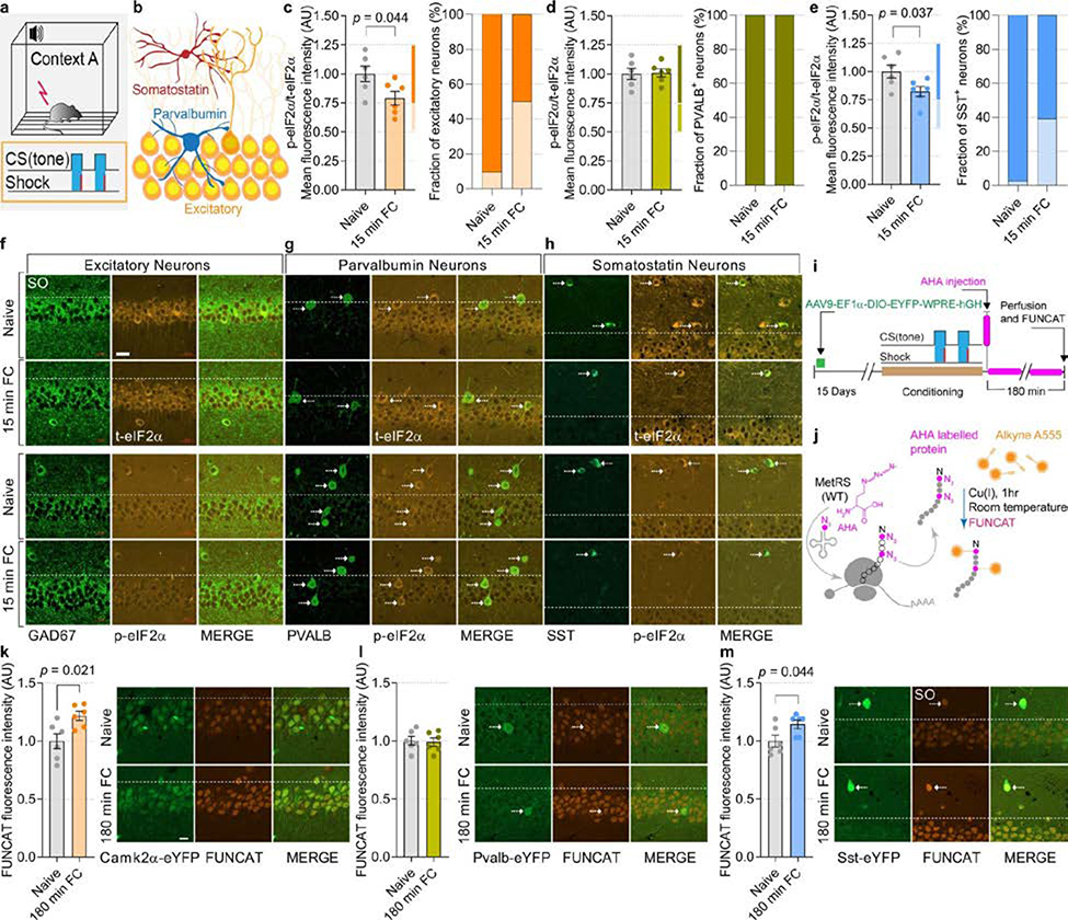
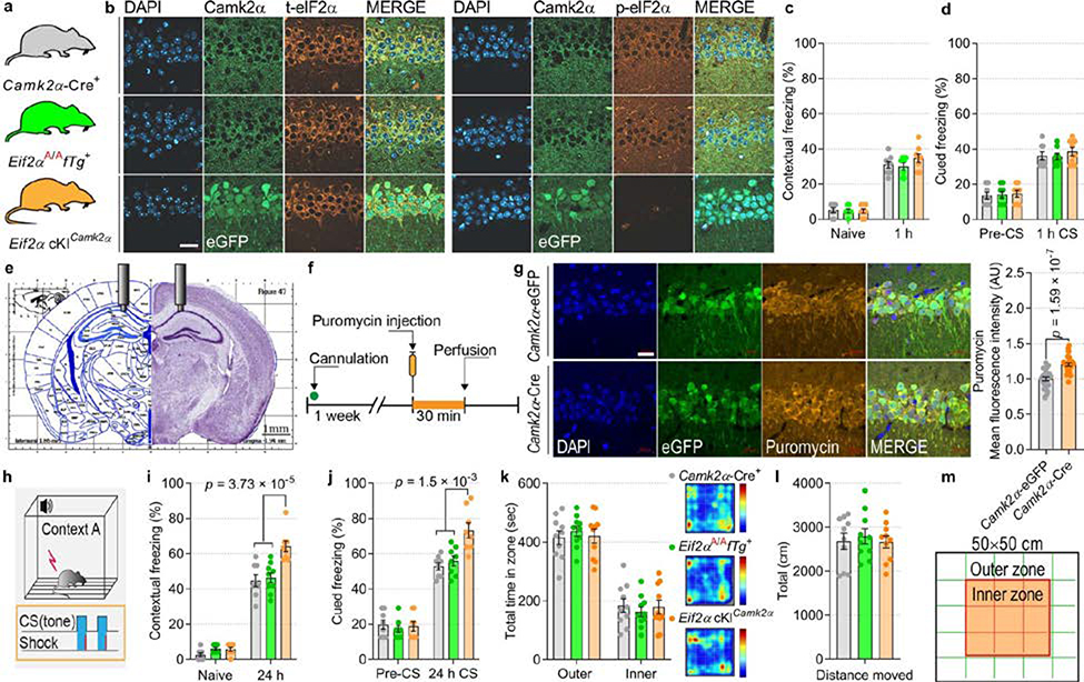
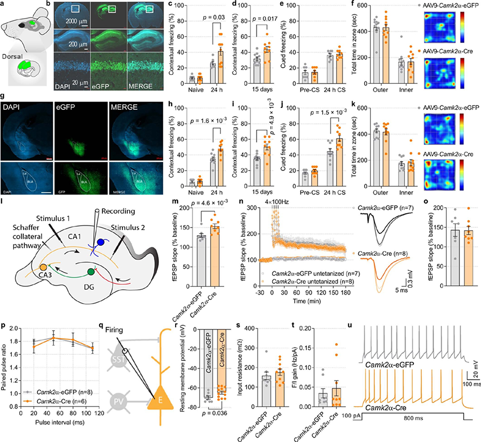
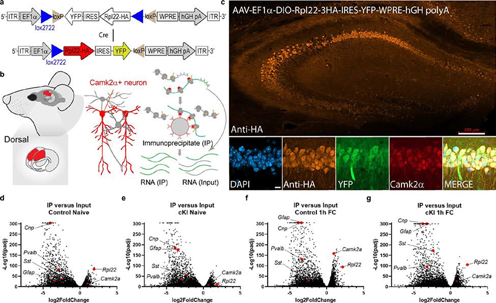
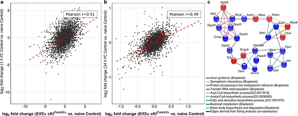
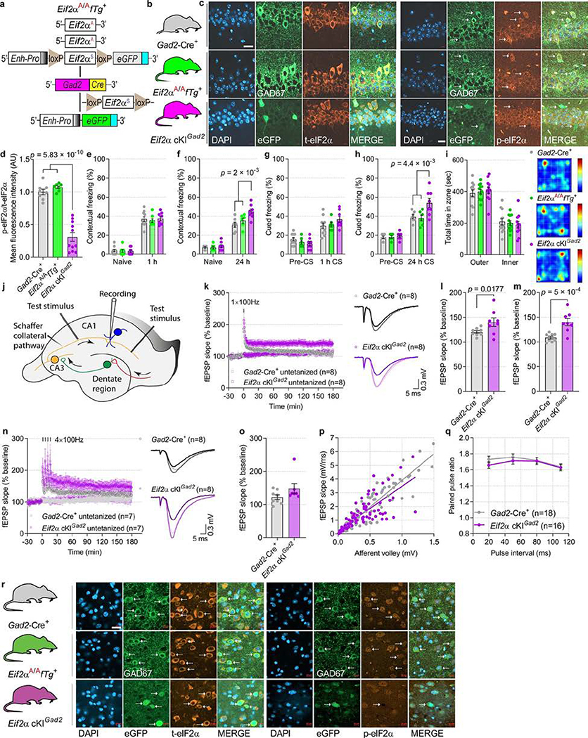
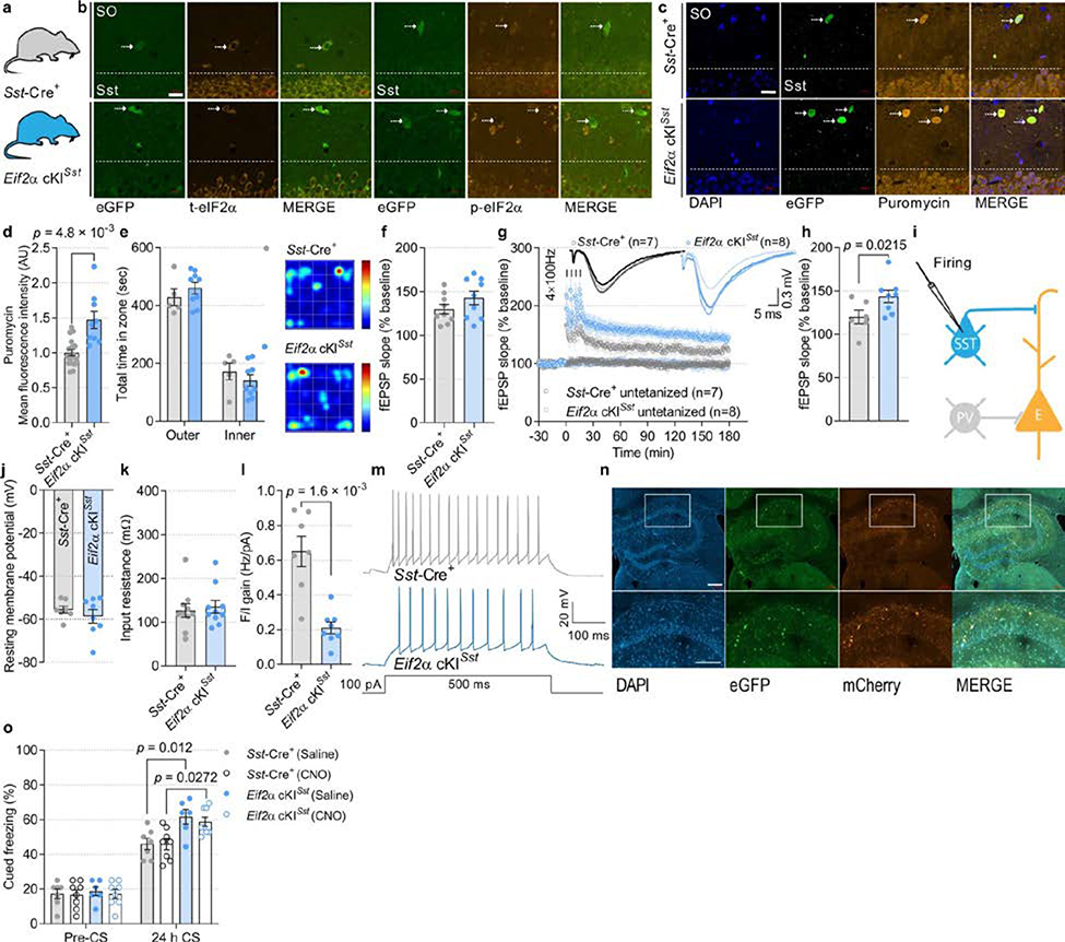
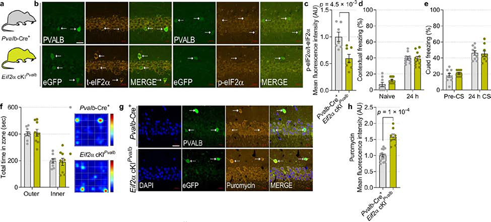
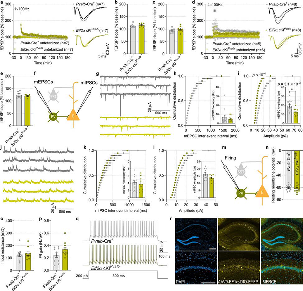
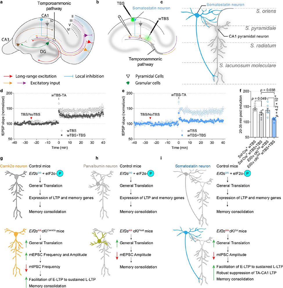
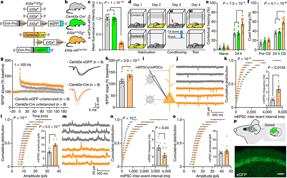
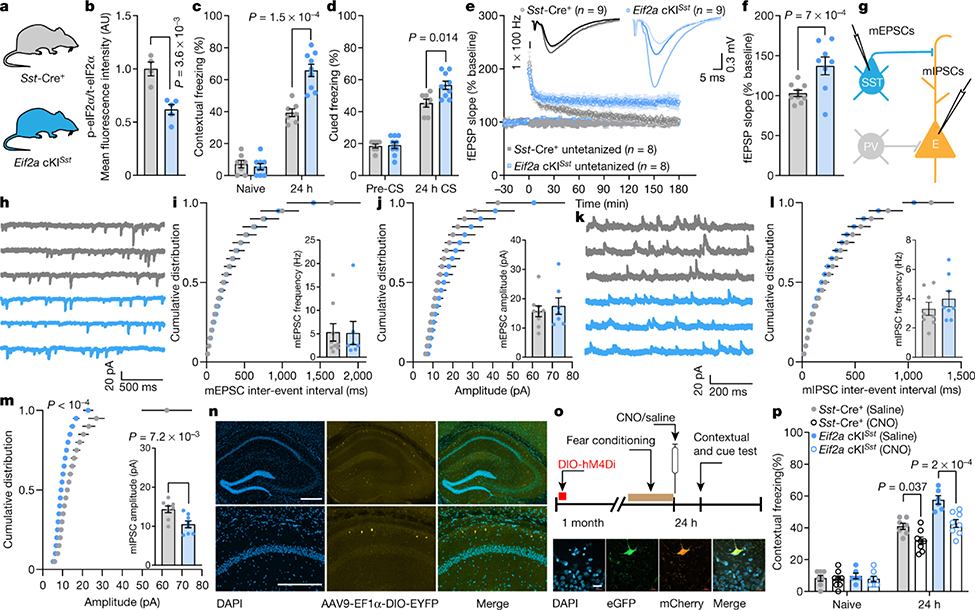
An important tenet of learning and memory is the notion of a molecular switch that promotes the formation of long-term memory1-4. The regulation of proteostasis is a critical and rate-limiting step in the consolidation of new memories5-10. One of the most effective and prevalent ways to enhance memory is by regulating the synthesis of proteins controlled by the translation initiation factor eIF211. Phosphorylation of the α-subunit of eIF2 (p-eIF2α), the central component of the integrated stress response (ISR), impairs long-term memory formation in rodents and birds11-13. By contrast, inhibiting the ISR by mutating the eIF2α phosphorylation site, genetically11 and pharmacologically inhibiting the ISR kinases14-17, or mimicking reduced p-eIF2α with the ISR inhibitor ISRIB11, enhances long-term memory in health and disease18. Here we used molecular genetics to dissect the neuronal circuits by which the ISR gates cognitive processing. We found that learning reduces eIF2α phosphorylation in hippocampal excitatory neurons and a subset of hippocampal inhibitory neurons (those that express somatostatin, but not parvalbumin). Moreover, ablation of p-eIF2α in either excitatory or somatostatin-expressing (but not parvalbumin-expressing) inhibitory neurons increased general mRNA translation, bolstered synaptic plasticity and enhanced long-term memory. Thus, eIF2α-dependent mRNA translation controls memory consolidation via autonomous mechanisms in excitatory and somatostatin-expressing inhibitory neurons.


Conflict of interest statement

Ethics declarations

Competing interests

The authors declare no competing interests.

Figures

Extended Data Fig. 1
Extended Data Fig. 1. Learning-induced reduction of p-eIF2α levels in excitatory and in the SST-expressing sub-population of GABAergic neurons.
a, Schematics of the fear conditioning experiment. b, Illustration of different neuronal subtypes in the CA1 region of the hippocampus. c, Quantitative analyses of immunofluorescence images depicting levels of p-eIF2α in CA1 excitatory neurons 15 min after fear conditioning. In the CA1 region, fear conditioning significantly reduces p-eIF2α in excitatory neurons (t 9.8 = 2.31; n = 6, 6, 12 neurons per 67,600 μm2 of mouse CA1). Graph on the right depicts a fraction of excitatory neurons showing a decrease in p-eIF2α (between 25–50% decrease) after fear conditioning. d, The p-eIF2α levels in CA1 PVALB+ neurons remain unchanged 15 min after fear conditioning (t 9.17 = 0.112; n = 6, 6, 10–11 neurons per 84,500 μm2 of mouse CA1). e, The fear conditioning significantly reduces p-eIF2α in the CA1 SST+ neurons (t 9.44 = 2.43; n = 6, 6, 6–7 neurons per 84,500 μm2 of mouse CA1). Graph on the right shows that in 39.02% of CA1 SST+ neurons, fear conditioning causes 25–50% decrease in p-eIF2α levels. fh, Representative images of p-eIF2α levels in excitatory (GAD67-negative neurons in CA1 pyramidal layer) and GABAergic neurons (PVALB- and SST-expressing) in naive and fear conditioning mice. Three independent experiments showed similar results. i, Timeline for fluorescent labelling of specific neuronal subtypes in CA1, fear conditioning paradigm and AHA injections to identify newly synthesized proteins. j, To visualize AHA-labelled proteins, the azide group (pink) of AHA is covalently bonded with fluorescent alkyne group (orange) through Cu(I) mediated fluorescent non-canonical amino acid tagging (FUNCAT). k, A 21.67 ± 3.91% increase in protein synthesis was observed in the CA1 excitatory neurons of fear conditioning compared to naive mice using FUNCAT (t 8.14 = 2.85; n = 6, 6, 12 neurons per mouse). Representative images of CA1 excitatory neurons showing FUNCAT in naive and fear conditioning mice. Two independent experiments showed similar results. l, Fear conditioning does not affect general protein synthesis in CA1 PVALB+ neurons (t 9.99 = 0.23; n = 6, 6, 6 neurons per mouse). Representative images of GABAergic PVALB+ neurons with FUNCAT. Two independent experiments showed similar results. m, In the CA1 SST+ neurons, fear conditioning causes 14.33 ± 3.64% increase in AHA labelling and FUNCAT signal intensity (t 9.09 = 2.33; n = 6, 6, 5 neurons per mouse). Representative images of CA1 GABAergic SST-expressing neurons with FUNCAT in naive and fear conditioning mice. Two independent experiments showed similar results. Stratum oriens (S.O.). Data are presented as mean ± s.e.m. in ce, km. p-values by two-tailed unpaired t-test with Welch’s correction in c, e, k and m are indicated. Points represent individual mice. Scale bars: 20 μm.
Extended Data Fig. 2
Extended Data Fig. 2. Eif2a cKICamk2a mice show enhanced protein synthesis and consolidation of contextual and auditory fear memory.
a, b, Immunofluorescent labelling of excitatory neurons shows a decrease in p-eIF2α levels in Eif2a cKICamk2a compared to control mice. Two independent experiments showed similar results. c, Short-term contextual memory is not altered in Eif2a cKICamk2a mice (F 2, 40 = 0.97; n = 8, 7, 8). d, Short-term auditory fear memory is not altered in Eif2a cKICamk2a mice (F 2, 42 = 0.46; n = 8, 8, 8). e, Illustration of cannulation site for unilateral injections of puromycin into the CA1 region. f, Experimental design of iSUnSET assay. g, Immunofluorescent images of puromycin in Eif2aA/AfTg+ mice injected with AAV9.Camk2a0.4.Cre.SV40 (Camk2a-Cre) shows enhanced protein synthesis (t 50.43 = 6.08; n = 26, 27, points represent group means). Five independent experiments showed similar results. h, Schematic representation of strong contextual and auditory fear conditioning. i, Contextual memory is enhanced in Eif2a cKICamk2a mice following a strong training protocol (F 2, 44 = 12.97; n = 8, 9, 8). j, Auditory fear memory is enhanced in Eif2a cKICamk2a mice following a strong training protocol (F 2, 42 = 7.64; n = 8, 8, 8). k, In an open field test, all groups spent a similar amount of time in the outer and inner zones (F 2, 54 = 5.6 × 10⁻¹²; n = 10, 10, 10). Representative heat-map of travelled path in an open field arena. l, The mean total distance moved (in cm) in the open field was similar in all groups (F 2, 27 = 0.19; n = 10, 10, 10). m, Open field test arena with illustration of outer and inner zones. Data are presented as mean ± s.e.m. in c, d, g, il. p-values by two-tailed unpaired t-test with Welch’s correction in g and by two-way ANOVA in i and j followed by Tukey’s multiple comparisons post hoc test are indicated. Points represent individual mice. Scale bars: 20 μm.
Extended Data Fig. 3
Extended Data Fig. 3. CA1- or amygdala-specific reduction of p-eIF2α facilitates consolidation of contextual fear memory.
a, Representative illustration of target area for p-eIF2α reduction in the dorsal hippocampus. Two independent experiments showed similar results. b, Immunohistochemistry for eGFP reporter demonstrates that deletion of WT Eif2a fTg is restricted to the CA1 region of the hippocampus. Two independent experiments showed similar results. c, d, Eif2aA/AfTg+ mice were injected with AAV9.Camk2a0.4.Cre.SV40 (AAV9-Camk2a-Cre) or control AAV9.Camk2a0.4.eGFP.WPRE.rBG (AAV9-Camk2a-eGFP) targeting the CA1. Contextual memory was significantly enhanced in AAV9-Camk2a-Cre-injected mice as compared to AAV9-Camk2a-eGFP-injected mice tested 24 h (F 1, 30 = 5.19; n = 7, 10) and 15 days (t 13.19 = 2.72; n = 10, 9) after training. e, Auditory fear memory remains unaffected (F 1, 28 = 0.18; n = 8, 8). f, There was no difference in the time spent in the outer and inner zone in an open field test (F 1, 36 = 3.63 × 10⁻⁹; n = 10, 10). Heat-map represents the group-average of path travelled in an open field arena, red = more time, blue = less time. g, Representative immunohistochemistry images demonstrating the amygdala-specific injections of the virus. Scale bars: 500 μm. Two independent experiments showed similar results. h, i, Contextual memory was significantly enhanced in Eif2aA/AfTg+ mice injected with a virus expressing AAV9-Camk2a-Cre in the amygdala tested 24 h (F 1, 32 = 11.92; n = 9, 9) and 15 days (t 12.2 = 3.42; n = 10, 9) after training. j, Auditory fear memory is also enhanced (F 1, 32 = 12.01; n = 9, 9). k, There was no difference in the time spent in the outer versus inner zone in an open field test (F 1, 36 = 8.3 × 10⁻⁹; n = 10, 10). Heat-map represents the group-average of path travelled in an open field arena. l, Experimental scheme in acute hippocampal slices: Schaffer collateral fibres were stimulated in two independent pathways with extracellular electrodes and fEPSPs were recorded in CA1 stratum radiatum. m, A single high-frequency train (1 × HFS for 1 s) elicited early LTP in slices from AAV9.Camk2a0.4.eGFP.WPRE.rBG (Camk2a-eGFP)-injected mice (E-LTP 30 min post-HFS, F 1, 7 = 8.2; n = 8, 8). n, o, L-LTP induced by four tetanic trains at 100 Hz is similar in slices from Camk2a-eGFP- and Camk2a-Cre-injected mice (o; F 1, 7 = 0.071; n = 7, 8). p, Paired-pulse facilitation (PPF) is defined as the ratio of the amplitude of the 2nd versus the 1st fEPSPs responses over increasing time intervals. The decay rates of PPF did not differ between Camk2a-eGFP- and Camk2a-Cre-injected mice, indicating intact short-term plasticity (F 1, 48 = 0.041; nmice = 8, 6, points represent group means). q, Diagram of experimental arrangement for recording intrinsic and firing properties in CA1 excitatory neurons. r, Resting membrane potential was significantly increased in Camk2a-Cre-injected mice (t 16.59 = 2.28; n = 8, 12). s, Input resistance is not affected in CAMK2α-eGFP and CAMK2α-Cre positive neurons (t 14.63 = 0.72; n = 8, 10). t, The number of action potential in response to incremental somatic depolarization (F/I gain relationship) is not different between CAMK2α-eGFP+ and CAMK2α-Cre+ excitatory neurons (t 13 = 0.56; n = 8, 9). u, Examples of traces obtained in response to 100 pA current injection in CAMK2α-eGFP+ or the CAMK2α-Cre+ excitatory neurons. Data are presented as mean ± s.e.m. in cf, hk, mp, rt. p-values by two-way ANOVA in c, h, j, followed by Sidak’s multiple comparisons post hoc test; two-tailed unpaired t-test with Welch’s correction in d, i, r; two-way ANOVA (repeated measurements) in m followed by Sidak’s multiple comparisons post hoc test are indicated. Points represent individual mice unless stated otherwise.
Extended Data Fig. 4
Extended Data Fig. 4. RiboTag.
a, Schematic representation of the AAV-EF1α-DIO-Rpl22–3HA-IRES-YFP-WPRE-hGH polyA (AAV-DIO-RiboTag) viral vector. We developed a Cre-dependent RiboTag viral vector based on AAV-DIO-RiboTag41. In the presence of Cre recombinase under a Camk2a promoter, the Rpl22-HA-IRES-YFP cassette is rearranged to the ON configuration that allows the Rpl22-HA expression under the Eef1a promoter in CAMK2α-positive excitatory neurons. b, Representative illustration of target area for bilateral AAV-DIO-RiboTag injection in the dorsal hippocampus. Tagged ribosomes from Rpl22-HA-expressing CAMK2α-positive excitatory neurons were recovered from total lysate by immunoprecipitation (IP) with magnetic beads conjugated to anti-HA antibodies. RNAs were extracted from the IP and total lysate. c, Immunohistochemistry for HA staining demonstrating that expression of AAV-DIO-RiboTag viral vector is restricted to the CAMK2α-positive excitatory neurons. Two independent experiments showed similar results. Scale bars: 200 μm. dg, Comparison of RNA-Seq from IP to input showed an enrichment for excitatory neurons marker Camk2a and depletion of oligodendrocytes marker 2′, 3′-cyclic-nucleotide 3′ phosphodiesterase (CNPase, Cnp), astrocytes marker (Gfap) and markers of inhibitory neurons Sst and Pvalb, indicating a successful immunoprecipitation from excitatory neurons.
Extended Data Fig. 5
Extended Data Fig. 5. Analysis of excitatory neurons-specific translational landscape and proteomics from hippocampi of Eif2a cKICamk2a mice and following fear conditioning learning.
a, Pairwise Pearson correlation coefficient between log2 fold changes in translation efficiency comparing the genotype effect (naive Eif2a cKICamk2a vs. naive control) and learning effect (1 h fear conditioning control vs. naive control) using all translated mRNAs (11,805 transcripts). b, Pairwise Pearson correlation coefficient between log2 fold changes in protein levels comparing the genotype effect (naive Eif2a cKICamk2a vs. naive control) and learning effect (24 h fear conditioning control vs. naive control) using all CA1 expressed proteins (5,237 proteins). Pearson correlation coefficients were significant in both cases (P < 0.0001), dots marked red denote transcripts/proteins differentially translated/expressed. c, Functional enrichment in protein subset similarity differentially expressed between proteomes of the two genotypes (52 proteins), and between proteomes of treatment vs. naive mice from both genotypes (27 proteins). GO ontologies, BioPlanet pathways and Reactome pathways showing significant enrichment (FDR adjusted P < 0.1) are presented. Nodes represent proteins, edges represent pathway/ontology relatedness. Black edges are links found by String analysis and represent co-expression evidence. Blue nodes denote reduced level of expression; red nodes denote increased level of expression. * denote proteins differentially expressed following 24 h fear conditioning in both mouse genotypes, but not between naive mice.
Extended Data Fig. 6
Extended Data Fig. 6. Consolidation of contextual and auditory fear memory and L-LTP induction is facilitated in Eif2a cKIGad2 mice.
a, b, Schematic of the breeding strategy to generate Eif2a cKIGad2 mice by crossing Eif2aA/AfTg+ mice with a Gad2-Cre+ transgenic line and the three genotypes of mice used in experiments. c, Immunofluorescent labelling of GAD67-positive neurons shows a decrease in p-eIF2α but no change in t-eIF2α in the CA1 region of Eif2a cKIGad2 compared to control mice. Two independent experiments showed similar results. eGFP expression indicates the successful Cre recombinase-mediated deletion of WT Eif2a fTg in GAD2+ GABAergic inhibitory neurons. d, Quantitative analyses of p-eIF2α levels in control and Eif2a cKIGad2 mice (F 2, 23 = 61.6; n = 8, 7, 11, points represent means per mouse). In Eif2a cKIGad2 mice, deletion of the floxed transgene significantly reduced p-eIF2α levels. e, Short-term contextual fear memory is not enhanced in Eif2a cKIGad2 mice (F 2, 44 = 0.084; n = 10, 7, 8). f, Long-term contextual fear memory is enhanced in Eif2a cKIGad2 mice (F 2, 36 = 7.41; n = 7, 6, 8). g, Short-term auditory fear memory is not altered in Eif2a cKIGad2 mice (F 2, 42 = 0.24; n = 10, 6, 8). h, Long-term auditory fear memory is enhanced in Eif2a cKIGad2 mice (F 2, 36 = 6.32; n = 7, 6, 8). i, In an open field test, all groups spent a similar amount of time in the outer versus inner zones (F 2, 64 = 9.8 × 10⁻¹²; n = 12, 12, 11). Representative group-average heat-map of travelled path in an open field arena. j, Experimental scheme in acute hippocampal slices: Schaffer collateral fibres were stimulated in two independent pathways with extracellular electrodes and fEPSPs were recorded in CA1 stratum radiatum. km, A single high-frequency train elicited E-LTP and generated a sustained L-LTP in Eif2a cKIGad2 slices (F 1, 7 = 6.9; n = 8, 8). no, L-LTP induced by four high frequency trains is similar in Gad2-Cre+ and Eif2a cKIGad2 slices (o, L-LTP, F 1, 7 = 3.03; n = 8, 7). p, fEPSP versus fibre volley were fitted similarly by linear regression (R2 = 0.84 for Gad2-Cre+ and R2 = 0.58 for Eif2a cKIGad2 slices). q, PPF did not differ between Gad2-Cre+ and Eif2a cKIGad2 slices (two-way ANOVA, F 1, 128 = 1.54; nmice = 18, 16, points represent group means). r, Immunofluorescent labelling of inhibitory interneurons in amygdala showing a decrease in p-eIF2α levels. Two independent experiments showed similar results. Data are presented as mean ± s.e.m. in di, kq. p-values by one-way ANOVA followed by Tukey’s multiple comparisons post hoc test in d; two-way ANOVA in f and h followed by Tukey’s multiple comparisons post hoc test; two-way ANOVA (repeated measurements) in l and m followed by Sidak’s multiple comparisons post hoc test are indicated. Points represent individual mice unless stated otherwise. Scale bars: 20 μm. Calibration: 0.3 mV, 5 ms.
Extended Data Fig. 7
Extended Data Fig. 7. Somatostatin-specific reduction of p-eIF2α facilitates L-LTP.
a, b, Immunofluorescent labelling of total and p-eIF2α in SST+ neurons in the CA1 region shows reduced p-eIF2α levels in cKI mice. Two independent experiments showed similar results. c, d, iSUnSET shows enhanced protein synthesis in Eif2a cKISst mice (t 10.11 = 3.6; n = 18, 9, points represent means per mouse). Two independent experiments showed similar results. e, Both groups spent a similar amount of time in the outer and inner zones during an open field test (F 1, 24 = 7.1 × 10⁻¹⁰; n = 5, 9). Representative group-average heat-map of travelled path in the test arena. f, A single high-frequency train (1 × HFS for 1 s) induces similar E-LTP (30 min post-HFS) in Sst-Cre+ and Eif2a cKISst slices. g, h, Four tetanic trains at 100 Hz induce a facilitated L-LTP in Eif2a cKISst slices (h, L-LTP, F 1, 7 = 9.531; n = 6, 8). i, Diagram of experimental arrangement for recording intrinsic and firing properties in somatostatin neuron. j, Resting membrane potential (t 10.43 = 0.86; n = 7, 8). k, Input resistance (t 17.92 = 0.38; n = 10, 10). l, The frequency-current (F/I gain) relationship is reduced in Eif2a cKISst mice (t 7.825 = 4.7; n = 7, 8). m, Example of traces obtained in response to 100 pA current injection in somatostatin-expressing interneurons in Sst-Cre+ and Eif2a cKISst mice. n, Representative illustration of target area for the injection of AAV9-EF1α-DIO-EYFP-WPRE-hGH to label SST-expressing neurons in the dorsal hippocampus. Two independent experiments showed similar results. o, Overview image from an Eif2a cKISst mouse in which the CA1 was injected with the Cre-dependent AAV9-DIO-hM4D(Gi)-mCherry (hM4Di) virus. Two independent experiments showed similar results. p, No effect on cued freezing by silencing CA1 SST+ neurons following fear conditioning in control and Eif2a cKISst mice (F 3, 50 = 4.38; n = 7, 8, 6, 8). Data are presented as mean ± s.e.m. in dh, jl, p. p-values by two-tailed unpaired t-test with Welch’s correction in d and l; and by mixed-effects model REML in h followed by Sidak’s multiple comparisons post hoc test are indicated; two-way ANOVA in p followed by Sidak’s multiple comparisons post hoc test. Points represent individual mice unless stated otherwise. Scale bars: 200 μm. Calibration: 0.3 mV, 5 ms.
Extended Data Fig. 8
Extended Data Fig. 8. Eif2a cKIPvalb mice show no change in consolidation of contextual and auditory fear memory.
a, Diagram depicts the genotypes of mice used in the experiments. b, c, Immunofluorescent labelling of total and p-eIF2α in PVALB+ neurons shows reduced p-eIF2α levels in Eif2a cKIPvalb mice (t 11.86 = 3.5; n = 7, 7, points represent means per mouse). Two independent experiments showed similar results. d, Long-term contextual fear memory is not changed in Eif2a cKIPvalb mice (F 1, 34 = 0.85; n = 9, 10). e, Long-term auditory fear memory remains intact in Eif2a cKIPvalb mice (F 1, 32 = 0.124; n = 9, 9). f, In an open field test, both groups spent a similar amount of time in the outer and inner zones (F 1, 32 = 6.6 × 10⁻¹²; n = 9, 9). Representative heat-map of travelled path in an open field arena. g, h, Immunohistochemistry of puromycin in Eif2a cKIPvalb shows enhanced protein synthesis (t 10.67 = 5.85; n = 15, 8, points represent means per mouse). Two independent experiments showed similar results. Data are presented as mean ± s.e.m. in cf, h. p-values by two-tailed unpaired t-test with Welch’s correction in c and h are indicated. Points represent individual mice unless stated otherwise. Scale bars: 20 μm.
Extended Data Fig. 9
Extended Data Fig. 9. No change in L-LTP threshold in Eif2a cKIPvalb mice.
ac, A 1 × HFS induced short-lasting E-LTP in Pvalb-Cre+ and Eif2a cKIPvalb hippocampal slices (E-LTP, F 1, 6 = 1.917; n = 7, 7). d, e, L-LTP induced by four tetanic trains at 100 Hz is similar in Pvalb-Cre+ and Eif2a cKIPvalb slices (e; L-LTP, t 7.542 = 0.6136; n = 6, 5). f, Diagram of experimental arrangement with whole cell recording of mEPSCs (in PVALB+) and mIPSCs (in excitatory neurons). g, Sample traces of mEPSCs. h, Cumulative distribution of mEPSC inter-event intervals (nmice = 13, 11, points represent group means). Inset shows similar mEPSC frequency in Pvalb-Cre+ and Eif2a cKIPvalb (t 15.41 = 0.84; n = 13, 11). i, Cumulative distribution of mEPSC amplitudes (nmice = 13, 11, points represent group means). Bar graph in inset shows reduced mEPSC amplitude in Eif2a cKIPvalb (t 20.34 = 3.35; n = 13, 11). j, Sample traces of mIPSCs. k, Cumulative distribution of mIPSC inter-event intervals (nmice = 10, 10, points represent group means). Inset shows similar mIPSC frequency (t 17.83 = 0.52; n = 10, 10). l, Cumulative distribution of mIPSC amplitudes (nmice = 10, 10, points represent group means). Inset, mIPSC amplitude is not changed between the groups (t 13.70 = 2.04; n = 10, 10). m, Diagram of experimental arrangement for recording intrinsic and firing properties from parvalbumin neuron. n, Resting membrane potential (t 17.55 = 0.79; n = 10, 10). o, Input resistance (t 17.92 = 0.38; n = 10, 10). p, The F/I gain relationship is similar in the Pvalb-Cre+ and Eif2a cKIPvalb mice (t 18 = 1.72; n = 10, 10). q, Representative traces obtained in response to 200 pA current injection in the parvalbumin-expressing interneurons from Pvalb-Cre+ and Eif2a cKIPvalb mice. r, Representative illustration of target area for the injection of AAV9-EF1α-DIO-EYFP-WPRE-hGH to label PVALB-expressing neurons in the dorsal hippocampus. Two independent experiments showed similar results. Data are presented as mean ± s.e.m. in ae, h, i, k, l, np. p-values by Kolmogorov–Smirnov test in i and by two-tailed unpaired t-test with Welch’s correction in i(inset) are indicated. Points represent individual mice unless stated otherwise. Scale bars: 200 μm. Calibration: 0.3 mV, 5 ms.
Extended Data Fig. 10
Extended Data Fig. 10. TBS-induced LTP in CA1 oriens/alveus (O/A) somatostatin-expressing interneurons exerts a more robust suppression of TA-CA1 LTP in Eif2a cKISst mice.
ac, Schematic of experimental setup in acute hippocampal slices: Temporoammonic afferents from the entorhinal cortex were stimulated by a weak TBS (wTBS) in presence or absence of prior TBS in CA1 oriens/alveus (O/A) region and fEPSPs were recorded in stratum lacunosum-moleculare of CA1. d, In Sst-Cre+ hippocampal slices, TBS in CA1-O/A region suppresses LTP of TA-CA1 field excitatory postsynaptic potentials (fEPSPs) when compared to slices with No-TBS in CA1-O/A region. e, In Eif2a cKISst hippocampal slices, TBS in CA1-O/A region also reduces TA-LTP relative to slices with No-TBS in CA1-O/A region. f, Summary plots of normalized LTP magnitude in TA-CA1 pathway. TBS at CA1-O/A supress TA-CA1 pathway in Sst-Cre+ (F 3, 24 = 11.85; n = 6, 8, points represent individual mice) and Eif2a cKISst (F 3, 24 = 11.85; n = 7, 7, points represent individual mice). However, the magnitude of suppression is larger in Eif2a cKISst than in Sst-Cre+ mice (F 3, 24 = 11.85; n = 8, 7, points represent individual mice). g, Reduction of p-eIF2α in CAMK2α-expressing excitatory neurons facilitates L-LTP and excitatory synaptic transmission (amplitude and frequency of mEPSCs), reduces inhibitory synaptic transmission (frequency of mIPSCs) and enhances memory consolidation (fear conditioning). h, Ablation of p-eIF2α in parvalbumin-expressing neurons causes a reduction in mEPSC amplitude without affecting threshold for L-LTP induction or consolidation of fear memory. i, Depletion of p-eIF2α in somatostatin-expressing neurons facilitates L-LTP and consolidation of fear memory by reducing the amplitude of mIPSCs in pyramidal neurons and enhancing suppression of LTP in the TA pathway. Data are presented as mean ± s.e.m. in df. p-values by one-way ANOVA followed by Tukey’s multiple comparisons post hoc test in f are indicated.
Fig. 1:
Fig. 1:. Reduction of p-eIF2α in excitatory neurons facilitates memory consolidation, LTP and excitatory synaptic transmission, and reduces inhibitory synaptic transmission.
a, The genetic components of the Eif2aA/AfTg+ mice. eGFP expression in the Eif2a cKICamk2a mice indicates successful Cre recombinase-mediated deletion of the wild-type Eif2a floxed transgene in excitatory neurons. b, The three genotypes of mice used in these experiments. c, Quantitative analyses of immunofluorescence images showing p-eIF2α as a proportion of total (t-)eIF2α in control and Eif2a cKICamk2a mice (F2,45 = 493.75, n = 13, 17, 18, points represent means per mouse). In Eif2a cKICamk2a mice, p-eIF2α levels are reduced owing to the deletion of the floxed transgene. d, Schematic of weak contextual and auditory fear conditioning. e, Long-term contextual memory is enhanced in Eif2a cKICamk2a mice (F2,40 = 8.66, n = 7, 8, 8). f, Long-term auditory fear memory is enhanced in Eif2a cKICamk2a mice (F2,40 = 26.81, n = 7, 8, 8). g, h, A single high-frequency train (1 × HFS for 1 s) generated sustained L-LTP in slices from mice injected with Camk2a–Cre (h, L-LTP 180 min post-HFS, F1,7 = 8.2, n = 8, 8). i, Whole-cell recording for mEPSCs and mIPSCs and firing. PV, PVALB+ neuron. j, Representative traces of mEPSCs from CAMK2α–eGFP+ and CAMK2α–Cre+ neurons. k, Cumulative distribution of mEPSC inter-event intervals recorded from CA1 excitatory neurons in mice injected with Camk2a–eGFP or Camk2a–Cre (nmice = 8, 8, points represent group means). Inset, increased mean frequency of mEPSCs in mice injected with Camk2a–Cre (t7.59 = 3.2, n = 8, 8). l, Cumulative distribution plots showing mEPSC amplitude (nmice = 8, 7, points represent group means). Inset, mEPSC amplitude is increased in Camk2a–Cre-injected mice (t12.19 = 3.64, n = 8, 7). m, Representative mIPSC traces. n, Cumulative distribution of mIPSC inter-event intervals recorded from CA1 excitatory neurons in mice injected with Camk2a–eGFP or Camk2a–Cre (nmice = 9, 11, points represent group means). Inset, mIPSC frequency was reduced in mice injected with Camk2a–Cre (t16.71 = 2.22, n = 9, 11). o, Cumulative distribution of mIPSC amplitudes (nmice = 9, 11, points represent group means). Inset, average mIPSC amplitude (t12.69 = 0.75, n = 9, 11). p, Target area for injection of Camk2a–eGFP and Camk2a–Cre and representative image of GFP-labelled CAMK2α+ excitatory neurons in CA1 of dorsal hippocampus; representative of five independent experiments. Mean ± s.e.m. (c, eh, k, l, n, o). One-way ANOVA (c), two-way ANOVA (e, f) followed by Tukey’s multiple comparisons post-hoc test; two-way ANOVA (repeated measures) (g, h) with Sidak’s multiple comparisons post-hoc test; Kolmogorov–Smirnov test (k, l, n); two-tailed unpaired t-test with Welch’s correction (insets in k, l, n). Points represent individual mice unless stated otherwise. Scale bar, 100 μm.
Fig. 2:
Fig. 2:. Reduction in p-eIF2α in SST+ neurons facilitates memory consolidation and LTP, and reduces inhibitory synaptic transmission onto CA1 pyramidal neurons.
a, The two genotypes of mice used. b, Quantification of total and p-eIF2α in CA1 SST+ neurons shows reduced p-eIF2α in Eif2a cKISst mice (t5.79 = 4.73, n = 4, 5, points represent means per mouse). c, d, Long-term contextual memory (c; F1, 28 = 19.19, n = 7, 9) and long-term auditory fear memory (d; F 1, 28 = 6.86, n = 7, 9) are enhanced in Eif2a cKISst mice. e, f, A single high-frequency train (1 × HFS for 1 s) leads to sustained LTP in Eif2a cKISst slices 180 min later (f; L-LTP, F1, 8 = 8.134, n = 9, 8). g, Experimental arrangement for whole-cell recording of mEPSCs (from SST+ neurons) and mIPSCs (from excitatory neurons). h, Representative traces of mEPSCs recorded from Sst-Cre+ and Eif2a cKISst mice. i, Cumulative distribution of mEPSC inter-event intervals (nmice = 9, 7, points represent group means). Inset, mEPSC frequency (t11.85 = 0.05, n = 9, 7). j, Cumulative distribution of mEPSC amplitudes (nmice = 9, 7, points represent group means). Inset, average mEPSC amplitude (t10.69 = 0.52, n = 9, 7). k, Sample traces of mIPSCs recorded from CA1 excitatory neurons in Sst-Cre+ and Eif2a cKISst mice. l, Cumulative distribution of mIPSC inter-event intervals (nmice = 8, 8, points represent group means). Inset, mIPSC frequency (t13.57 = 0.98, n = 8, 8). m, Cumulative distribution of mIPSC amplitudes (nmice = 8, 8, points represent group means). Inset, mIPSC amplitude was reduced in Eif2a cKISst mice (t14 = 3.14, n = 8, 8). n, Representative illustration of target area for the injection of AAV9-EF1α -DIO-EYFP-WPRE-hGH. Image shows labelled SST-expressing neurons in the dorsal hippocampus. Four independent experiments showed similar results. o, Top, timeline of DREADD experiments in fear conditioning paradigm immediately followed by injection of saline (control) or clozapine-N-oxide (CNO (silenced)). Bottom, images of brain section from an Eif2a cKISst mouse in which CA1 was injected with Cre-dependent AAV9-DIO-hM4D(Gi)-mCherry (hM4Di). Representative of two independent experiments. p, Chemogenetic inhibition of SST+ neurons in CA1 by CNO administration immediately after training attenuates consolidation of contextual fear memory in control and Eif2a cKISst mice (F3,50 = 13.7, n = 7, 8, 6, 8). Mean ± s.e.m. Two-tailed unpaired t-test with Welch’s correction (b, insets in i, j, l, m); two-way ANOVA (c, d, p) followed by Sidak’s multiple comparisons post-hoc test; mixed-effects model (restricted maximum likelihood (REML); e, f) followed by Sidak’s multiple comparisons post-hoc test; Kolmogorov–Smirnov test (m). Points represent individual mice unless stated otherwise. Scale bars, 200 μm.

Comment in

References

    1. Santini E, Huynh TN & Klann E Mechanisms of translation control underlying long-lasting synaptic plasticity and the consolidation of long-term memory. Prog. Mol. Biol. Transl. Sci 122, 131–167 (2014). - PMC - PubMed
    1. Gal-Ben-Ari S et al. Consolidation and translation regulation. Learn. Mem 19, 410–422 (2012). - PMC - PubMed
    1. Costa-Mattioli M, Sossin WS, Klann E & Sonenberg N Translational control of long-lasting synaptic plasticity and memory. Neuron 61, 10–26 (2009). - PMC - PubMed
    1. Costa-Mattioli M & Walter P The integrated stress response: from mechanism to disease. Science 368, eaat5314 (2020). - PMC - PubMed
    1. Sonenberg N, Morgan MA, Merrick WC & Shatkin AJ A polypeptide in eukaryotic initiation factors that crosslinks specifically to the 5’-terminal cap in mRNA. Proc. Natl Acad. Sci. USA 75, 4843–4847 (1978). - PMC - PubMed

Publication types

MeSH terms