. 2020 Sep 21:2020:2650873.
doi: 10.1155/2020/2650873. eCollection 2020.

Secondary Vitrectomy with Internal Limiting Membrane Plug due to Persistent Full-Thickness Macular Hole OCT-Angiography and Microperimetry Features: Case Series

Affiliations

Secondary Vitrectomy with Internal Limiting Membrane Plug due to Persistent Full-Thickness Macular Hole OCT-Angiography and Microperimetry Features: Case Series

Dominika Wrzesińska et al. J Ophthalmol. .

Abstract

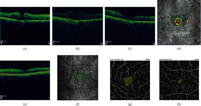
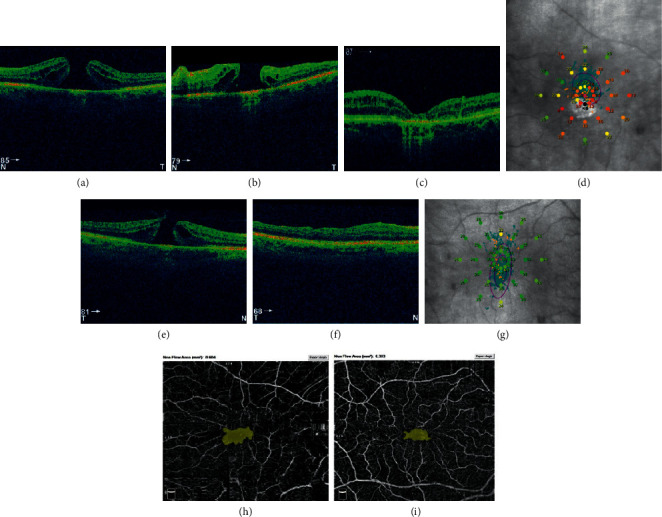
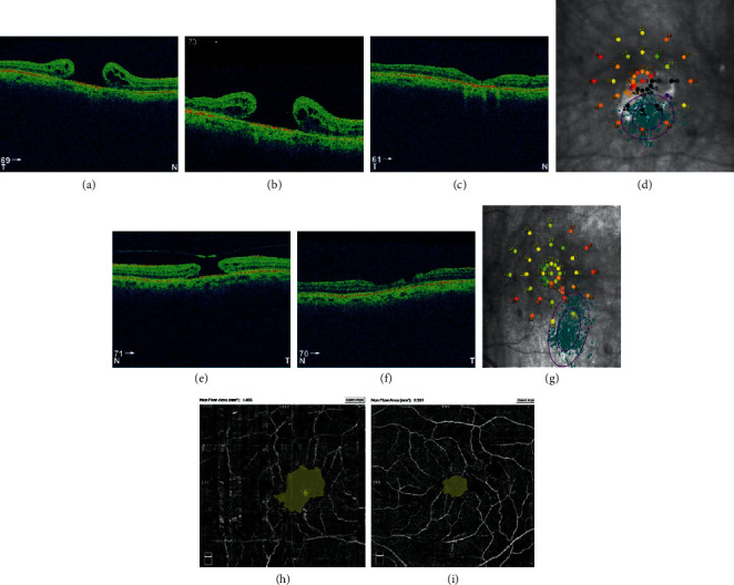
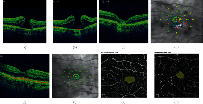
Purpose: To study the features in OCT-angiography and microperimetry in eyes with persistent full-thickness macular hole (FTMH) closed with the secondary plana vitrectomy (PPV) with autologous internal limiting membrane (ILM) plug.

Methods: Secondary PPV was performed with closing the persistent FTMH with ILM plug, C3F8 tamponade, and face-down positioning. Four patients were followed for 6 months with best corrected visual acuity (BCVA) measurement, SD-OCT and OCT-A, and microperimetry. The results were compared with the fellow eye; in two patients, it was the healthy eye, and in two remaining eyes, successfully closed FTMH after primary PPV.

Results: ILM flap was integrated in all cases with V-shape of closure, and atrophy was found in one case, with the largest diameter of FTMH. BCVA improved in two cases and remained the same in two cases. In OCT-A, the area of foveal avascular zone (FAZ) was larger, and foveal vessel density (FVDS) was smaller in eyes after secondary PPV in comparison to fellow eyes. In microperimetry, retinal sensitivity was lower in eyes after secondary PPV, and eccentric fixation was found in 2 of 4 patients.

Conclusion: Although the anatomical results of repeated surgeries of FTMH with ILM plug are favorable, visual function results may be limited. Secondary closure of FTMH with ILM plug may lead to atrophy, changes in the macular vasculature, and eccentric fixation. The trial is registered with NCT03701542.


Conflict of interest statement

The authors declare that they have no conflicts of interest.

Figures

Figure 1
Figure 1
(a) Full-thickness macular hole (FTMH) in the right eye before surgery. Hole diameter equal to 475 µm. (b) Unclosed FTMH in the right eye 1 month after vitrectomy. Poor image quality caused by the rest of SF6 gas. Hole diameter equal to 452 µm. (c) Closed FTMH 3 months after secondary vitrectomy in the right eye. (d) Postoperative microperimetric results of the right eye. (e) OCT image of macula of the healthy, left eye. (f) Normal microperimetry result of the left eye. Postoperative FAZ measurement using a nonflow software. Foveal avascular zone (FAZ) of the right eye (g) twice operated, increased significantly compared to FAZ of the healthy, left eye (h).
Figure 2
Figure 2
Left eye of the patient no. 2. (a) FTMH before surgery. Diameter equal to 485 µm. (b) Unclosed FTMH after vitrectomy with diameter 765 µm. (c) Closed FTMH with intraretinal cysts, 6 months after secondary PPV. (d) Postoperative microperimetric results. FTMH of the right eye of patient no. 2 before (e) and after primary, successful pars plana vitrectomy (PPV) (f). (g) Postoperative microperimetric results of the right eye. (h) Postoperative FAZ of left eye, operated twice. Nonflow area: 0.604 mm2, BCVA 0.1. (i) Postoperative foveal avascular zone (FAZ) of the right eye after one vitrectomy, nonflow area: 0.363 mm2, BCVA equal to 0.8.
Figure 3
Figure 3
(a) Preoperative FTMH of the right eye of patient no. 3 with diameter 765 µm. (b) Unclosed FTMH after primary vitrectomy, 940 µm of diameter. (c) After secondary vitrectomy with atrophy. (d) Postoperative microperimetric results, BCVA 0.05. FTMH of the left eye with diameter 660 µm before (e) and after primary, successful vitrectomy (f). (g) Postoperative microperimetric results, BCVA 0.1. (h) Postoperative foveal avascular zone (FAZ) of the right eye, nonflow area: 1.856 mm. (i) Postoperative FAZ of the left eye, nonflow area: 0.557 mm2.
Figure 4
Figure 4
(a) Preoperative FTMH of the left eye of patient no. 4 with diameter 588 µm. (b) Unclosed FTMH 1 month after vitrectomy, hole diameter equal to 726 µm. (c) V-shaped closure of FTMH after secondary vitrectomy. (d) Postoperative microperimetric results of the left eye. (e) OCT image of macula of the healthy, right eye. (f) Normal microperimetry result of the right eye. (g) Postoperative FAZ of the left eye, nonflow area: 0.818 mm2. (h) FAZ of the right eye, nonflow area: 0.653 mm2.

References

    1. Wrzesińska D., Nowomiejska K., Nowakowska D., et al. Vertical and horizontal m-charts and microperimetry for assessment of the visual function in patients after vitrectomy with ilm peeling due to stage 4 macular hole. Journal of Ophthalmology. 2019;2019 doi: 10.1155/2019/4975973.4975973 - DOI - PMC - PubMed
    1. Reibaldi M., Rizzo S., Avitabile T., et al. Iatrogenic retinal breaks in 25-gauge vitrec to my under air compared with the standard 25-gauge system for macular diseases. Retina. 2014;34(8):1617–1622. doi: 10.1097/iae.0000000000000112. - DOI - PubMed
    1. Reid G. A., McDonagh N., Wright D. M., Yek J. T. O., Essex R. W., Lois N. First failed macular hole surgery or reopening of a previously closed hole. Retina. 2020;40(1):1–15. doi: 10.1097/iae.0000000000002564. - DOI - PMC - PubMed
    1. Kelly N. E., Wendel R. T. Vitreous surgery for idiopathic macular holes. Archives of Ophthalmology. 1991;109(5):654–659. doi: 10.1001/archopht.1991.01080050068031. - DOI - PubMed
    1. Ma F. Y., Xi R. J., Chen P. F., Hao Y. H. Free autologous internal limiting membrane transplantation in the treatment of large macular hole. International Journal of Ophthalmology. 2019;12(5):848–851. doi: 10.18240/ijo.2019.05.24. - DOI - PMC - PubMed

Associated data