Functionalized Scaffold and Barrier Membrane with Anti-BMP-2 Monoclonal Antibodies for Alveolar Ridge Preservation in a Canine Model
- PMID: 33029518
- PMCID: PMC7530509
- DOI: 10.1155/2020/6153724
Functionalized Scaffold and Barrier Membrane with Anti-BMP-2 Monoclonal Antibodies for Alveolar Ridge Preservation in a Canine Model
Abstract
Introduction: The aim of this study was to investigate the ability of anti-bone morphogenetic protein 2 monoclonal antibody (anti-BMP-2 mAb) to functionalize scaffolds to mediate bone regeneration in a canine model.
Materials and methods: The mandibular right premolar 4 (PM4) was extracted in eight beagle dogs and grafted with anti-BMP-2 mAb+anorganic bovine bone mineral with 10% collagen (ABBM-C) and porcine bilayer native collagen membrane (CM). The ABBM-C and CM were functionalized with either anti-BMP-2 mAb (test group) or an isotype matched control mAb (control group). Animals were euthanized at 12 weeks for radiographic, histologic, and histomorphometric analyses. Outcomes were compared between groups.
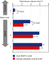
Results: 3D imaging using cone beam computed tomography (CBCT) revealed that sites treated with ABBM-C and CM functionalized with anti-BMP-2 mAb exhibited significantly more remaining bone width near the alveolar crest, as well as buccal bone height, compared with control groups. Histologic and histomorphometric analyses demonstrated that in anti-BMP-2 mAb-treated sites, total tissue volume was significantly higher in the coronal part of the alveolar bone crest compared with control sites. In anti-BMP-2 mAb-treated sites, bone formation was observed under the barrier membrane.
Conclusion: Functionalization of the ABBM-C scaffold and CM appeared to have led to bone formation within healing alveolar bone sockets.
Copyright © 2020 Seiko Min et al.
Conflict of interest statement
The authors declare no conflicts of interest.
Figures


Similar articles
-
Collagen sponge functionalized with chimeric anti-BMP-2 monoclonal antibody mediates repair of nonunion tibia defects in a nonhuman primate model: An exploratory study.J Biomater Appl. 2017 Oct;32(4):425-432. doi: 10.1177/0885328217733262. J Biomater Appl. 2017. PMID: 28992803
-
Alveolar ridge dimensional changes following ridge preservation procedure using SocketKAP(™) : exploratory study of serial cone-beam computed tomography and histologic analysis in canine model.Clin Oral Implants Res. 2016 Sep;27(9):1144-51. doi: 10.1111/clr.12711. Epub 2015 Dec 13. Clin Oral Implants Res. 2016. PMID: 26660705
-
Collagen Sponge Functionalized with Chimeric Anti-BMP-2 Monoclonal Antibody Mediates Repair of Critical-Size Mandibular Continuity Defects in a Nonhuman Primate Model.Biomed Res Int. 2017;2017:8094152. doi: 10.1155/2017/8094152. Epub 2017 Mar 16. Biomed Res Int. 2017. PMID: 28401163 Free PMC article.
-
Antibody Mediated Osseous Regeneration (AMOR) in Conjunction With Decortication in Rabbit Calvaria Model.J Biomed Mater Res B Appl Biomater. 2025 Jul;113(7):e35609. doi: 10.1002/jbm.b.35609. J Biomed Mater Res B Appl Biomater. 2025. PMID: 40605532
-
Biomechanical analysis of engineered bone with anti-BMP2 antibody immobilized on different scaffolds.J Biomed Mater Res B Appl Biomater. 2016 Oct;104(7):1465-73. doi: 10.1002/jbm.b.33492. Epub 2015 Aug 7. J Biomed Mater Res B Appl Biomater. 2016. PMID: 26252572 Free PMC article.
References
-
- Schropp L., Wenzel A., Kostopoulos L., Karring T. Bone healing and soft tissue contour changes following single-tooth extraction: a clinical and radiographic 12-month prospective study. The International Journal of Periodontics & Restorative Dentistry. 2003;23(4):313–323. - PubMed
MeSH terms
Substances
LinkOut - more resources
Full Text Sources

