

Tomography-based definition of keratoconus for Down syndrome patients
- PMID: 33029546
- PMCID: PMC7534157
- DOI: 10.1186/s40662-020-00215-1
Tomography-based definition of keratoconus for Down syndrome patients
Abstract
Background: To assess the diagnostic ability of Pentacam HR (Oculus Optikgeräte, GmbH, Wetzlar, Germany) tomographic indices in discriminating keratoconus (KC) and KC suspect (KCS) in 10- to 30-year-old patients with Down syndrome (DS).
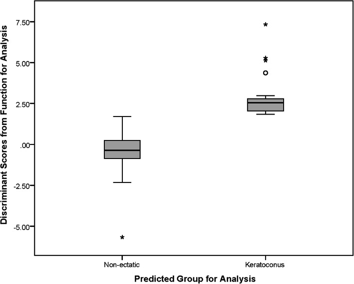
Methods: In this study, DS patients were enrolled through special needs schools, the National Down Syndrome Society, and relevant non-profit organizations. Diagnoses were made independently by two experienced specialists. Forty Pentacam indices related to corneal thickness, volume, density, keratometry, power, shape, aberration, and elevation were extracted. For each index, the accuracy for KC and KCS diagnosis was evaluated using discriminant analysis and the area under receiver operating characteristic curve (AUROC). From each enrolled case, data from only one eye was entered in the analyses.
Results: Analyses were performed on data from 25 KC, 46 KCS, and 154 non-ectatic DS eyes. The best discriminants for KC were anterior higher order aberrations (HOA) (cutoff > 0.643, AUROC = 0.879), posterior vertical coma (cutoff > 0.0702 μm, AUROC = 0.875), anterior vertical coma (cutoff > 0.4124 μm, AUROC = 0.868), and total HOA (cutoff > 0.608, AUROC = 0.867). The difference between AUROCs were not statistically significant (all P > 0.05). For KCS, the best discriminants were minimum corneal thickness (cutoff ≤ 480.0 μm, AUROC = 0.775), corneal volume (cutoff ≤ 55.3 μm, AUROC = 0.727) and Belin Ambrosio display-total deviation (BAD-D) (cutoff > 2.23, AUROC = 0.718) with no significant difference between AUROCs (all P > 0.05).
Conclusions: In this sample of DS patients, best KC discriminators were HOA and coma which showed good diagnostic ability. For KCS, best predictors were minimum corneal thickness, corneal volume, and BAD-D with relatively good diagnostic ability. Defining a new set of KC diagnostic criteria for DS patients is suggested.
Keywords: Diagnostic criteria; Discriminant analysis; Down syndrome; Keratoconus; Tomography.
© The Author(s) 2020.
Conflict of interest statement
Competing interestsThe authors declare that they have no competing interests.
Figures

Similar articles
-
Scheimpflug Tomographic Indices for Classifying Normal, Down Syndrome and Clinical Keratoconus in Pediatric Patients.Diagnostics (Basel). 2024 Sep 2;14(17):1932. doi: 10.3390/diagnostics14171932. Diagnostics (Basel). 2024. PMID: 39272718 Free PMC article.
-
Comparison of a Scheimpflug imaging with other screening indices in diagnosing keratoconus and keratoconus suspect.Sci Rep. 2024 Oct 5;14(1):23187. doi: 10.1038/s41598-024-74497-z. Sci Rep. 2024. PMID: 39369097 Free PMC article.
-
Accuracy of the posterior corneal elevation values of Pentacam HR from different reference surfaces in early ectasia diagnosis.Int Ophthalmol. 2021 Feb;41(2):629-638. doi: 10.1007/s10792-020-01618-8. Epub 2020 Oct 23. Int Ophthalmol. 2021. PMID: 33095345
-
To what extent is pregnancy-induced keratoconus progression reversible? A case-report and literature review.Eur J Ophthalmol. 2023 Jan;33(1):NP37-NP41. doi: 10.1177/11206721211045187. Epub 2021 Sep 17. Eur J Ophthalmol. 2023. PMID: 34533408 Review.
-
The Underlying Relationship between Keratoconus and Down Syndrome.Int J Mol Sci. 2022 Sep 16;23(18):10796. doi: 10.3390/ijms231810796. Int J Mol Sci. 2022. PMID: 36142709 Free PMC article. Review.
Cited by
-
Systemic Associations with Keratoconus.Life (Basel). 2023 Jun 10;13(6):1363. doi: 10.3390/life13061363. Life (Basel). 2023. PMID: 37374145 Free PMC article. Review.
-
Scheimpflug Tomographic Indices for Classifying Normal, Down Syndrome and Clinical Keratoconus in Pediatric Patients.Diagnostics (Basel). 2024 Sep 2;14(17):1932. doi: 10.3390/diagnostics14171932. Diagnostics (Basel). 2024. PMID: 39272718 Free PMC article.
-
Anterior pituitary, sex hormones, and keratoconus: Beyond traditional targets.Prog Retin Eye Res. 2022 May;88:101016. doi: 10.1016/j.preteyeres.2021.101016. Epub 2021 Nov 2. Prog Retin Eye Res. 2022. PMID: 34740824 Free PMC article.
-
Corneal characteristics in Down syndrome patients with normal and keratoconic cornea.Front Med (Lausanne). 2022 Sep 16;9:985928. doi: 10.3389/fmed.2022.985928. eCollection 2022. Front Med (Lausanne). 2022. PMID: 36186827 Free PMC article.
-
Down syndrome: a review of ocular manifestations.Ther Adv Ophthalmol. 2022 Jun 30;14:25158414221101718. doi: 10.1177/25158414221101718. eCollection 2022 Jan-Dec. Ther Adv Ophthalmol. 2022. PMID: 35795721 Free PMC article. Review.
References
LinkOut - more resources
Full Text Sources
Research Materials
