Review
. 2021 Apr;50(4):645-663.
doi: 10.1007/s00256-020-03632-4. Epub 2020 Oct 7.

Edema-like marrow signal intensity: a narrative review with a pictorial essay

Affiliations
Review

Edema-like marrow signal intensity: a narrative review with a pictorial essay

Davide Maraghelli et al. Skeletal Radiol. 2021 Apr.

Abstract

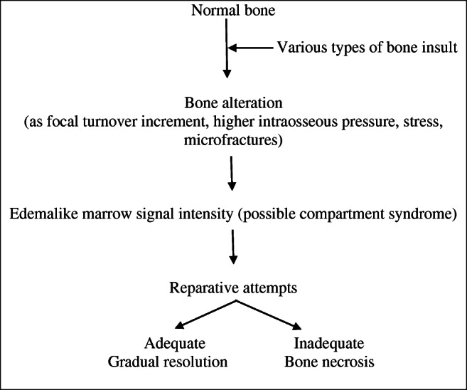
The term edema-like marrow signal intensity (ELMSI) represents a general term describing an area of abnormal signal intensity at MRI. Its appearance includes absence of clear margins and the possibility of exceeding well-defined anatomical borders (for example, physeal scars). We can define "ELMSI with unknown cause" an entity where the characteristic MR appearance is associated with the absence of specific signs of an underlying condition. However, it is more often an important finding indicating the presence of an underlying disease, and we describe this case as "ELMSI with known cause." It presents a dynamic behavior and its evolution can largely vary. It initially corresponds to an acute inflammatory response with edema, before being variably replaced by more permanent marrow remodeling changes such as fibrosis or myxomatous connective tissue that can occur over time. It is important to study ELMSI variations over time in order to evaluate the activity state and therapeutic response of an inflammatory chronic joint disease, the resolution of a trauma, and the severity of an osteoarthritis. We propose a narrative review of the literature dealing with various subjects about this challenging topic that is imaging, temporal evolution, etiology, differential diagnoses, and possible organization, together with a pictorial essay.

Keywords: AVN; Edema-like marrow signal intensity; MRI; Rheumatoid arthritis; SIFK.


Conflict of interest statement

The authors declare that they have no conflict of interest.

Figures

Fig. 1
Fig. 1
a, b Degenerative. Modic changes type I in a 78-year-old woman with back pain. MRI shows the SI alterations in D11 and D12 endplates (arrows), that appear hypointense on T1w (a) and hyperintense on T2w (b) imaging. These alterations correspond to vertebral body edema and hypervascularity
Fig. 2
Fig. 2
a, b Degenerative. Modic changes type II in a 53-year-old man. The hyperintense SI on T1w imaging of two lumbar bordering endplates (arrows in a) and hyperintense SI on T2w imaging (arrows in b) reflects fatty replacements of the red bone marrow, characterizing Modic changes type II
Fig. 3
Fig. 3
a, b, c Degenerative. Modic changes type III in a 53-year-old woman. The hypointense SI areas (arrows) both on T1w (A) and T2w (B) imaging reflect a subchondral bone sclerosis. The lumbosacral spine x-ray of the same patient (C) allows to better observe the bone sclerosis (arrow) of the caudal lumbar spine (courtesy of Giacomo Aringhieri, MD, University of Pisa School of Medicine, Italy)
Fig. 4
Fig. 4
Bone insult cascade. Adapted from references [50, 51]
Fig. 5
Fig. 5
a, b, c, d Primary. A 53-year-old male patient with pain, swelling, and stiffness of the left knee. STIR imaging from the first MRI exam (a, b) highlights edema (A single asterisk (*) in a and arrowheads in b) in the medial condyle; degenerative phenomena affecting meniscuses, articular cartilage and ligaments are not present. STIR imaging performed after two months (c, d) shows that edema (*), from the medial compartment of the knee, has moved into the lateral compartment of the same knee, while all the other findings observed at the first examination remained unchanged. Every possible etiology has been excluded: this is an example of migratory primary ELMSI
Fig. 6
Fig. 6
a, b, c, d Traumatic. A 17-year-old female with a traumatic ELMSI after a contusion of the right knee. STIR coronal imaging (a, b) demonstrates the presence of the hyperintense ELMSI (*) in the anterior side of the lateral femoral condyle (a) and in the lateral side of the tibial plateau (b). The presence of an abundant marrow signal alteration is often correlated with a ligament injury: in this case, the anterior cruciate ligament (ACL) is scarcely recognizable (arrows) both in the T1w (c) and T2w (d) sequences: the finding is compatible with a high-grade ACL injury
Fig. 7
Fig. 7
a, b, c, d Traumatic (CRPS) and iatrogenic. A 58-year-old woman that underwent surgery for left hallux valgus correction. All the figures are STIR images. Panels a and b come from the first exam, few weeks after surgery, where it is possible to observe an ELMSI involving the first metatarsal head (arrow in a) compatible with the post-surgery (iatrogenic) ELMSI; no ELMSI of the tarsal bones (b). After 6 months, the patient continued to feel pain in the left foot and MRI was repeated. MRI shows the almost total remission of the ELM in the surgical site (arrow in c), confirming the alteration was a simple post-surgery (iatrogenic) ELMSI, but its presence with “patchy” distribution in the cuneiform bones, in the cuboid, in the talus, in the calcaneus, associated with effusion in the tarsal and ankle joints (d), led to the diagnosis of CRPS
Fig. 8
Fig. 8
a, b, c, d Inflammatory. A 56-year-old rheumatoid arthritis (RA) male patient with pain and swelling in the left wrist. TWIST Angio 3D MIP imaging (a) clearly shows the inflammation with hypervascularization and edema (arrowheads) affecting the left wrist. Contrast-enhanced T1w imaging (b) demonstrates an abundant intra-articular effusion in the distal radioulnar joint (*) with thickening and enhancement of synovia (arrow), a condition referable to inflammation in the active phase. STIR coronal imaging (c) highlights the ELMSI affecting part of the ulna, lunate, triquetrum, hamate and capitate bones, and the effusion in radioscaphoid and radioulnar joints. Finally, the X-rays of the hand (d) taken at the same time shows a “soft-tissue sign” near the ulnar styloid process (arrow). but absence of erosions, while MRI is strongly positive for active inflammation [courtesy of Giovanni D'Elia, MD, Careggi University Hospital, Florence, Italy]
Fig. 9
Fig. 9
a, b, c, d Inflammatory. A 49-year-old woman with swelling and pain in the left sternoclavicular joint and history of psoriasis. MRI highlights the ELMSI of the joint (arrows), which is hyperintense in STIR (a–c) and hypointense in T1w (b–d) imaging. This is a very particular condition and can be referred to either an early SAPHO (synovitis-acne-pustulosis-hyperostosis-osteitis) syndrome, which is often connected with a special form of psoriasis (pustulosis palmoplantaris) and has an inflammatory involvement of the sternoclavicular joint, or a Tietze syndrome, a painful inflammation that can affect the costochondral, costosternal, or sternoclavicular joints
Fig. 10
Fig. 10
a, b, c, d, e, f, g, h AVN. A 53-year-old woman with pain during mobilization of the right knee; MRI has raised the suspicion of AVN (bone infarct) and we can note the evolution of the findings over 4 years. T1w (a) and proton density fat–suppressed (PD-FS, b) imaging show two areas of altered signal in the distal end of the femur and in the proximal end of the tibia with irregular morphology and inhomogeneous structure (*), with a peripheral rim, hypointense in T1w (arrows) and hyperintense in PD-FS (arrows) imaging corresponding to the reactive interface along the margin of infarct. Three months after the previous MRI (c, T1w and d, PD-FS), a further increase in the interface (arrows), a reduction in the ischemic area and signs of regeneration of the cancellous bone are observed. While the MRI findings are well evident, the CT performed in the same period (e) shows only a limited rarefaction (*) of the cancellous bone. One year later (f, T2w-FS) the infarct outcome area is still evident but smaller compared to previous examinations. Four years later (g, PD and h, STIR) the infarct zone appears less extensive and more shaded, but still present; the interface appears larger. ELMSI had not been detected for all 4 years and the patient progressively underwent an improvement in the clinical situation, with the disappearance of the pain. It is important to underline that ELMSI represents a fundamental indicator of a subchondral fracture and a progression towards epiphyseal collapse, which in this case did not occur
Fig. 11
Fig. 11
a, b, c, d Infectious. Spondylodiscitis involving L4–L5 in a 52-year-old man with fever and low back pain. MRI shows a large ELMSI (*) of the vertebral bodies, appearing hypointense in T1w imaging (a), hyperintense in T2w (b), STIR (c), and contrast-enhanced T1w imaging (d). The interposed disc is also involved in the inflammatory process, appearing hyperintense on T2w, STIR and contrast-enhanced T1w sequences
Fig. 12
Fig. 12
a, b, c Benign neoplastic (osteoid osteoma). A 8-year-old male patient with pain in the end of the left thigh worsening at night. T1w imaging (a) shows a hypointense lesion with well-defined margins (arrowhead) surrounded by a slightly hypointense halo (*). STIR imaging (b) demonstrates the edematous nature of the halo (*) and well highlights the great extension of the perilesional ELMSI itself. Contrast-enhanced T1w imaging (c) shows the enhancement of the nidus (arrowhead) and the slight enhancement of the perilesional marrow signal alterations (*)
Fig. 13
Fig. 13
a, b, c, d Benign and malignant neoplastic. ELMSI from an osteoblastoma of the left hemisoma of S1 in a 36-year-old male patient (a–b). STIR coronal image (a) allows to detect an extensive ELMSI (*) involving the left hemisomas of S1 and S2 and the left posterior-lower corner of L5 (arrowhead). In STIR axial image (b) we can see both the ELMSI (*) and the benign nodule of S1 (arrowheads). Marrow alterations from a periosteal osteosarcoma of the proximal diaphysis of the right femur in a 74-year-old woman (c–d). STIR coronal image (c) shows the exophytic development of the tumor (arrow) and the extensive edemigenous reaction of the femoral spongiosa (arrowheads); in d, T1w image, on the axial plane, we can see the tumor (arrows) and the hypointense ELMSI (*) occupying the entire medullary canal
Fig. 14
Fig. 14
a, b, c Iatrogenic. A 73-year-old male patient with recent radiotherapy of the lumbar spine for the treatment of a non-Hodgkin lymphoma. T1w (a) and STIR (b) images demonstrate the presence of an abundant ELMSI (*) and the ADC map (c) shows the absence of diffusion restriction in L3, that was previously affected by the disease

Similar articles

Cited by

References

    1. Gorbachova T, Amber I, Beckmann NM, Bennett DL, Chang EY, Davis L, et al. Nomenclature of subchondral nonneoplastic bone lesions. Am J Roentgenol. 2019:1–20. 10.2214/ajr.19.21571. - PubMed
    1. Solomon L. Bone-marrow oedema syndrome. J Bone Joint Surg. 1993;75B:175–176. - PubMed
    1. Thiryayi WA, Thiryayi SA, Freemont AJ. Histopathological perspective on bone marrow oedema reactive bone change and haemorrhage. Eur J Radiol. 2008;67:62–67. - PubMed
    1. Lang P, Genant HK, Majumadar S. In: Bone marrow disorders. MRI of the musculoskeletal system. Chan WP, Lang P, Genant HK, editors. Philadelphia: WB Saunders; 1994. pp. 445–485.
    1. Vogler JB, III, Murphy WA., Jr . In: Diffuse marrow diseases. MRI of the musculoskeletal system. Berquest TH, editor. Philadelphia: Lippincott, Williams & Wilkins; 2001. pp. 979–1028.