. 2020 Oct;2(10):605-615.
doi: 10.1002/acr2.11176. Epub 2020 Oct 7.

TSG-6 Is Weakly Chondroprotective in Murine OA but Does not Account for FGF2-Mediated Joint Protection

Affiliations

TSG-6 Is Weakly Chondroprotective in Murine OA but Does not Account for FGF2-Mediated Joint Protection

Linyi Zhu et al. ACR Open Rheumatol. 2020 Oct.

Abstract

Objective: Tumor necrosis factor α-stimulated gene 6 (TSG-6) is an anti-inflammatory protein highly expressed in osteoarthritis (OA), but its influence on the course of OA is unknown.

Methods: Cartilage injury was assessed by murine hip avulsion or by recutting rested explants. Forty-two previously validated injury genes were quantified by real-time polymerase chain reaction in whole joints following destabilization of the medial meniscus (DMM) (6 hours and 7 days). Joint pathology was assessed at 8 and 12 weeks following DMM in 10-week-old male and female fibroblast growth factor 2 (FGF2)-/- , TSG-6-/- , TSG-6tg (overexpressing), FGF2-/- ;TSG-6tg (8 weeks only) mice, as well as strain-matched, wild-type controls. In vivo cartilage repair was assessed 8 weeks following focal cartilage injury in TSG-6tg and control mice. FGF2 release following cartilage injury was measured by enzyme-linked immunosorbent assay.

Results: TSG-6 messenger RNA upregulation was strongly FGF2-dependent upon injury in vitro and in vivo. Fifteeen inflammatory genes were significantly increased in TSG-6-/- joints, including IL1α, Ccl2, and Adamts5 compared with wild type. Six genes were significantly suppressed in TSG-6-/- joints including Timp1, Inhibin βA, and podoplanin (known FGF2 target genes). FGF2 release upon cartilage injury was not influenced by levels of TSG-6. Cartilage degradation was significantly increased at 12 weeks post-DMM in male TSG-6-/- mice, with a nonsignificant 30% reduction in disease seen in TSG-6tg mice. No differences were observed in cartilage repair between genotypes. TSG-6 overexpression was unable to prevent accelerated OA in FGF2-/- mice.

Conclusion: TSG-6 influences early gene regulation in the destabilized joint and exerts a modest late chondroprotective effect. Although strongly FGF2 dependent, TSG-6 does not explain the strong chondroprotective effect of FGF2.


Figures

Figure 1
Figure 1
Summary of experiment design.
Figure 2
Figure 2
Tumor necrosis factor α–stimulated gene 6 (TSG‐6) is regulated in an fibroblast growth factor 2 (FGF2)‐dependent manner. A, Regulation of TSG‐6 gene expression by recutting injury or FGF2 stimulation of murine cartilage explants. Murine hip cartilage (from male animals) was avulsed (avulsion injury) directly into serum‐free Dulbecco's Modified Eagle's Medium (DMEM) and incubated for 48 hours (48 h). Rested cartilage was either recut (recut injury) or treated with FGF2 (250 ng/ml) in fresh DMEM and left for 4 hours prior to RNA extraction. Each point represents the messenger RNA (mRNA) from four hips pooled. Tsg6 levels were expressed relative to Gapdh and normalized to the control. B, RNA was extracted from male wild‐type (WT) or FGF2−/− mouse joints (n = 3) at specified times after destabilization of the medial meniscus. Tsg6 mRNA levels were expressed relative to 18s and normalized to the WT 0 h control. Statistical significance was determined by analysis of variance with Sidak post hoc testing, comparing treated compared with rested controls (A) and WT with FGF2−/− (B) for each time point.
Figure 3
Figure 3
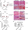
Increased cartilage degradation in male tumor necrosis factor α–stimulated gene 6 (TSG‐6−/−) mice 12 weeks after destabilization of the medial meniscus (DMM). Histological chondropathy scores and representative histologic sections of knee cartilage 8 or 12 weeks after DMM in male (A) and female (B) wild‐type (WT; C57BL/6) and TSG‐6−/− mice. Black arrows in the images indicate cartilage defects. Scale bar = 200 µm. Statistical significance was determined by Mann‐Whitney U tests. Mean osteophyte scores: size (C) and maturity (D) in WT (C57BL/6) and TSG‐6−/− male mice 8 weeks post‐DMM. Also shown are contralateral (CL) joint controls. NS, not significant.
Figure 4
Figure 4
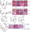
Joint pathology in tumor necrosis factor α–stimulated gene 6 (TSG)tg mice at 8 and 12 weeks after destabilization of the medial meniscus (DMM). Histological chondropathy scores (left) and representative histologic sections (right) of knee cartilage 8 and 12 weeks after sham or DMM in male (A) and female (B) TSG‐6tg and wild‐type (WT; Balb/c) mice. Black arrows in the images indicate cartilage defects. Scale bar = 200 µm. Statistical significance was determined by Mann‐Whitney U tests. There were no significant differences seen between any of the female experimental groups by two‐way analysis of variance (ANOVA). C, Fold increase of Tsg6 over WT transgene (by quantitative reverse transcription polymerase chain reaction) was plotted against summed cartilage score for male mice 12 weeks post‐DMM. R 2 correlation statistical analysis was performed. Mean osteophyte scores, size (D) and maturity (E), were carried out in WT (Balb/c) and TSG‐6tg male mice 8 weeks after DMM. Histological scores (left) (F) and representative histologic sections (right) (G) of knee cartilage 8 weeks after DMM in male WT(C57BL6), fibroblast growth facror 2 (FGF2)−/−;TSG‐6tg, and FGF2−/−;TSG‐6WT mice. Balb/c histology scores are derived from Figure 4A. Statistical significance was determined by one‐way ANOVA with Sidak post hoc testing. H, Level of Tsg6 expression was plotted against summed cartilage score for male FGF2−/−;TSG‐6tg mice 8 weeks post‐DMM. R2 correlation statistical analysis was performed.
Figure 5
Figure 5
Tumor necrosis factor α–stimulated gene 6 (TSG‐6)tg mice do not have enhanced cartilage repair capacity. A, fibroblast growth factor 2 (FGF2) levels were measured by enzyme‐linked immunosorbent assay (MSD PLEX bFGF assay) in conditioned medium collected from injured mouse hips (a mixture of male and female) from wild‐type (WT; Balb/c), TSG‐6tg, TSG‐6+/‐ (heterozygotes), and TSG‐6−/− (knockout, homozygotes) mice. B, Transverse section of the joint showing the position of patella groove. C, Cartilage repair scores (left) and representative histological images of Safranin O–stained sections (right) 8 weeks after focal cartilage injury in male WT (Balb/c mice), TSG‐6tg, and C57BL/6 mice, n = 6 to 12 mice per group. D, Cartilage repair scores after focal cartilage injury in female WT (Balb/c) and TSG‐6tg mice. Scale bar = 200 µm. Mann‐Whitney U tests were used to determine statistical significance.

References

    1. Lee TH, Lee GW, Ziff EB, Vilcek J. Isolation and characterization of eight tumor necrosis factor‐induced gene sequences from human fibroblasts. Mol Cell Biol 1990;10:1982–8. - PMC - PubMed
    1. Milner CM, Day AJ. TSG‐6: a multifunctional protein associated with inflammation. J Cell Sci 2003;116:1863–73. - PubMed
    1. Lee TH, Wisniewski HG, Vilček J. A novel secretory tumor necrosis factor‐inducible protein (TSG‐6) is a member of the family of hyaluronate binding proteins, closely related to the adhesion receptor CD44. J Cell Biol 1992;116:545–57. - PMC - PubMed
    1. Tammi MI, Day AJ, Turley EA. Hyaluronan and homeostasis: a balancing act. J Biol Chem 2002;277:4581–4. - PubMed
    1. Day AJ, Milner CM. TSG‐6: A multifunctional protein with anti‐inflammatory and tissue‐protective properties. Matrix Biol 2019;78–79:60–83. - PubMed