

The Histopathology of Oral Cancer Pain in a Mouse Model and a Human Cohort
- PMID: 33030108
- PMCID: PMC8173346
- DOI: 10.1177/0022034520961020
The Histopathology of Oral Cancer Pain in a Mouse Model and a Human Cohort
Abstract
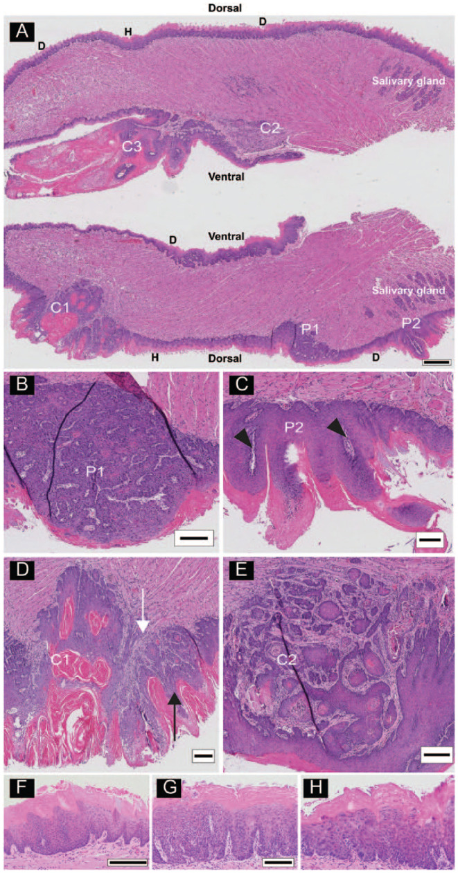
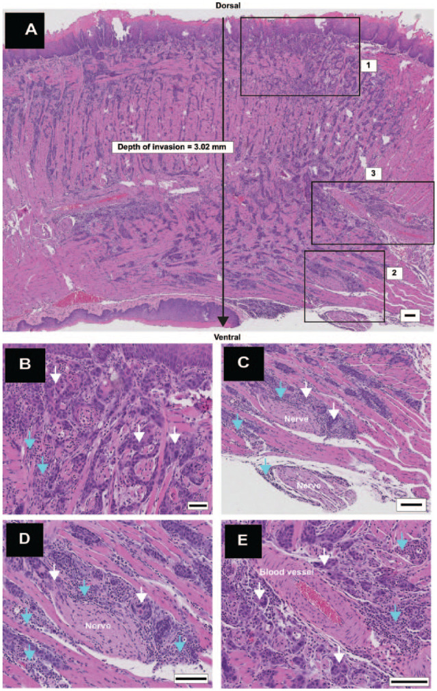
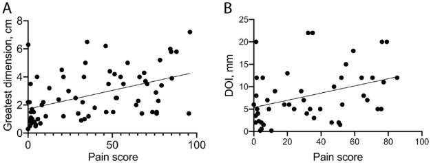
Oral cancer patients often have severe, chronic, and mechanically induced pain at the site of the primary cancer. Oral cancer pain is initiated and maintained in the cancer microenvironment and attributed to release of mediators that sensitize primary sensory nerves. This study was designed to investigate the histopathology associated with painful oral cancers in a preclinical model. The relationship of pain scores with pathologic variables was also investigated in a cohort of 72 oral cancer patients. Wild-type mice were exposed to the carcinogen, 4-nitroquinoline 1-oxide (4NQO). Nociceptive (pain) behavior was measured with the dolognawmeter, an operant device and assay for measuring functional and mechanical allodynia. Lesions developed on the tongues and esophagi of the 4NQO-treated animals and included hyperkeratoses, papillomas, dysplasias, and cancers. Papillomas included lesions with benign and dysplastic pathological features. Two histologic subtypes of squamous cell carcinomas (SCCs) were identified-SCCs with exophytic and invasive components associated with papillary lesions (pSCCs) and invasive SCCs without exophytic histology (iSCCs). Only the pSCC subtype of tongue cancer was associated with nociceptive behavior. Increased tumor size was associated with greater nociceptive behavior in the mouse model and more pain experienced by oral cancer patients. In addition, depth of invasion was associated with patient-reported pain. The pSCC histology identifies 4NQO-induced tongue cancers that are expected to be enriched for expression and release of nociceptive mediators.
Keywords: 4-nitroquinoline 1-oxide; nociception; nociceptive pain; oral SCC; oral cancer pathology; oral carcinogenesis.
Conflict of interest statement
Figures

Similar articles
-
Oral cavity and esophageal carcinogenesis modeled in carcinogen-treated mice.Clin Cancer Res. 2004 Jan 1;10(1 Pt 1):301-13. doi: 10.1158/1078-0432.ccr-0999-3. Clin Cancer Res. 2004. PMID: 14734483
-
Oral-specific ablation of Klf4 disrupts epithelial terminal differentiation and increases premalignant lesions and carcinomas upon chemical carcinogenesis.J Oral Pathol Med. 2015 Nov;44(10):801-9. doi: 10.1111/jop.12307. Epub 2015 Jan 21. J Oral Pathol Med. 2015. PMID: 25605610
-
Oral mucosal carcinogenesis in SENCAR mice.Anticancer Res. 2002 Sep-Oct;22(5):2733-40. Anticancer Res. 2002. PMID: 12529989
-
4-nitroquinoline-1-oxide induced experimental oral carcinogenesis.Oral Oncol. 2006 Aug;42(7):655-67. doi: 10.1016/j.oraloncology.2005.10.013. Epub 2006 Jan 30. Oral Oncol. 2006. PMID: 16448841 Review.
-
The 4-NQO mouse model: An update on a well-established in vivo model of oral carcinogenesis.Methods Cell Biol. 2021;163:197-229. doi: 10.1016/bs.mcb.2020.09.004. Epub 2020 Nov 12. Methods Cell Biol. 2021. PMID: 33785166 Review.
Cited by
-
Assessing Orofacial Pain Behaviors in Animal Models: A Review.Brain Sci. 2023 Feb 24;13(3):390. doi: 10.3390/brainsci13030390. Brain Sci. 2023. PMID: 36979200 Free PMC article. Review.
-
Near-infrared fluorescence imaging with an MET-targeting probe for biopsy site selection in patients with oral potentially malignant disorders.Cell Rep Med. 2025 Mar 18;6(3):101978. doi: 10.1016/j.xcrm.2025.101978. Epub 2025 Feb 24. Cell Rep Med. 2025. PMID: 39999837 Free PMC article. Clinical Trial.
-
Oral cancer induced TRPV1 sensitization is mediated by PAR2 signaling in primary afferent neurons innervating the cancer microenvironment.Sci Rep. 2022 Mar 8;12(1):4121. doi: 10.1038/s41598-022-08005-6. Sci Rep. 2022. PMID: 35260737 Free PMC article.
-
The impact of tumor immunogenicity on cancer pain phenotype using syngeneic oral cancer mouse models.Front Pain Res (Lausanne). 2022 Sep 12;3:991725. doi: 10.3389/fpain.2022.991725. eCollection 2022. Front Pain Res (Lausanne). 2022. PMID: 36172037 Free PMC article.
-
Measurement of the Association of Pain with Clinical Characteristics in Oral Cancer Patients at Diagnosis and Prior to Cancer Treatment.J Pain Res. 2024 Feb 2;17:501-508. doi: 10.2147/JPR.S423318. eCollection 2024. J Pain Res. 2024. PMID: 38328017 Free PMC article.
References
-
- Abbey LM, Kaugars GE, Gunsolley JC, Burns JC, Page DG, Svirsky JA, Eisenberg E, Krutchkoff DJ, Cushing M. 1995. Intraexaminer and interexaminer reliability in the diagnosis of oral epithelial dysplasia. Oral Surg Oral Med Oral Pathol Oral Radiol Endod. 80(2):188–191. - PubMed
-
- Allen-Ayodabo CO, Eskander A, Davis LE, Zhao H, Mahar AL, Karam I, Singh S, Gupta V, Bubis LD, Moody L, et al.. 2019. Symptom burden among head and neck cancer patients in the first year after diagnosis: association with primary treatment modality. Oral Oncol. 99:104434. - PubMed
-
- Arora A, Husain N, Bansal A, Neyaz A, Jaiswal R, Jain K, Chaturvedi A, Anand N, Malhotra K, Shukla S. 2017. Development of a new outcome prediction model in early-stage squamous cell carcinoma of the oral cavity based on histopathologic parameters with multivariate analysis: the aditi-nuzhat lymph-node prediction score (ANLPS) system. Am J Surg Pathol. 41(7):950–960. - PubMed
-
- Bapat AA, Hostetter G, Von Hoff DD, Han H. 2011. Perineural invasion and associated pain in pancreatic cancer. Nat Rev Cancer. 11(10):695–707. - PubMed
Publication types
MeSH terms
Substances
Grants and funding
LinkOut - more resources
Full Text Sources
Medical
Research Materials
