. 2020 Dec;17(6):887-900.
doi: 10.1007/s13770-020-00288-y. Epub 2020 Oct 8.

Comparative Study on Bone Marrow-Versus Adipose-Derived Stem Cells on Regeneration and Re-Innervation of Skeletal Muscle Injury in Wistar Rats

Affiliations

Comparative Study on Bone Marrow-Versus Adipose-Derived Stem Cells on Regeneration and Re-Innervation of Skeletal Muscle Injury in Wistar Rats

Manal H Moussa et al. Tissue Eng Regen Med. 2020 Dec.

Abstract

Background: Skeletal muscle injuries are frequent clinical challenges due to associated fibrosis and disability. Regenerative medicine is an emerging promising strategy for such cases. The aim of this study was to compare between the effects of bone marrow-mesenchymal stem cells (BM-MSCs) versus adipose tissue stromal cells (ADSCs) on regeneration and re-innervation of skeletal muscle laceration injury in Wistar rats at different time intervals.

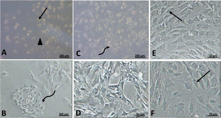
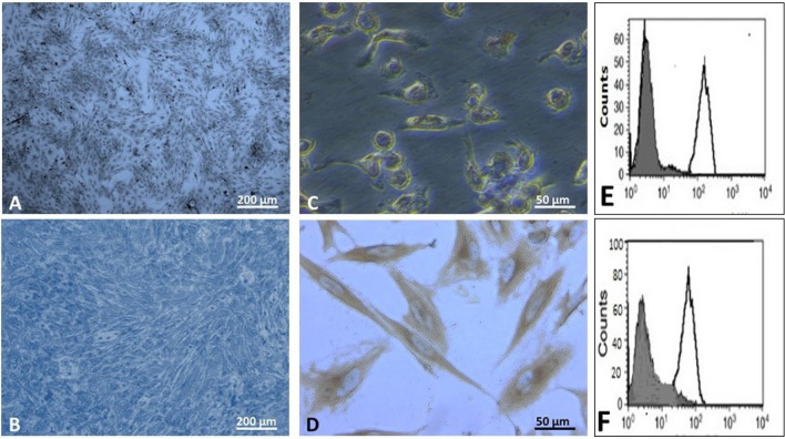
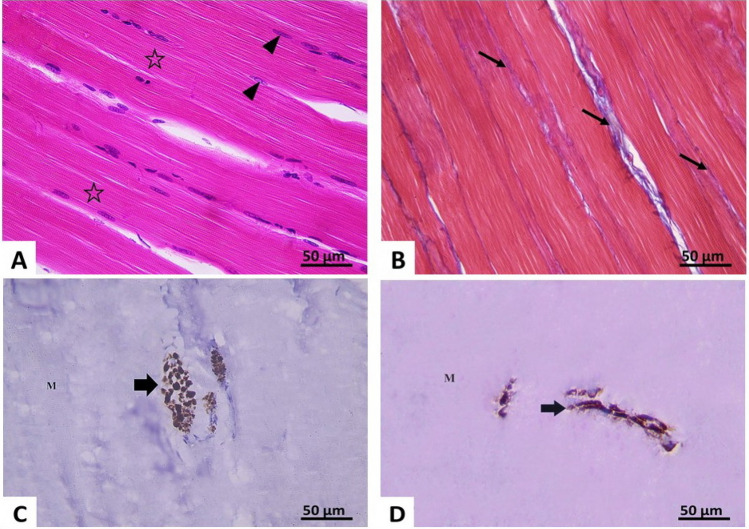
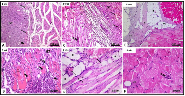
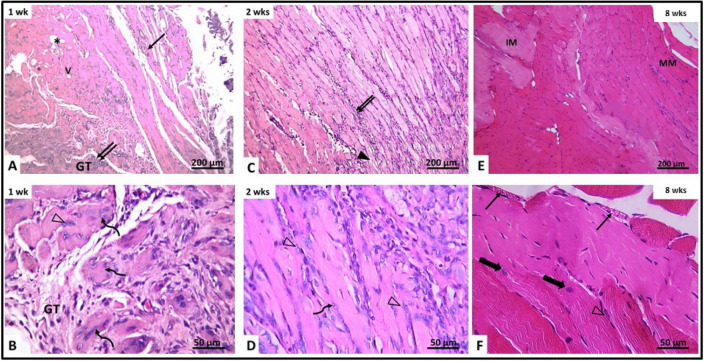
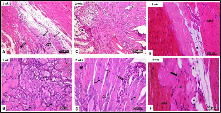
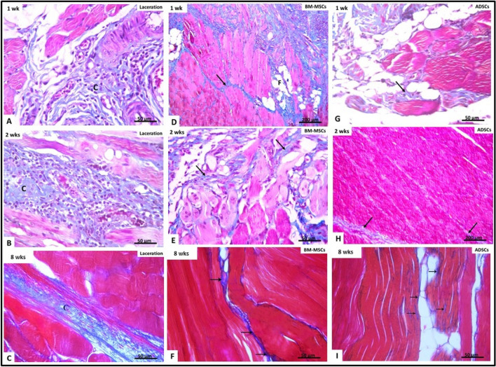
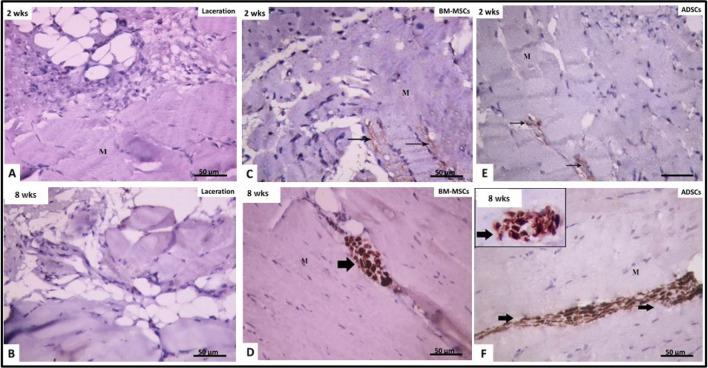
Methods: Six young male rats were used as a source of allogenic MSCs. Eighty-four adult female rats were divided into: Group I (control), Group II (Untreated Laceration): right gluteal muscle was lacerated and left for spontaneous healing, Group III (BM-MSCs): right gluteal muscle was lacerated with concomitant local intramuscular injection of 1 × 10⁶ BM-MSCs in the lacerated muscle, Group IV (ADSCs): right gluteal muscle was lacerated with concomitant local intramuscular injection of 1 × 10⁶ ADSCs in lacerated muscle. Rats were sacrificed after one, two and eight weeks. Muscles were processed to prepare sections stained with H&E, Mallory's trichrome and immune-histochemical staining (neurofilament light chain).

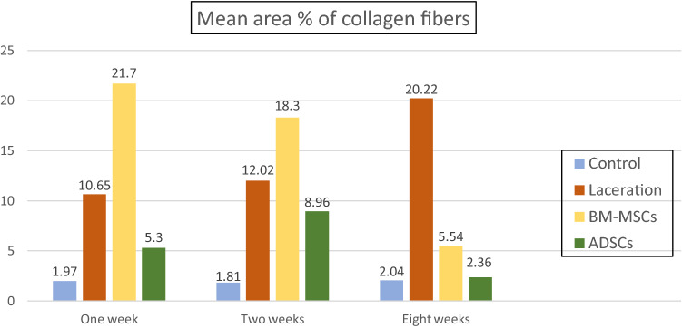
Results: A significant increase in collagen fibers and failure of re-innervation were noticed in untreated laceration group. BM-MSCs-treated groups showed regeneration of muscle fibers but with increased collagen fibers. Meanwhile, ADSCs showed better regenerative effects evidenced by significant increase in the number of myotubes and significant decrease in collagen deposition. Re-innervation was noticed in MSCs-injected muscles after 8 weeks of laceration.

Conclusion: Both BM-MSCs and ADSCs improved regeneration of skeletal muscle laceration injury at short- and long-term durations. However, fibrosis was less in ADSCs-treated rats. Effective re-innervation of injured muscles occurred only at the long-term duration.

Keywords: Adipose tissue-derived stem cells; Bone marrow-derived mesenchymal stem cells; Muscle laceration; Neurofilament light chain; Re-innervation.


Conflict of interest statement

The authors have no conflicting financial or competing interests.

Figures

Fig. 1
Fig. 1
Phase contrast micrographs rat BM-MSCs rat ADSCs. A, B day four of primary culture rat BM-MSCs showing a large number of attached cells of different shapes. Some cells appeared spindle-shaped (↑), and others appeared polygonal (▲). Notice the presence of many unattached cells. Rat ADSCs showing whorl shaped appearance of a starting colony (curved arrow). C, D day six of primary culture rat BM-MSCs showing further increased in the number of attached cells. The attached cells appeared of variable shapes. There is the evidence of start of colony formation (curved arrow). ADSCs culture showing further increased in the number of attached cells. Notice that the cells show multiple interdigitating cell processes. E Rat BM-MSCs on the 5th day of passage one showing the whorl appearance of a colony formed of spindle-shaped cells (↑). F Rat ADSCs on the 3rd day of passage one showing colonies of spindle-shaped attached cells (↑) with polygonal cells in between
Fig. 2
Fig. 2
A, B phase contrast micrographs of Giemsa stain X 100, in the third passage, showing colonies of spindle-shaped cells that are seen in rat BM-MSCs on the 4th day rat ADSCs on 2nd day. C, D phase contrast micrographs of CD133 X400 rat BM-MSCs showing weak brownish cytoplasmic reaction in some cultured cell rat ADSCs showing strong positive brown cytoplasmic reaction in all cells. E, F Flow cytometric analysis of cell-surface antigens, showing most cells positive for CD90 rat BM-MSCs rat ADSCs
Fig. 3
Fig. 3
Photomicrographs of section of right gluteal muscle of the control group X400 A H&E-stained section showing well-defined cross striations (*) and peripheral elongated nuclei (▲).B Mallory’s trichrome: thin bundles of collagen fibers (↑) are seen in the interstitium between the muscle fibers. C, D Immune-histochemical reaction for neurofilament light chain: showing positive expression (↑) in transverse (C) and longitudinal nerves (D) between muscle fibers (M)
Fig. 4
Fig. 4
H&E-stained section of gluteal muscle from laceration group (group II) at different time intervals. A, B After 1 week (IIa) (A) showing granulation tissue (GT) containing mononuclear inflammatory cells, congested blood vessels (↑) and disrupted muscle fibers (▲). The granulation tissue (GT) contains fragmented muscles with pyknotic nuclei (thick arrow). Other muscles contain neutrophils (Δ). C, B After 2 weeks (IIb) showing disrupted vacuolated muscles (V), congested blood vessels () and inflammatory cellular infiltration (↑↑). D Showing congested blood vessels (), fat cells (*) and regenerating myotubes containing centrally located nuclei (curved arrow). E, F After 8 weeks (IIc) showing the area of injury containing fibrous tissue and fat cells (*). Minimal inflammatory cellular infiltration (↑↑) and fibrous tissue (FT) are seen between muscle fibers
Fig. 5
Fig. 5
Showing the mean number of myotubes/field in different groups
Fig. 6
Fig. 6
Photomicrographs of right gluteal muscle from laceration group treated with BM-MSCs (group III). A, B After 1 week (IIIa) showing vacuolated muscles (V), cellular granulation tissue (GT), congested blood vessels (↑) and inflammatory cellular infiltration (↑↑). The granulation tissue (GT) is seen containing myotubes with centrally located nuclei (curved arrow) and activated cells with vesicular nuclei (Δ). C, D After 2 weeks (IIIb), disrupted muscle fibers are seen with apparent splitting and loss of striations (▲). Inflammatory cellular infiltration (↑↑) is also noticed. Showing regenerating myotubes containing centrally located vesicular nuclei and basophilic cytoplasm (curved arrow). Activated cells with vesicular nuclei (Δ) are also seen. E, F After 8 weeks (IIIc) showing mature muscle fibers (MM) and immature muscle fibers (IM) at area of injury showing congested blood vessels (↑) and muscle fibers with central nuclei and incomplete striations (thick arrow). Active cells with vesicular nuclei are seen at the periphery of some muscle fibers (Δ)
Fig. 7
Fig. 7
Photomicrographs of right gluteal muscle from laceration group treated with ADSCs (group IV). A, B After 1 week (IVa) granulation tissue (GT) contains blood vessels (↑) and inflammatory cellular infiltration (↑↑). Showing vacuolated muscles (V) and regenerating myotubes with central vesicular nuclei (curved arrow). Activated cells with vesicular nuclei (Δ) are also seen. C, D After 2 weeks (IVb) showing fat cells (*) at area of injury showing myotubes with basophilic cytoplasm and centrally located chain of vesicular nuclei (curved arrow). E, F Active cells with vesicular nuclei (Δ) and inflammatory cellular infiltration (↑↑) are seen after 8 weeks (IVc) showing pale-stained muscle fibers near mature muscle fibers (MM) and fat cells (*) muscle fibers with well-defined striations (MM), immature muscle fibers with ill-defined striations (IM) and muscle fibers with central nuclei and incomplete striations (thick arrow) are seen
Fig. 8
Fig. 8
Mallory’s trichrome-stained sections in different groups. (A, D, G) after 1 week (B, E, H) after 2 weeks (C, F, I) after 8 weeks. Laceration group (group II) D–F BM-MSCs-treated group (group III) ADSCs-treated group (group IV). Collagen fibers (↑) are seen between muscle fibers. At every time point, the amount of collagen fibers decreased in stem cell-treated groups (III, IV) compared to untreated laceration group (II). Also notice minimal amounts of collagen fibers in ADSCs-treated group (IV) compared to BM-MSCs-treated groups (III) at each time point. Fat cells (*) are seen between muscle fibers
Fig. 9
Fig. 9
Showing the mean area percentage of collagen fibers in different groups
Fig. 10
Fig. 10
Immune-histochemical reaction for neurofilament light chain × 400: at 2 weeks (A, C, E) and 8 weeks (B, D, F). A, B Laceration group (II): showing negative reaction for neurofilament tight chain between muscle fibers (M). C, D BM-MSCs-treated group (III): showing faint positive immune reaction (↑) at 2 weeks and strong positive reaction after 8 weeks. E, F ADSCs-treated group (IV): showing faint positive reaction for neurofilament light chain after 2 weeks and strong reaction after 8 weeks

References

    1. Beldjilali-Labro M, Garcia A, Farhat F, Bedoui F, Grosset J, Dufresne M, Legallais C. Biomaterials in tendon and skeletal muscle tissue engineering: current trends and challeng. Materials (Basel) 2018;11:1116. - PMC - PubMed
    1. Pereira T, Gärtner A, Amorim I, Armada-da-Silva P, Gomes R, Pereira C, et al. Biomaterials and stem cell therapies for injuries associated to skeletal muscle tissues. Adv Biomater Sci Biomed Appl. 2013;23:329–363.
    1. Peçanha R, Bagno LL, Ribeiro MB, Robottom Ferreira AB, Moraes MO, Zapata-Sudo G, et al. Adipose-derived stem-cell treatment of skeletal muscle injury. J Bone Joint Surg Am. 2012;94:609–17. - PubMed
    1. Tian ZL, Jiang SK, Zhang M, Wang M, Li JY, Zhao R, et al. a7nAChR is expressed in satellite cells at different myogenic status during skeletal muscle wound healing in rats. J Mol Histol. 2015;46:499–509. - PubMed
    1. Filareto A, Rinaldi F, Arpke RW, Darabi R, Belanto JJ, Toso EA, et al. Pax3-induced expansion enables the genetic correction of dystrophic satellite cells. Skelet Muscle. 2015;5:36. - PMC - PubMed

LinkOut - more resources