

Primary isolated hepatic tuberculosis mimicking small hepatocellular carcinoma: A case report
- PMID: 33031307
- PMCID: PMC7544287
- DOI: 10.1097/MD.0000000000022580
Primary isolated hepatic tuberculosis mimicking small hepatocellular carcinoma: A case report
Abstract
Rationale: Mycobacterium tuberculosis (TB) remains a serious threat in developing countries. Primary isolated hepatic tuberculosis is extremely rare. Because of its non-specific imaging features, noninvasive preoperative imaging diagnosis of isolated hepatic tuberculoma remains challenging.
Patient concerns: A 48-year-old man was admitted to our hospital due for suspected liver neoplasm during health examination.
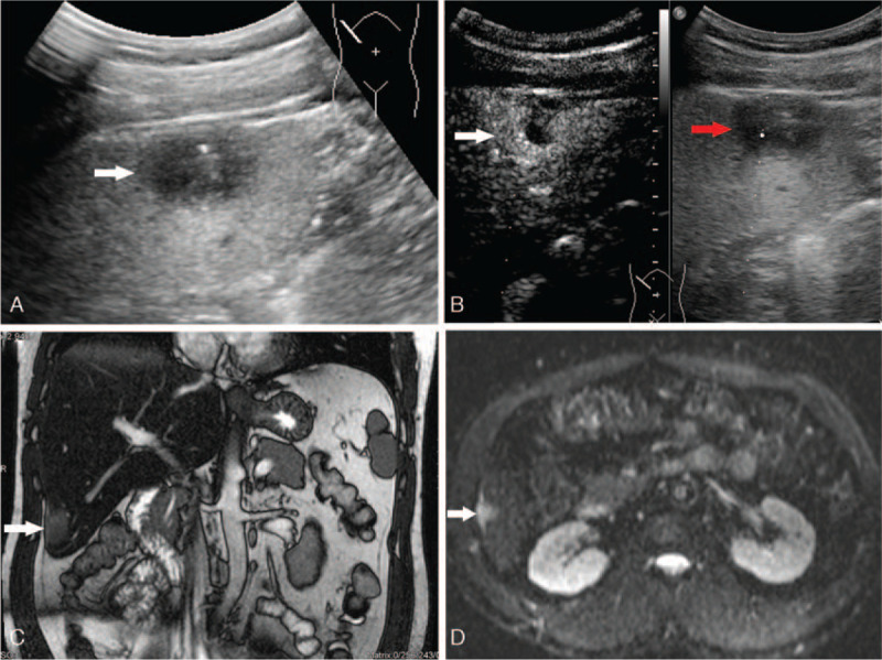
Diagnoses: The tests for blood, liver function, and tumor markers were within normal range. Preoperative ultrasonography (US) showed a hypoechoic lesion with a longitudinal diameter of 2.5 cm in segment six of liver. It exhibited early arterial phase hyperenhancement and late arterial phase rapid washout in contrast-enhanced US. It demonstrated hyperintensity in T2-weighted magnetic resonance imaging and partly restricted diffusion in diffusion-weighted imaging. For this nodule, the preoperative diagnosis was small hepatocellular carcinoma (HCC).
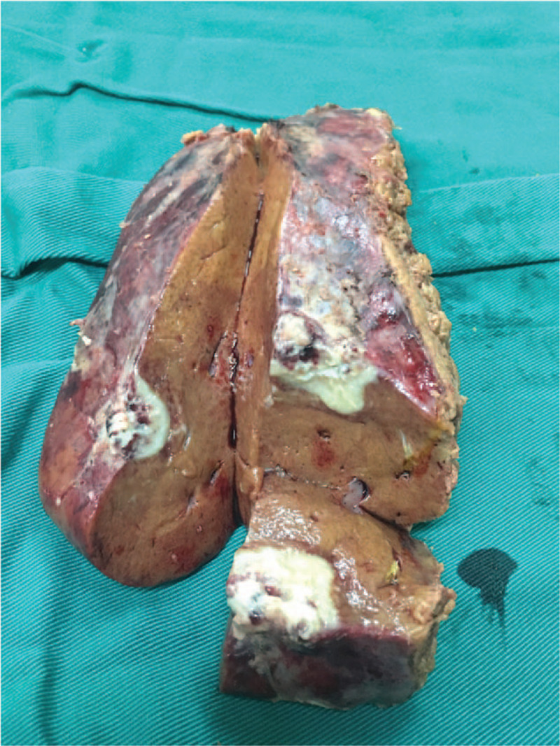
Interventions: Laparoscopic hepatectomy was performed. Intraoperative extensive adhesion in the abdominal cavity and liver was found. The lesion had undergone expansive growth.
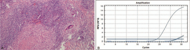
Outcomes: Microscopically, a granuloma with some necrosis was detected. With both acid-fast staining and TB fragment polymerase chain reaction showing positive results, TB was the final histology diagnosis. After surgery, the patient declined any anti-TB medication. During the follow-up, he had no symptoms. In the sixth month after surgery, he underwent an upper abdominal US. It showed no lesions in the liver.
Lessons: Because of non-specific imaging findings and non-specific symptoms, a diagnosis of isolated hepatic TB is difficult to make, especially for small lesions. A diagnosis of HCC should be made cautiously when small isolated lesions in the liver are encountered, especially in patients without a history of hepatitis and with negative tumor markers.
Figures
References
-
- Singh D, Singh S, Raut SB, et al. . Isolated liver tuberculosis: a case report. Pediatr Surg Int 2004;20:727–8. - PubMed
-
- Mert A, Ozaras R, Tabak F, et al. . Localized hepatic tuberculosis. Eur J Intern Med 2003;14:511–2. - PubMed
-
- Setime M, Rwegerera GM, Chowdhury W, et al. . Isolated tubercular hepatic abscess with diffuse pattern mimicking hepatocellular carcinoma in HIV positive patient: a case report. Health Res 2014;16:333–6. - PubMed
Publication types
MeSH terms
Substances
LinkOut - more resources
Full Text Sources
