. 2020 Oct 20;119(8):1481-1488.
doi: 10.1016/j.bpj.2020.09.005. Epub 2020 Sep 16.

Mechanisms of Light-Induced Deformations in Photoreceptors

Affiliations

Mechanisms of Light-Induced Deformations in Photoreceptors

K C Boyle et al. Biophys J. .

Abstract

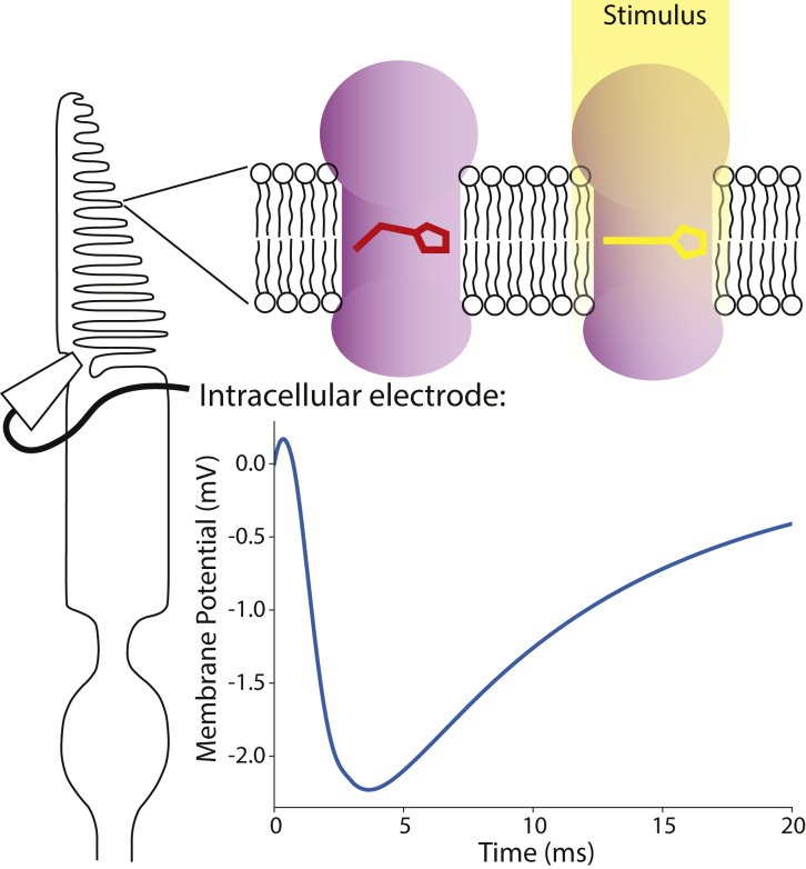
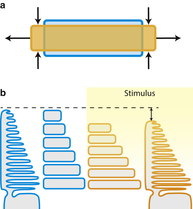
Biological cells deform on a nanometer scale when their transmembrane voltage changes, an effect that has been visualized during the action potential using quantitative phase imaging. Similar changes in the optical path length have been observed in photoreceptor outer segments after a flash stimulus via phase-resolved optical coherence tomography. These optoretinograms reveal a fast, millisecond-scale contraction of the outer segments by tens of nanometers, followed by a slow (hundreds of milliseconds) elongation reaching hundreds of nanometers. Ultrafast measurements of the contractile response using line-field phase-resolved optical coherence tomography show a logarithmic increase in amplitude and a decreasing time to peak with increasing stimulus intensity. We present a model that relates the early receptor potential to these deformations based on the voltage-dependent membrane tension-the mechanism observed earlier in neurons and other electrogenic cells. The early receptor potential is caused by conformational changes in opsins after photoisomerization, resulting in the fractional shift of the charge across the disk membrane. Lateral repulsion of the ions on both sides of the membrane affects its surface tension and leads to its lateral expansion. Because the volume of the disks does not change on a millisecond timescale, their lateral expansion leads to an axial contraction of the outer segment. With increasing stimulus intensity and the resulting tension, the area expansion coefficient of the disk membrane also increases as thermally induced fluctuations are pulled flat, resisting further expansion. This leads to the logarithmic saturation observed in measurements as well as the peak shift in time. This imaging technique therefore relates the structural changes in the photoreceptor to the underlying neurological function of transducing light into electrical signals. Such label-free optical monitoring of neural activity using fast interferometry may be applicable not only to optoretinography but also to neuroscience in general.


Figures

Figure 1
Figure 1
OPL changes in the photoreceptor outer segment in response to light stimuli. A brief flash (in this case 1 ms at 6.11 × 106 ph/μm2) elicits a fast, millisecond-scale contraction (negative deformation), which is then overtaken by a hundreds-of-milliseconds-long elongation. To see this figure in color, go online.
Figure 2
Figure 2
Intracellular electrical recording of the cone outer segment demonstrates the transmembrane potential transient after a 1 ms flash caused by the conformation change of the opsins embedded in the disk membranes. Each photoisomerization induces a 10−10 V potential change, leading to a characteristic potential change up to a few mV in the outer segment, with its time course shaped by the time constants of the various steps in the phototransduction cycle. To see this figure in color, go online.
Figure 3
Figure 3
The deformation model of the photoreceptor outer segments assumes (a) the membrane expansion and subsequent flattening of the fixed-volume disks after the stimulus, which result in (b) the shrinkage of the outer segment that contains a stack of approximately 1000 disks. To see this figure in color, go online.
Figure 4
Figure 4
Saturation of the deformations due to the increasing expansion modulus of the disk membranes. (a) At low tensions, the disk membranes contain thermally induced fluctuations that require little force to pull out, depending on the smallest bending radius as labeled, but at higher forces, the intermolecular bonds of the membrane begin to dominate. (b) The resulting area expansion coefficient increases with tension, and in the small range of tensions relevant to the outer segment disks, indicated by the red arrow, can be approximated as a linear function of tension. (c) The relative area expansion thus saturates logarithmically over a 100-μN/m tension change. The rate of saturation depends on mechanical parameters of the membrane, notably including the minimal bending radius illustrated here, where 5 nm is the size of an individual lipid head. The larger minimal radii could be defined by the much larger opsins embedded in the membrane. To see this figure in color, go online.
Figure 5
Figure 5
The time-dependent deformation model fits the observations well. (a) The combined model is comprised of two components: the fast contraction caused by the ERP and the linearly rising slow expansion that represents both the LRP and the osmotic swelling of the outer segment. (b) The model parameters are fitted to observations at two stimulus intensities; then the same parameters are used in (c) to predict the peak deformation as a function of stimulus intensity from additional measurements. The error bars denote +/- 1 standard deviation from 4 repeat measurements. To see this figure in color, go online.

References

    1. Müller W., Töpke H. The early receptor potential (ERP) Doc. Ophthalmol. 1987;66:35–74. - PubMed
    1. Hodgkin A.L., Obryan P.M. Internal recording of the early receptor potential in turtle cones. J. Physiol. 1977;267:737–766. - PMC - PubMed
    1. Smith N.P., Lamb T.D. The a-wave of the human electroretinogram recorded with a minimally invasive technique. Vision Res. 1997;37:2943–2952. - PubMed
    1. Zhang F., Kurokawa K., Miller D.T. Cone photoreceptor classification in the living human eye from photostimulation-induced phase dynamics. Proc. Natl. Acad. Sci. USA. 2019;116:7951–7956. - PMC - PubMed
    1. Hillmann D., Spahr H., Hüttmann G. In vivo optical imaging of physiological responses to photostimulation in human photoreceptors. Proc. Natl. Acad. Sci. USA. 2016;113:13138–13143. - PMC - PubMed

Publication types

LinkOut - more resources