

Review
doi: 10.5152/dir.2020.19482.
Magnetic resonance lymphangiography: with or without contrast?
Affiliations
- PMID: 33032980
- PMCID: PMC7664753
- DOI: 10.5152/dir.2020.19482
Item in Clipboard
Review
Magnetic resonance lymphangiography: with or without contrast?
Diagn Interv Radiol.
2020 Nov.
Abstract
Lymphedema is an important medical issue around the world, caused by an anomalous collection of fluid in soft tissue due to congenital malformations or stenosis or obstruction of lymphatic vessels. Magnetic resonance lymphangiography (MRL) is an emerging technique focused on noninvasive or minimally invasive imaging of lymphatics with the goal to diagnose and treat lymphedema. This review will briefly discuss lymphatic imaging starting with lymphography and radionuclide lymphoscintigraphy up to the newest methods, focusing on MRL, a rising technique, and highlighting the technical aspects fundamental for achieving high-resolution MRL.
Conflict of interest statement
The authors declared no conflicts of interest.
Figures
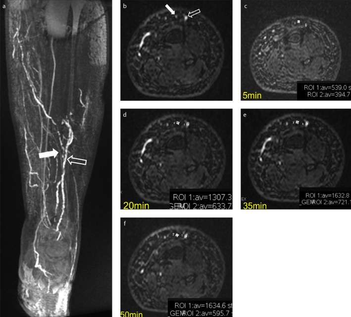
Clinical example of the kinetic of enhancement (ROI 1 = lymphatic; empty arrow; ROI 2= vein, white arrow). Coronal and axial 3D T1 gradient-echo sequences with SPECtral inversion at Lipid (FSPGR) show different morphology and diameter of lymphatic vessels (empty arrow) and veins (white arrow)
(a, b). Taking into account the kinetic of enhancement at 5, 20, 35, 50 minutes after subcutaneous injection of contrast media, there is a clear increase in the region of interest (ROI) values in the affected lymphatic vessel, while in the vein, an uptrend followed by a down trend is observed (c–f).

Noninvasive MRL performed through heavily T2-weighted sequences without subcutaneous contrast media injection allow lymphatics visualization; however, in case of severe lymphedema, the hyperintensity of epifascial areas could prevent the visualization of the underlying lymphatics (a). T2 sequences, if used alone, are not useful to give functional information about the run-time of enhancement of lymphatics. A balanced sequence ECG-triggered (3D steady-state free precession, SSFP) can show both the epifascial distribution of lymphedema and a map of veins (b, arrow), whereas a post-contrast T1-weighted 3D with fat saturation sequence can visualize both lymphatics (arrowhead) and veins (arrows), allowing functional information as well (c).
References
-
- Zuther JE, Norton S. Lymphedema management: the comprehensive guide for practitioners. 3rd ed. Vol. 3. New York: Thieme Medical Publishers; 2005.
Publication types
MeSH terms
Substances
LinkOut - more resources
Full Text Sources
Medical
