. 2020 Dec;216(4):1009-1022.
doi: 10.1534/genetics.120.303494. Epub 2020 Oct 8.

Post-translational Regulation of DNA Polymerase η, a Connection to Damage-Induced Cohesion in Saccharomyces cerevisiae

Affiliations

Post-translational Regulation of DNA Polymerase η, a Connection to Damage-Induced Cohesion in Saccharomyces cerevisiae

Pei-Shang Wu et al. Genetics. 2020 Dec.

Abstract

Double-strand breaks that are induced postreplication trigger establishment of damage-induced cohesion in Saccharomyces cerevisiae, locally at the break site and genome-wide on undamaged chromosomes. The translesion synthesis polymerase, polymerase η, is required for generation of damage-induced cohesion genome-wide. However, its precise role and regulation in this process is unclear. Here, we investigated the possibility that the cyclin-dependent kinase Cdc28 and the acetyltransferase Eco1 modulate polymerase η activity. Through in vitro phosphorylation and structure modeling, we showed that polymerase η is an attractive substrate for Cdc28 Mutation of the putative Cdc28-phosphorylation site Ser14 to Ala not only affected polymerase η protein level, but also prevented generation of damage-induced cohesion in vivo We also demonstrated that Eco1 acetylated polymerase η in vitro Certain nonacetylatable polymerase η mutants showed reduced protein level, deficient nuclear accumulation, and increased ultraviolet irradiation sensitivity. In addition, we found that both Eco1 and subunits of the cohesin network are required for cell survival after ultraviolet irradiation. Our findings support functionally important Cdc28-mediated phosphorylation, as well as post-translational modifications of multiple lysine residues that modulate polymerase η activity, and provide new insights into understanding the regulation of polymerase η for damage-induced cohesion.

Keywords: Cdc28/Cdk1; Eco1; cohesin; damage-induced cohesion; polymerase eta.


Figures

Figure 1
Figure 1
Polη is a substrate of the Cdc28 kinase in vitro, supported by in silico modeling. (A) Polη contains three CDK consensus motifs (in blue). The putative phosphorylation sites are bold, marked with asterisks and shown with numbers; (i) complete CDK consensus motif, (ii) partial CDK consensus motif. (B) Cdc28-Polη in vitro kinase assay. Purified recombinant Polη-GST or Polη-3A-GST (Polη with the three putative phosphorylation sites mutated to alanine) were incubated with purified Cdc28/Clb2, in the presence or absence of 32P-γATP, for 15 or 30 min (indicated with filled blue circles). Recombinant human H1 was used as positive control. The kinase assay was repeated three times. (C) In silico modeling of Polη in complex with Cdc28. Protein docking of a Cdc28 homology model (green) and Polη (yellow), with bound DNA (pink/violet). Inset: Close-up of the Cdc28 active site showing Polη-S14 positioned close to the modeled ATP’s γ-phosphate (modeled ATP in gray with standard atom coloring). This minimized theoretical heterodimer model indicates that the Polη-K17 can interact with the Cdc28 phosphor-T169, crucial for kinase activity. CDK, cyclin-dependent kinase.
Figure 2
Figure 2
The Polη-S14A mutation affects protein level, but not TLS activity. (A) Protein levels of Myc-tagged Polη-phosphorylation mutants in G2/M phase. Cdc11 was used as loading control. (B) UV spot assay of Polη single and triple phosphorylation mutants. Tenfold serial dilutions of midlog phase cells were spotted on YEPD plates and exposed to 35 J/m2 UVC. Plates were documented after 3 days. YEPD, YEP media with glucose.
Figure 3
Figure 3
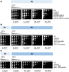
Eco1, cohesin, and Polη are required for cell survival after UV irradiation. (A–C) Tenfold serial dilutions of midlog phase cells were spotted on YEPD, with or without follow-on UVC exposures. Semipermissive temperatures for the temperature-sensitive cohesin loader and cohesin subunits mutants were used: 30° for scc2-4 and scc1-73, and 32° for smc1-259. Plates were documented on the second or third day. One representative experiment from two independent assays performed is shown. RT, room temperature.
Figure 4
Figure 4
Polη is an Eco1 substrate in vitro. (A) In vitro acetylation assay. Purified recombinant Polη was incubated with or without purified recombinant Eco1, in the presence or absence of 14C-acetyl-coenzyme A (14C-CoA), and acetylation was visualized by autoradiography. (B) Mass spectrometry detected in vitro (Eco1-mediated) and in vivo (DSB-specific) acetylation sites are shown in green and blue, respectively. DSB, double-strand break; NLS, nuclear localization sequence; PAD, polymerase-associated domain; PIP, PCNA-interacting protein motif; UBZ, ubiquitin-binding/zinc-finger motif.
Figure 5
Figure 5
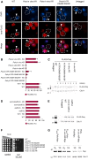
Polη-KR mutations affect nuclear accumulation and total protein level. (A) Representative in situ immunofluorescence images of indicated WT or mutated FLAG–Polη, stained with anti-FLAG and counterstained with DAPI. Nuclear accumulation of Polη after break induction in G2/M phase was determined by colocalization of anti-FLAG and DAPI signals, indicated by white arrows. Nonnuclear staining with anti-FLAG (Polη) is indicated by gray arrows. (B) Percent of cells that expressed FLAG-Polη at detectable level and showed nuclear accumulation, with and without break induction in G2/M phase. Quantified from in situ stained cells. Means ± SD from at least two independent experiments are shown. (C) Protein levels of FLAG-Polη with indicated KR mutations (asynchronized cells). Numbers one and two indicate two different transformation clones. Cdc11 was used as loading control. (D) Polη nuclear accumulation in the absence of Eco1 and/or Wpl1. Quantified as in B. Means ± SD from at least two independent experiments are shown. (E) Protein levels of FLAG-Polη in G2/M phase, in the presence or absence of Eco1 and/or Wpl1. Cdc11 was used as loading control. (F) Spot assay to monitor UV sensitivities of the Polη-KR mutants. Tenfold serial dilutions of midlog phase cells were spotted on YEPD plates, subsequently exposed to 35 J/m2 UVC, and documented after 3 days. (G) Protein levels of Polη-Myc and Polη-in vivo KR-Myc, expressed from the endogenous or the constitutive strong ADH promoter. The samples were from the same gel, but the image was cropped to show selected samples. Cdc11 was used as loading control. 1G, 1 hr break induction by addition of galactose (PGAL-HO); G2, G2/M arrested cells; HO, homothallic switching endonuclease; YEPD, YEP media supplemented with glucose.
Figure 6
Figure 6
Polη-S14 is important for establishment of damage-induced cohesion. (A) A schematic experimental outline for a typical damage-induced cohesion experiment. Cells expressing the temperature-sensitive allele smc1-259 (smc1 ts) are synchronized in G2/M by addition of benomyl. Galactose is then added for expression of ectopic PGAL-SMC1 (Smc1 WT) and induction of DSBs on chromosome III (PGAL-HO). After 1 hr, the temperature is raised to 35°, restrictive for smc1-259, whereby the cohesion established during S phase (blue rings) is destroyed. The Tet-O/TetR-GFP system (green dots) is used to monitor damage-induced cohesion (red rings) on chromosome V. (B) Damage-induced cohesion assay of PADH-Polη-in vivo KR. Means ± SD from at least two independent experiments are shown. (C) Damage-induced cohesion assay of indicated single Polη-phosphorylation mutants (S/T to A). Means ± SD from at least two independent experiments are shown. Statistically significant differences were analyzed with one-way ANOVA, Scheffe post hoc test (P < 0.05). (D) Protein levels of Polη-Myc and Polη-S14A-Myc, expressed from the ADH promoter. Cdc11 was used as loading control. (E) Damage-induced cohesion assay of PADH-Polη-S14A. Means ± SD from at least two independent experiments are shown. 1G, 1 hr break induction by addition of galactose (PGAL-HO); DSBs, double-strand breaks; G2, G2/M arrested cells; HO, homothallic switching endonuclease.

References

    1. Acharya N., Manohar K., Peroumal D., Khandagale P., Patel S. K. et al. , 2019. Multifaceted activities of DNA polymerase eta: beyond translesion DNA synthesis. Curr. Genet. 65: 649–656. 10.1007/s00294-018-0918-5 - DOI - PubMed
    1. Alt A., Lammens K., Chiocchini C., Lammens A., Pieck J. C. et al. , 2007. Bypass of DNA lesions generated during anticancer treatment with cisplatin by DNA polymerase eta. Science 318: 967–970. 10.1126/science.1148242 - DOI - PubMed
    1. Bertoletti F., Cea V., Liang C. C., Lanati T., Maffia A. et al. , 2017. Phosphorylation regulates human poleta stability and damage bypass throughout the cell cycle. Nucleic Acids Res. 45: 9441–9454. 10.1093/nar/gkx619 - DOI - PMC - PubMed
    1. Billon P., Li J., Lambert J. P., Chen Y., Tremblay V. et al. , 2017. Acetylation of PCNA sliding surface by Eco1 promotes genome stability through homologous recombination. Mol. Cell 65: 78–90. 10.1016/j.molcel.2016.10.033 - DOI - PubMed
    1. Brands A., and Skibbens R. V., 2008. Sister chromatid cohesion role for CDC28-CDK in Saccharomyces cerevisiae. Genetics 180: 7–16. 10.1534/genetics.108.092288 - DOI - PMC - PubMed

Publication types

MeSH terms