. 2020 Oct 8;10(1):16787.
doi: 10.1038/s41598-020-73693-x.

Hydrojet-based delivery of footprint-free iPSC-derived cardiomyocytes into porcine myocardium

Affiliations

Hydrojet-based delivery of footprint-free iPSC-derived cardiomyocytes into porcine myocardium

Marbod Weber et al. Sci Rep. .

Abstract

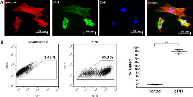
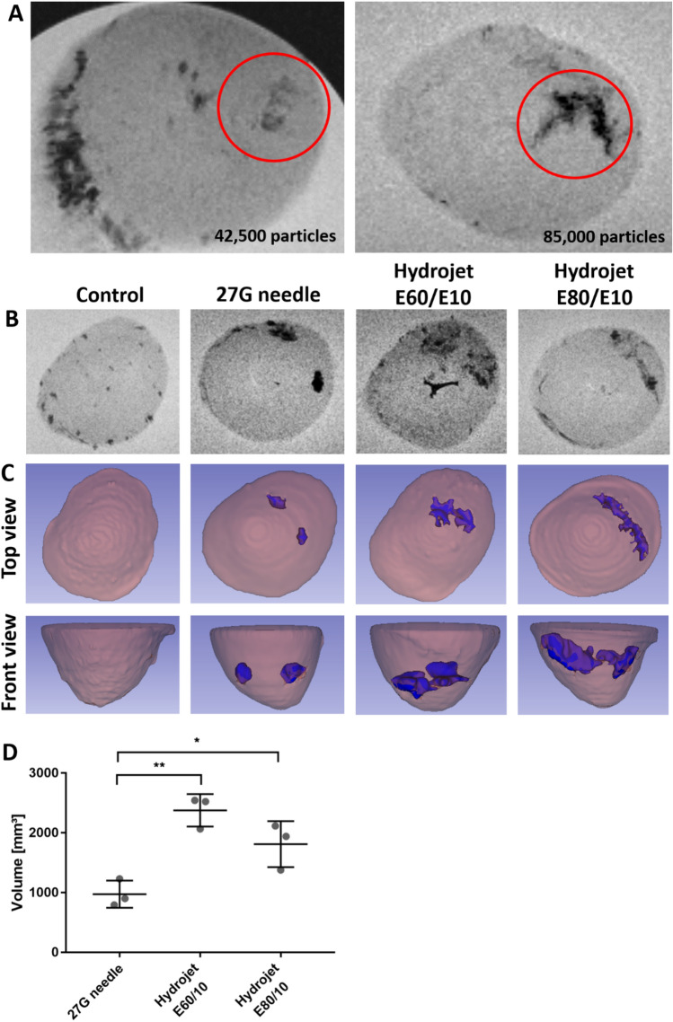
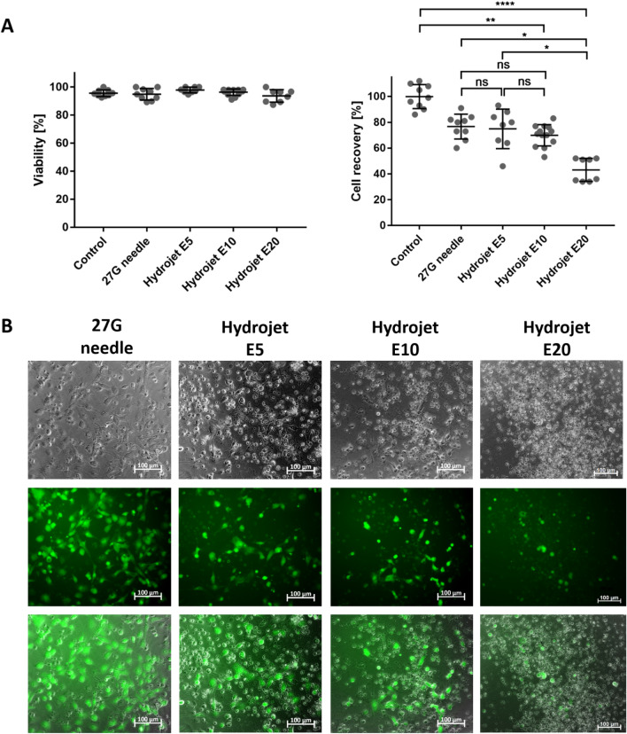
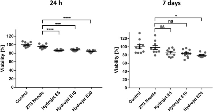
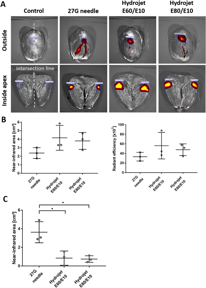
The reprogramming of patient´s somatic cells into induced pluripotent stem cells (iPSCs) and the consecutive differentiation into cardiomyocytes enables new options for the treatment of infarcted myocardium. In this study, the applicability of a hydrojet-based method to deliver footprint-free iPSC-derived cardiomyocytes into the myocardium was analyzed. A new hydrojet system enabling a rapid and accurate change between high tissue penetration pressures and low cell injection pressures was developed. Iron oxide-coated microparticles were ex vivo injected into porcine hearts to establish the application parameters and the distribution was analyzed using magnetic resonance imaging. The influence of different hydrojet pressure settings on the viability of cardiomyocytes was analyzed. Subsequently, cardiomyocytes were delivered into the porcine myocardium and analyzed by an in vivo imaging system. The delivery of microparticles or cardiomyocytes into porcine myocardium resulted in a widespread three-dimensional distribution. In vitro, 7 days post-injection, only cardiomyocytes applied with a hydrojet pressure setting of E20 (79.57 ± 1.44%) showed a significantly reduced cell viability in comparison to the cells applied with 27G needle (98.35 ± 5.15%). Furthermore, significantly less undesired distribution of the cells via blood vessels was detected compared to 27G needle injection. This study demonstrated the applicability of the hydrojet-based method for the intramyocardial delivery of iPSC-derived cardiomyocytes. The efficient delivery of cardiomyocytes into infarcted myocardium could significantly improve the regeneration.


Conflict of interest statement

LJ, WL, AF, LB, MDE are employees of Erbe Elektromedizin GmbH, Tuebingen. All other authors declare no competing financial and non-financial interests relevant to the submitted work.

Figures

Figure 1
Figure 1
Analysis of cells obtained after cardiac differentiation of iPSCs. (A) Fluorescence microscopic images of cTNT and α-actinin positive cells after cardiomyocyte differentiation. Nuclei were stained with DAPI. (B) Detection of cTNT positive cells after the cardiac differentiation by flow cytometry. Results are shown as mean ± SD (n = 3). Statistical differences were determined using paired t-test (**p < 0.01).
Figure 2
Figure 2
Detection of microparticle distribution in the porcine hearts using magnetic resonance imaging (MRI). (A) Determination of detectable microparticle amount in porcine hearts using 45,000 or 85,000 microparticles. A single injection of particles was performed using the new hydrojet system with tissue penetration pressure of E80 and injection pressure of E10 (E80/E10). The microparticles in the heart are highlighted by a red encircled region. (B) MRI of microparticles after the application of 85,000 microparticles (two injections) using 27G needle or the hydrojet system with tissue penetration pressures of E80 or E60 and an injection pressure of E10 (E80/E10 or E60/E10). (C) 3D reconstruction of particle distribution in porcine hearts using 3D Slicer software. (D) Comparison of microparticle distribution volume in porcine hearts. Results are shown as mean ± SD (n = 3). Statistical differences were determined using one-way ANOVA followed by Bonferroni’s multiple comparison test (*p < 0.05; **p < 0.01).
Figure 3
Figure 3
Analysis of the viability and recovery rates of cardiomyocytes immediately after the injection using hydrojet or 27G needle and calcein AM staining of injected cells 24 h after cultivation. (A) Determination of cell viability and cell recovery by trypan blue staining and counting of cells immediately after the injection of 1 × 106 cardiomyocytes in CMM using a 27G needle or the hydrojet system with different injection pressure settings (E5, E10, or E20). Results are shown as mean ± SD (control, 27G needle (n = 9), E5 and E20 (n = 8), E10 (n = 15). Statistical differences were determined using one-way ANOVA followed by Bonferroni’s multiple comparison test or Kruskal–Wallis test followed by Dunn's multiple comparison test (*p < 0.05; **p < 0.01; ****p < 0.0001, ns: non-significant). (B) Representative images of calcein AM stained cardiomyocytes 24 h after the cultivation of injected cells in cell culture plates for 24 h.
Figure 4
Figure 4
Analysis of the viability of cardiomyocytes 24 h and 7 days after the application of cells. Determination of cell viability using PrestoBlue assay 24 h and 7 days after the seeding of injected cardiomyocytes into cell culture plates. The viability of cardiomyocytes without injection (control) was set to 100% and the viability of cardiomyocytes injected by 27G needle or hydrojet was expressed relative to these cells. Results are shown as mean ± SEM [control, E5, E10, and E20 (n = 10), and 27G needle (n = 9)]. Statistical differences were determined using one-way ANOVA followed by Bonferroni’s multiple comparison test (*p < 0.05, ***p < 0.001, ****p < 0.0001).
Figure 5
Figure 5
Distribution of cardiomyocytes in porcine hearts after the application with hydrojet system. 100 µl CMM without or with XenoLight DiR fluorescent dye-labeled 1 × 106 cardiomyocytes was applied using 27G needle or hydrojet system with tissue penetration pressures of E80 or E60 and injection pressure of E10 (E80/E10 or E60/E10) into porcine hearts. (A) IVIS images of the apex region of the hearts from outside and inside of the myocardium. The intersection of the apex is schematically indicated as a white line. (B) Comparison of the radiant efficiency and near-infrared area after the application of cardiomyocytes into the myocardium. (C) Detection of the NIR-labeled area of blood vessels containing cardiomyocytes. Results are shown as mean ± SD (n = 3). Statistical differences were determined using one-way ANOVA followed by Bonferroni’s multiple comparison test (*p < 0.05).

Similar articles

Cited by

References

    1. Wong ND. Epidemiological studies of CHD and the evolution of preventive cardiology. Nat. Rev. Cardiol. 2014;11(5):276. doi: 10.1038/nrcardio.2014.26. - DOI - PubMed
    1. Cahill TJ, Choudhury RP, Riley PR. Heart regeneration and repair after myocardial infarction: Translational opportunities for novel therapeutics. Nat. Rev. Drug Discov. 2017;16(10):699. doi: 10.1038/nrd.2017.106. - DOI - PubMed
    1. Li Z, Guan J. Hydrogels for cardiac tissue engineering. Polymers. 2011;3(2):740–761. doi: 10.3390/polym3020740. - DOI
    1. Lázár E, Sadek HA, Bergmann O. Cardiomyocyte renewal in the human heart: Insights from the fall-out. Eur. Heart J. 2017;38(30):2333–2342. doi: 10.1093/eurheartj/ehx343. - DOI - PMC - PubMed
    1. Talman V, Ruskoaho H. Cardiac fibrosis in myocardial infarction—From repair and remodeling to regeneration. Cell Tissue Res. 2016;365(3):563–581. doi: 10.1007/s00441-016-2431-9. - DOI - PMC - PubMed

Publication types