. 2020 Oct 8;10(1):16837.
doi: 10.1038/s41598-020-74003-1.

Femtosecond electronic structure response to high intensity XFEL pulses probed by iron X-ray emission spectroscopy

Affiliations

Femtosecond electronic structure response to high intensity XFEL pulses probed by iron X-ray emission spectroscopy

Roberto Alonso-Mori et al. Sci Rep. .

Abstract

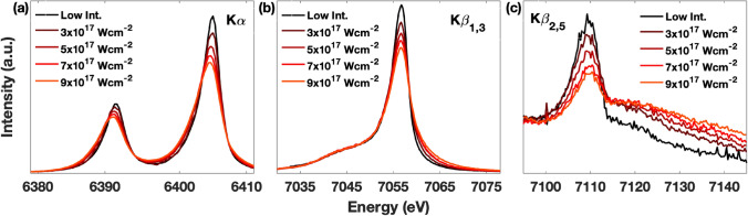
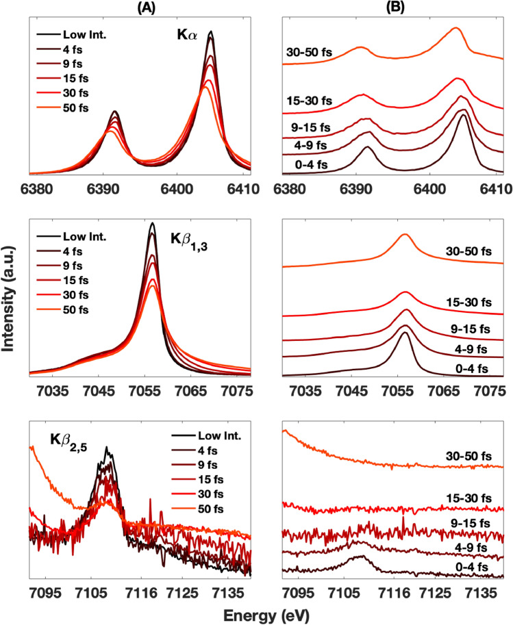
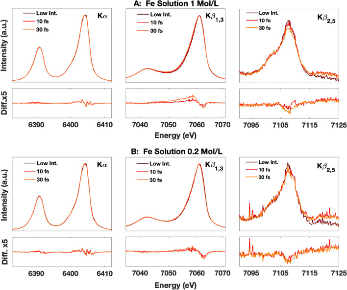
We report the time-resolved femtosecond evolution of the K-shell X-ray emission spectra of iron during high intensity illumination of X-rays in a micron-sized focused hard X-ray free electron laser (XFEL) beam. Detailed pulse length dependent measurements revealed that rapid spectral energy shift and broadening started within the first 10 fs of the X-ray illumination at intensity levels between 1017 and 1018 W cm-2. We attribute these spectral changes to the rapid evolution of high-density photoelectron mediated secondary collisional ionization processes upon the absorption of the incident XFEL radiation. These fast electronic processes, occurring at timescales well within the typical XFEL pulse durations (i.e., tens of fs), set the boundary conditions of the pulse intensity and sample parameters where the widely-accepted 'probe-before-destroy' measurement strategy can be adopted for electronic-structure related XFEL experiments.


Conflict of interest statement

The authors declare no competing interests.

Figures

Figure 1
Figure 1
Fe emission spectra (a) Kα, (b)1,3, and (c)2,5 measured with 10 fs X-ray pulses at different intensity levels up to ~ 9 × 1017 W cm−2. All spectra are normalized to the shot-to-shot measurement of the incoming pulse energy.
Figure 2
Figure 2
Column A shows the Kα, Kβ1,3, and Kβ2,5 cumulative emission spectra (top, middle, bottom respectively) measured with different X-ray pulse durations at an average intensity of 3 × 1017 W cm-2. Their respective deconvolved spectral evolution are shown on column B. The deconvolved traces are vertically offset by the amounts proportional to their timing.
Figure 3
Figure 3
K emission spectra of FeSO4 with a concentration of 1 mol/L (A) and 0.2 mol/L (B) at different beam conditions (A and B TOP) and the difference spectra (with a 5 × zoom) compared to the unmodified spectrum acquired at low intensity (A and B BOTTOM). The 10 fs and 30 fs spectra were collected with a 2 μm size beam and intensities of 9 × 1017 and 5 × 1017 W cm−2 respectively.

References

    1. Neutze R, Wouts R, van der Spoel D, Weckert E, Hajdu J. Potential for biomolecular imaging with femtosecond X-ray pulses. Nature. 2000;406:752–757. doi: 10.1038/35021099. - DOI - PubMed
    1. Seibert MM, et al. Single mimivirus particles intercepted and imaged with an X-ray laser. Nature. 2011;470:78–81. doi: 10.1038/nature09748. - DOI - PMC - PubMed
    1. van der Schot G, et al. Imaging single cells in a beam of live cyanobacteria with an X-ray laser. Nat. Commun. 2015;6:5704. doi: 10.1038/ncomms6704. - DOI - PubMed
    1. Chapman HN, et al. Femtosecond X-ray protein nanocrystallography. Nature. 2011;470:73–U81. doi: 10.1038/nature09750. - DOI - PMC - PubMed
    1. Boutet S, et al. High-resolution protein structure determination by serial femtosecond crystallography. Science. 2012;337:362–364. doi: 10.1126/science.1217737. - DOI - PMC - PubMed