

Severe Acute Respiratory Syndrome Coronavirus 2 Nucleocapsid Protein in the Ocular Tissues of a Patient Previously Infected With Coronavirus Disease 2019
- PMID: 33034620
- PMCID: PMC7545349
- DOI: 10.1001/jamaophthalmol.2020.3962
Severe Acute Respiratory Syndrome Coronavirus 2 Nucleocapsid Protein in the Ocular Tissues of a Patient Previously Infected With Coronavirus Disease 2019
Abstract
Importance: Coronavirus disease 2019 (COVID-19) has been recognized as a pandemic by the World Health Organization. Whether severe acute respiratory syndrome coronavirus 2 (SARS-CoV-2) can also infect tissues besides the respiratory system, such as the ocular tissues, remains unclear.
Objective: To determine whether SARS-CoV-2 exists intracellularly in the ocular tissues of a patient previously infected with COVID-19.
Design, setting, and participants: This case study analyzed a patient previously infected with COVID-19 who had an acute glaucoma attack during her rehabilitation. Plasma samples and tissue specimens, including ones from the conjunctiva, anterior lens capsular, trabecular meshwork, and iris, were collected during phacoemulsification and trabeculectomy surgery. Specimens from another patient who had glaucoma but not COVID-19 were used as a negative control.
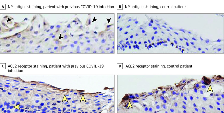
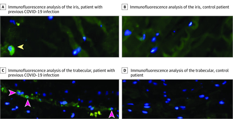
Main outcomes and measures: Specimens were analyzed using hematoxylin-eosin staining. The nucleocapsid protein antigen of SARS-CoV-2 was measured in the conjunctiva, trabecular meshwork, and iris using immunofluorescence and immunohistochemistry. The expression of angiotensin-converting enzyme 2 receptor on the conjunctiva was measured using immunohistochemistry.
Results: The patient with a previous COVID-19 infection was female and 64 years old, and the control patient without a COVID-19 infection history was male and 61 years old. The nucleocapsid protein antigen of SARS-CoV-2 was detected on the cells of the conjunctiva, trabecular, and iris of the patient infected with COVID-19 but not in the control participant, while angiotensin-converting enzyme 2 receptor proteins were detected on the conjunctiva of both patients.
Conclusions and relevance: The nucleocapsid protein antigen of SARS-CoV-2 existed intracellularly in the ocular tissues of a patient previously infected with COVID-19. Thus, SARS-CoV-2 can also infect ocular tissues in addition to the respiratory system.
Conflict of interest statement
Figures
References
-
- Zhou Y, Zeng Y, Tong Y, et al. Ophthalmologic evidence against the interpersonal transmission of 2019. novel coronavirus through conjunctiva. Published online February 12, 2020. https://www.medrxiv.org/content/10.1101/2020.02.11.20021956v1 - DOI
Publication types
MeSH terms
Substances
LinkOut - more resources
Full Text Sources
Medical
Miscellaneous
