

Epithelioid hemangioma of bone harboring FOS and FOSB gene rearrangements: A clinicopathologic and molecular study
- PMID: 33034932
- PMCID: PMC7739373
- DOI: 10.1002/gcc.22898
Epithelioid hemangioma of bone harboring FOS and FOSB gene rearrangements: A clinicopathologic and molecular study
Abstract
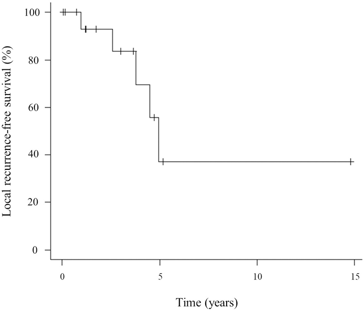
The diagnosis of epithelioid hemangioma (EH) remains challenging due to its rarity, worrisome histologic features, and locally aggressive clinical and radiographic presentation. Especially in the bone, EH can be misdiagnosed as a malignant vascular neoplasm due its lytic, often destructive or multifocal growth, as well as atypical morphology. The discovery of recurrent FOS and FOSB gene fusions in the pathogenesis of most EH has strengthened its stand-alone classification, distinct from other malignant epithelioid vascular lesions, such as epithelioid hemangioendothelioma or angiosarcoma. In this study we investigate a group of molecularly confirmed skeletal EH by the presence of FOS or FOSB gene rearrangements to better define its clinical and pathologic characteristics within a homogenous molecular subset. The cohort included 38 patients (25 males, 13 females), with a mean age at diagnosis of 38 years (range, 4-75). Regional, multifocal presentation was noted in 10 cases. Only six cases were correctly recognized as EH by the referring institutions, while most were misdiagnosed as other vascular tumors. Of the 17 patients with follow-up data available, five patients (29%) developed local recurrence after marginal en bloc excision (n = 3) or curettage (n = 2). Local recurrence-free survival rates were 84% at 3 years and 38% at 5 years. No metastasis or disease-related death was identified. Imaging studies exhibited no specific features, showing cortical bone destruction and soft-tissue extension in 14 (38%) cases. FOS gene rearrangements were detected in 28 (74%) of cases, while FOSB rearrangements in 10 (26%) cases. Our results highlight the significant challenges encountered in establishing a correct diagnosis exclusive of the molecular testing, mainly due to its overlap to other malignant epithelioid vascular tumors. Skeletal EH emerges as a genetically defined locally aggressive vascular neoplasm, with a high rate of local recurrence, but lacking the propensity for distant spread.
Keywords: FOS; FOSB; epithelioid hemangioma; fusions.
© 2020 Wiley Periodicals LLC.
Conflict of interest statement
Figures


References
-
- Fetsch JF, Sesterhenn IA, Miettinen M, Davis CJ Jr. Epithelioid hemangioma of the penis: a clinicopathologic and immunohistochemical analysis of 19 cases, with special reference to exuberant examples often confused with epithelioid hemangioendothelioma and epithelioid angiosarcoma. Am J Surg Pathol. 2004;28 (4):523–533. - PubMed
-
- Nielsen GP, Srivastava A, Kattapuram S, et al. Epithelioid hemangioma of bone revisited: a study of 50 cases. Am J Surg Pathol. 2009;33 (2):270–277. - PubMed
-
- Rosai J, Akerman LR. Intravenous atypical vascular proliferation. A cutaneous lesion simulating a malignant blood vessel tumor. Arch Dermatol. 1974;109 (5):714–717. - PubMed
-
- Castro C, Winkelmann RK. Angiolymphoid hyperplasia with eosinophilia in the skin. Cancer. 1974;34 (5):1696–1705. - PubMed
Publication types
MeSH terms
Substances
Grants and funding
LinkOut - more resources
Full Text Sources
Medical
Research Materials
Miscellaneous
