. 2020 Nov 19;5(22):e143213.
doi: 10.1172/jci.insight.143213.

Neutralizing antibody against SARS-CoV-2 spike in COVID-19 patients, health care workers, and convalescent plasma donors

Affiliations

Neutralizing antibody against SARS-CoV-2 spike in COVID-19 patients, health care workers, and convalescent plasma donors

Cong Zeng et al. JCI Insight. .

Abstract

Rapid and specific antibody testing is crucial for improved understanding, control, and treatment of COVID-19 pathogenesis. Herein, we describe and apply a rapid, sensitive, and accurate virus neutralization assay for SARS-CoV-2 antibodies. The assay is based on an HIV-1 lentiviral vector that contains a secreted intron Gaussia luciferase (Gluc) or secreted nano-luciferase reporter cassette, pseudotyped with the SARS-CoV-2 spike (S) glycoprotein, and is validated with a plaque-reduction assay using an authentic, infectious SARS-CoV-2 strain. The assay was used to evaluate SARS-CoV-2 antibodies in serum from individuals with a broad range of COVID-19 symptoms; patients included those in the intensive care unit (ICU), health care workers (HCWs), and convalescent plasma donors. The highest neutralizing antibody titers were observed among ICU patients, followed by general hospitalized patients, HCWs, and convalescent plasma donors. Our study highlights a wide phenotypic variation in human antibody responses against SARS-CoV-2 and demonstrates the efficacy of a potentially novel lentivirus pseudotype assay for high-throughput serological surveys of neutralizing antibody titers in large cohorts.

Keywords: COVID-19; Cellular immune response.


Conflict of interest statement

Conflict of interest: A patent application (#63/028,972) related to this work has been filed to the US Patent and Trademark Office, in which SLL, CZ, JPE, YMZ, and PQ are listed as the applicants.

Figures

Figure 1
Figure 1. inGluc-based HIV-1 lentiviral S pseudotypes bearing SARS-CoV-2 spikes.
293T cells seeded on 6-well plates were cotransfected with 0.8 μg HIV-1–NL4.3–inGluc vector plus 0.4 μg SARS-CoV-2 spike-coding plasmid. Forty-eight hours after transfection, viral supernatant was harvested and used to infect target cells. Unless otherwise indicated, 293T/ACE2 cells were used for infection. (A) Schematic representation of the pseudoviral production and infection. Note that Gluc activity can only be detected in virus-infected target cells — and not in the virus-producing cells — because of the presence of an intron inserted in the sense of the vector that splits the Gluc gene into 2 parts. (B and C) Titers of HIV-1 inGluc pseudotypes bearing the spikes of SARS-CoV (n = 6), SARS-CoV-2 (n = 6), or VSV-G (n = 3); absolute luciferase readouts at 48 hours after infection, and relative infectivity compared with the background, were plotted, respectively. (D) Indicated doses of viral supernatant were used to infect 293T/ACE2 cells seeded in 24-well plates, and 20 μL of supernatant of virus-infected cells were used to measure the Gluc activity as shown. The dashed line indicates the background of luciferase activity; n = 3. (E) Indicated amounts of culture media harvested from virus-infected cells were used to measure Gluc activity; n = 3. (F) Relative infectivity of HIV-inGluc pseudotypes bearing S proteins of SARS-CoV, SARS-CoV-2, or VSV-G in indicated target cells, with parental or those overexpressing ACE2; n = 6. Data were analyzed as mean ± SD.
Figure 2
Figure 2. Comparison of HIV-1 inGluc pseudotypes bearing C9-tagged spikes of SARS-CoV or WTs.
(A) Relative infectivity. Experiments were performed as described as Figure 1, B and C, except that either WT or C9-tagged spikes were used for virus production; n = 6. *P < 0.001, by 2-tailed t test. (B and C) Western blotting analysis of C9-tagged S protein expression in the virus-producing cells (B) and purified viral particles (C). Viral production was carried as described in Figure 1, and viral producer cells were lysed and analyzed by Western blotting using anti-C9, anti-p24, and/or anti–β-actin. (DF) Virus-producing cells were digested with PBS-5 mM EDTA and incubated with 10 μg/mL sACE2-Fc for 2 hours; cells were washed 3 times, incubated with FITC-labeled anti-human Fc for 45 minutes, and analyzed by flow cytometry. (D) Representative cell populations analyzed for SARS-CoV and SARS-CoV-2; the percentage of positive cells for FITC anti-human Fc was shown. (E) Histogram analysis of virus-producing cells for SARS-CoV and SARS-CoV-2. (F) Relative mean fluorescence intensities of cells expressing indicated spikes; n = 2; no statistical analysis was performed. Note that cells transfected with an empty vector pCIneo served as negative control, the fluorescence intensity of which was set to 1. Data were analyzed as mean ± SD.
Figure 3
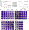
Figure 3. Neutralization of SARS-CoV-2 by COVID-19 patient sera.
Validation of inGluc-based lentiviral pseudotypes using an authentic SARS-CoV-2 US-WA-1 strain. Note that all samples tested here and throughout the studies were blinded before testing. (A) A group of 8 blinded patient sera was tested for neutralization of the SARS-CoV-2 lentiviral S pseudotypes bearing C9-tagged SARS-CoV-2 S or WT. Note that only patient serum sample (sample 8) was tested for the WT spike, the pattern of which almost perfectly overlaps with that of C9-tagged spike; n = 3. The ELISA OD450 values of 8 samples are indicated for each number (cutoff, 0.40). (B) Results of infectious SARS-CoV-2 plaque-reduction neutralization assay for testing of 8 blinded samples. Vero-E6 cells were infected for 3 days with infectious SARS-CoV-2, pretreated with or without the indicated diluted sera. Cells were fixed and stained with 0.25% crystal violet for visualization of plaques; n = 3. Note that the BEI guinea pig antiserum to SARS-CoV did not inhibit SARS-CoV-2 infection. (C) Types of serum or plasma samples did not appear to affect the neutralization pattern generated by inGluc-based lentiviral pseudotypes bearing SARS-CoV-2 spike. Serum, sodium citrate-treated plasma, and EDTA-treated plasma from the same patient were used for SARS-CoV-2 S pseudotype neutralization. Data were analyzed as mean ± SD.
Figure 4
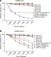
Figure 4. Examining the cross-reactivity of COVID-19 patient sera with SARS-CoV/SARS-CoV-2 S lentiviral pseudotypes.
(A and B) The neutralization assay was carried out as described in Figures 3 and 4, except that HIV-1 inGluc pseudotypes bearing SARS-CoV S (A) were used in parallel with that of SARS-CoV-2 (B). In addition to 5 patient sera (samples 1, 2, 3, 5, and 8 from Figure 3A), stocks of polyclonal (guinea pig and rabbit) or monoclonal (human) antibodies against SARS-CoV obtained from BEI Resources were also tested; the exact concentrations of these antibodies are unknown. A mouse monoclonal antibody 2B04 against SARS-CoV-2, with a stock concentration of 10 μg/mL, was diluted to the same extent as the patient sera and other antibodies and tested; n = 3. Data were analyzed as mean ± SD.
Figure 5
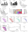
Figure 5. Evaluations of neutralizing antibody levels in COVID-19 hospitalized inpatients, ICU patients, health care workers, and convalescent plasma donors.
Blinded serum samples were serially diluted and tested for neutralizing activity against lentiviral pseudotypes bearing SARS-CoV-2 S. (AE) Ranges of neutralizing antibody titer IC50 in the 4 indicated groups (x axis); percent in each study group was plotted (y axis). (FJ) Neutralization curves of 4 different groups, as presented by the relative infectivity of SARS-CoV-2 S pseudotypes in the presence of indicated serum samples. The y axis indicates the relative viral infectivity by setting the viral infectivity without serum to 100%; the x axis indicates dilution fold of serum samples. (K) Correlational analysis of pseudovirus neutralization IC50 and N protein IgG antibody ELISA OD450 values; r = 0.4192, P = 0.0027 as indicated; n = 49. r = 0.4192, P = 0.0027, as indicated by correlation of XY analyses; n = 49. (L) Correlational analysis between pseudovirus neutralization IC50 and age; n = 30. Data were analyzed as mean ± SD.
Figure 6
Figure 6. A secreted Nluc–based lentiviral SARS-CoV-2 S neutralization assay with improved stability and sensitivity, and its application in measuring SARS-CoV-2 neutralizing antibody levels in COVID-19 patients.
293T cells were transfected with lentiviral vector (pNL4.3 inGluc or pNL4.3 secNluc) along with the SARS-CoV-2 S-C9 plasmid. Media was 48 hours after transfection and used to infect 293T/ACE2 cells; luciferase activity was measured at indicated times to determine the viral infectivity; n = 3 for all experiments. (A) Stability of inGluc and secNluc luciferase signals measured over time. A total of 20 μL of Gaussia luciferase substrate or 20 μL of Nano Luciferase substrate were added simultaneously, and luminescence measurements were then read every 2 minutes for 60 minutes. Plotted are the luminescence reads relative to the 0 minutes time point, which was set to 100%; secNluc exhibited a signal that was more stable than the inGluc virus infected cells. (B and C) Infectivity of SARS-CoV-2 S secNluc pseudotypes. (B) The Nano-luciferase activity of culture medium harvested from virus-infected cells at indicated times. (C) Relative viral infectivity was plotted by setting the mock infection to 1.0. (D) Indicated amounts of viral supernatant were used to infect 293T/ACE2 cells seeded in 24-well plates, and 20 μL of supernatant from virus-infected cells was used to measure the secNluc activity as shown. The dashed line indicates the background luminescence. (E) Experiment was performed as described in the legends of Figure 3A and Figure 4B, except secNluc lentiviral pseudotypes were used for infection. Data were analyzed as mean ± SD.

Update of

References

    1. [No authors listed]. Coronavirus Disease (COVID-19): Situation Report. World Health Organization. https://www.who.int/emergencies/diseases/novel-coronavirus-2019/situatio... Accessed October 15, 2020.
    1. Wu F, et al. A new coronavirus associated with human respiratory disease in China. Nature. 2020;579(7798):265–269. doi: 10.1038/s41586-020-2008-3. - DOI - PMC - PubMed
    1. doi: 10.1101/2020.04.30.2008567. Hachim A, et al. Beyond the Spike: identification of viral targets of the antibody response to SARS-CoV-2 in COVID-19 patients. medRxiv. Published May 22, 2020. Accessed October 15, 2020. - DOI
    1. Luchsinger LL, et al. Serological Assays Estimate Highly Variable SARS-CoV-2 Neutralizing Antibody Activity in Recovered COVID19 Patients [published ahead of print September 11, 2020]. J Clin Microbiol. https://doi.org 10.1128/JCM.02005-20. - PMC - PubMed
    1. doi: 10.1101/2020.05.13.092619. Robbiani DF, et al. Convergent antibody responses to SARS-CoV-2 infection in convalescent individuals. bioRxiv. Published May 22, 2020. Accessed October 15, 2020. - DOI - PMC - PubMed

Publication types

Substances