. 2020 Oct 15;183(2):335-346.e13.
doi: 10.1016/j.cell.2020.08.050. Epub 2020 Oct 8.

Single Residue Variation in Skeletal Muscle Myosin Enables Direct and Selective Drug Targeting for Spasticity and Muscle Stiffness

Affiliations

Single Residue Variation in Skeletal Muscle Myosin Enables Direct and Selective Drug Targeting for Spasticity and Muscle Stiffness

Máté Gyimesi et al. Cell. .

Abstract

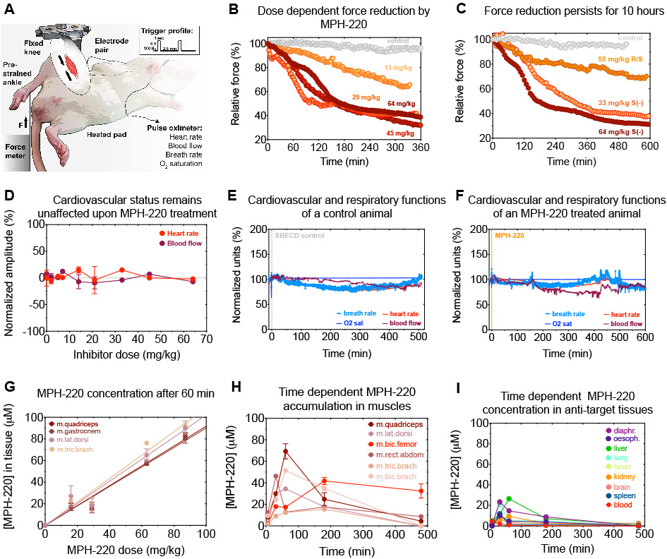
Muscle spasticity after nervous system injuries and painful low back spasm affect more than 10% of global population. Current medications are of limited efficacy and cause neurological and cardiovascular side effects because they target upstream regulators of muscle contraction. Direct myosin inhibition could provide optimal muscle relaxation; however, targeting skeletal myosin is particularly challenging because of its similarity to the cardiac isoform. We identified a key residue difference between these myosin isoforms, located in the communication center of the functional regions, which allowed us to design a selective inhibitor, MPH-220. Mutagenic analysis and the atomic structure of MPH-220-bound skeletal muscle myosin confirmed the mechanism of specificity. Targeting skeletal muscle myosin by MPH-220 enabled muscle relaxation, in human and model systems, without cardiovascular side effects and improved spastic gait disorders after brain injury in a disease model. MPH-220 provides a potential nervous-system-independent option to treat spasticity and muscle stiffness.

Keywords: artificial intelligence; blebbistatin; crystallography; deep learning; force; motor protein; musculoskeletal disorder; sarcomere; stroke; unmet medical need.


Conflict of interest statement

Declaration of Interests The authors declare the following competing interests: employment, A.M.-C. and M.G. are owners of Motorpharma, Ltd. and A.Á.R. and M.G. are part-time employed by Motorpharma, Ltd.; related patents, PCT/EP2017/051829, WO/2017/129782, HU1800129A2, PCT/HU2019/050017, WO/2019/202346A2, and WO/2019/202346A3; J.A.S. is a cofounder and member of the scientific advisory boards of Cytokinetics and MyoKardia, biotechnology companies developing small molecules that target the sarcomere for the treatment of various muscle diseases; K.M.R. is on the scientific advisory board at MyoKardia; and J.A.S., D.V.T., and K.M.R. are cofounders of Kainomyx Inc., a biotechnology company focused on developing small molecules to target tropical diseases.

Figures

Figure 1.
Figure 1.. MPH-220 inhibits skeletal muscle myosin with extreme selectivity independently of the nervous system.
(A) Current muscle relaxants target the central nervous system or act peripherally, whereas MPH-220 directly inhibits myosin. (B) Human myosin-2 isoforms contain phenylalanine (black F) at the beginning of the HP-helix between the switch-2 loop (red) and the relay region, except for fast skeletal myosin isoforms, where this position is leucine (blue L) (cf. Fig. S1). (C) MPH-220 was designed to bind into the blebbistatin-binding cavity of the motor domain, to enable inhibition in the actin-detached state. (D) Close-up view of the communication center of the functional regions (actin-binding cleft, switch-2 loop of the active site and the HP-relay-converter region) in the homology model of MyHC IIa with bound MPH-220 (orange) reveals interaction between the morpholino group and Leu476 in the HP-helix. (E) Actin-activated ATPase inhibition of three myosin-2 isoforms (cf. Table S1). (F) Actin-activated ATPase inhibition of NM2A, NM2B, NM2C and the NM2CF490L variant. (G) Actin-activated ATPase inhibition of expressed human β-cardiac myosin with MPH-220 and blebbistatin. (H) Actin-activated ATPase inhibition of human muscle myosin samples from biopsies of m. soleus, m. vastus lateralis and heart ventricle. In contrast to MPH-220, blebbistatin fully inhibited m. vastus lateralis sample and β-cardiac myosin samples. Mean ± SD are shown in all ATPase experiments, n=3-9.
Fig. 2.
Fig. 2.. Crystal structure of MPH-220-bound fast skeletal muscle myosin.
(A) Cartoon representation of the heavy chain (gray) of rabbit skeletal muscle myosin-2 in Mg.ADP.VO4 (spheres, blue) pre-powerstroke state with the essential light chain bound to the lever arm. MPH-220 binds to the bottom of the actin-binding cleft close to the HP-helix, with its morpholine ring in close proximity of Leu476. Blue mesh: electron density corresponding to MPH-220 at sigma 1.0. (B) Comparison of the MPH-220 (orange) binding site in MyHC IIb (gray/beige) and the blebbistatin (yellow) binding site in Dictyostelium myosin-2 (dark gray/brown) (1YV3.pdb (Allingham et al., 2005)). The chiral OH groups of both inhibitors interact with the same residues Gly248 and Leu270 (Gly240 and Leu262 in Dictyostelium). Mesh: surfaces based on Van-der-Walls radii, calculated with PyMol. (Bottom) Residues involved in compound binding in Ligplot+ v2.2 (Laskowski and Swindells, 2011) representation; U50: upper 50-kDa subdomain and L50: lower 50-kDa subdomain (gray); sw-2: switch-2 loop (red); HP: HP-helix (beige). Hydrogen bonds (up to 3.2 Å) are shown as green lines. (C) MPH-220 bound MyHC IIb structure with superimposed cardiac HP-helix illustrates the steric clash between the morpholine ring of MPH-220 and the cardiac HP-helix Phe473 (red) residue.
Fig. 3.
Fig. 3.. MPH-220 reduces skeletal muscle force without cardiovascular effects.
(A) Isometric force of rat hindleg was measured, while vital functions were detected by a non-invasive pulse oximeter. (B-C) Dose dependent force reduction after MPH-220 treatment persisted for more than 10 hours. (D) Heart rate (red circle) and pulse distention - reflecting local blood flow at the carotid artery - (maroon circle) show the maximal signal deflections during the measurements (60-600 min) on 1-4 animals/dose. (E-F) Cardiovascular and respiratory functions as a function of time after treatment with SBECD control or MPH-220. Note that permanent oxygen level is maintained by O2-supplemented isoflurane anesthesia. (G) Dose-dependent MPH-220 accumulation in rat muscle tissues 60 minutes after oral treatment. Mean ± SD are shown; n = 2-3. (H-I) Time-dependent MPH-220 accumulation in rat tissues after 35 mg/kg i.p treatment. Mean ± SD are shown; n = 2-11.
Fig. 4.
Fig. 4.. MPH-220 efficiently improves gait functions of spastic rats after brain injury.
(A) Position of pyramidal tract lesion (red dots). (B) Open-field gait analysis was performed by deep-learning algorithms coupled to sub-millimeter 3D movement detection. Dots represent the determined positions; right and left forelimbs (RF, LF), right and left hindlegs (RH, LH). (C) Fractional turn orientation of spastic rat before (gray) and after (orange) 15 mg/kg MPH-220 treatment. (D) Falling and spontaneous cramping rates normalized to the total distance travelled (upper) and the number of touches on the cylinder wall (lower) after 20 mg/kg MPH-220 treatment. (E) Paw position distribution of the healthy (upper) and treated (lower) animals before and after 20 mg/kg MPH-220 treatment in slow regime. (F) Averaged distances between the left (upper) and right (lower) fore- and hindlimbs in each frame of the 3D recordings relative to the healthy values. Mean ± SD are shown.
Fig. 5.
Fig. 5.. Principal component analysis of spastic rats reveals details of improved gait components after MPH-220 treatment.
(A and B) Box and whiskers (min to max) representation of global (A) and speed-specific (B) principal component (PC) analysis of gait parameters of healthy (gray) and operated (colored) animals before (darker) and after (lighter) MPH-220 treatment. (C) Parameter contributions to PC1 in the slow and medium regimes and to PC3 in the fast regimes (cf. Fig. S5). (D) Heatmap of major parameters contributing to PC1 (blue-orange) and PC3 (purple-orange) (cf. Fig. S5). (D) Schematic representation of MPH-220 effects on paw positions in slow and fast regimes.

References

    1. Allingham JS, Smith R, and Rayment I (2005). The structural basis of blebbistatin inhibition and specificity for myosin II. Nat Struct Mol Biol 12, 378–379. - PubMed
    1. Berman H, Henrick K, and Nakamura H (2003). Announcing the worldwide Protein Data Bank. Nat Struct Biol 10, 980. - PubMed
    1. Billington N, Wang A, Mao J, Adelstein RS, and Sellers JR (2013). Characterization of three full-length human nonmuscle myosin II paralogs. J Biol Chem 288, 33398–33410. - PMC - PubMed
    1. Bjornsdottir A, Gudmundsson G, Blondal H, and Olafsson E (2013). Incidence and prevalence of multiple system atrophy: a nationwide study in Iceland. Journal of neurology, neurosurgery, and psychiatry 84, 136–140. - PubMed
    1. Blair E, Langdon K, McIntyre S, Lawrence D, and Watson L (2019). Survival and mortality in cerebral palsy: observations to the sixth decade from a data linkage study of a total population register and National Death Index. BMC neurology 19, 111. - PMC - PubMed

Publication types

MeSH terms