Review
. 2020 Oct 6;8(4):23.
doi: 10.3390/jdb8040023.

C. elegans Apical Extracellular Matrices Shape Epithelia

Affiliations
Review

C. elegans Apical Extracellular Matrices Shape Epithelia

Jennifer D Cohen et al. J Dev Biol. .

Abstract

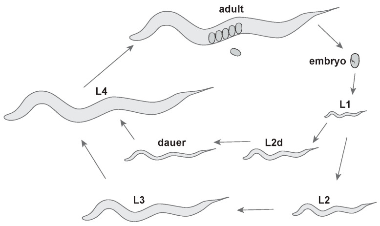
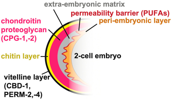
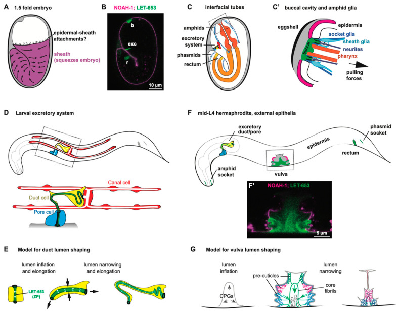
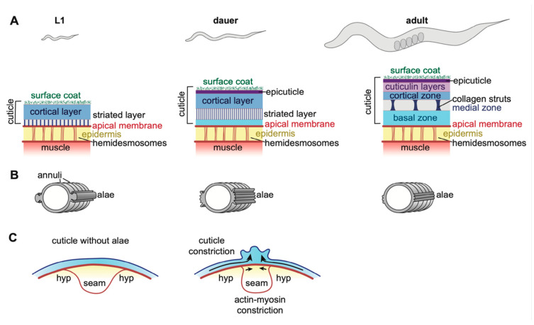
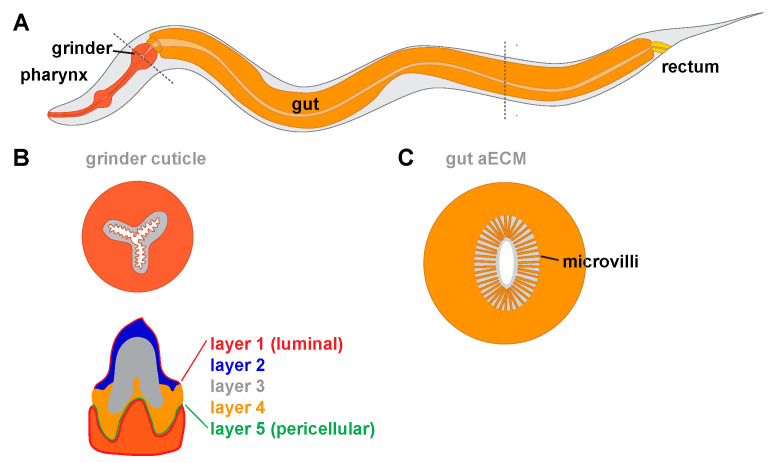
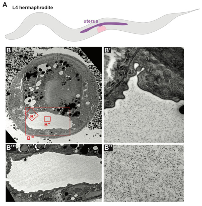
Apical extracellular matrices (aECMs) coat exposed surfaces of epithelia to shape developing tissues and protect them from environmental insults. Despite their widespread importance for human health, aECMs are poorly understood compared to basal and stromal ECMs. The nematode Caenorhabditis elegans contains a variety of distinct aECMs, some of which share many of the same types of components (lipids, lipoproteins, collagens, zona pellucida domain proteins, chondroitin glycosaminoglycans and proteoglycans) with mammalian aECMs. These aECMs include the eggshell, a glycocalyx-like pre-cuticle, both collagenous and chitin-based cuticles, and other understudied aECMs of internal epithelia. C. elegans allows rapid genetic manipulations and live imaging of fluorescently-tagged aECM components, and is therefore providing new insights into aECM structure, trafficking, assembly, and functions in tissue shaping.

Keywords: C. elegans; apical extracellular matrix; cuticle; eggshell; glycocalyx.


Conflict of interest statement

The authors declare no conflicts of interest. The funders had no role in the design of the study; in the collection, analyses, or interpretation of data; in the writing of the manuscript, or in the decision to publish the results.

Figures

Figure 1
Figure 1
C. elegans life cycle. Adults lay embryos that hatch into L1 larvae. Larvae molt into subsequent stages. Under stress (low food, high temperatures, and crowding), larvae can molt into an alternative L3 stage called dauer, which can resume reproductive development upon return to non-stressful conditions. After Wormatlas [28].
Figure 2
Figure 2
C. elegans eggshell. The outermost vitelline layer (black) contains the CBD-1/PERM-2/PERM-4 complex [31]. Next, the chitin-rich layer (yellow) is followed by the chondroitin proteoglycan layer (pink), which contains the chondroitin-proteoglycan proteins CPG-1 and CPG-2 [23]. The extra-embryonic matrix (gray) and the peri-embryonic matrix (orange), which line the embryo, are separated by the permeability barrier (red) [24,31].
Figure 3
Figure 3
C. elegans pre-cuticles shape developing epithelia. (A) Diagram of a 1.5 fold C. elegans embryo encased in the embryonic sheath. The sheath distributes actinomyosin-based forces that squeeze the embryo into a worm-shape [47]. (B) Confocal image of fluorescently-tagged ZP proteins LET-653 and NOAH-1 in a 1.5 fold embryo. LET-653 (green) primarily lines interfacial tubes, including the excretory duct and pore lumen, the rectum (r), and the buccal cavity (b) [48]. NOAH-1 (magenta) is present in the embryonic sheath [47]. (C) Cross section of a 1.5-fold embryo, showing interfacial tubes. (C’) The pharynx, glia, and neurites are pulled posteriorly while being anchored anteriorly by the pre-cuticle aECM [49,50,51]. Magenta represents NOAH-1-containing pre-cuticle, and green represents LET-653-containing pre-cuticle, as shown in in panel B. (D) The larval excretory system. The duct and pore tubes are lined by pre-cuticle and cuticle (green), while the canal tube contains a non-cuticular aECM. Black denotes junctions. (E) Model for duct lumen shaping by LET-653 and the pre-cuticle (adapted from [20]). LET-653 (green) promotes duct lumen inflation and resists morphogenetic stretching and squeezing forces (arrows) to maintain proper lumen diameter. (F) Diagram of a mid-L4 larva, showing tissues lined by pre-cuticle and cuticle. (F’) Confocal image of fluorescently-tagged ZP proteins LET-653 and NOAH-1 in the L4 vulva. (G) Model for vulva lumen shaping by the pre-cuticle (adapted from [48]). After initial lumen inflation by CPGs, distinct pre-cuticles form along the apical surfaces of different vulva cell types. Connections between these pre-cuticles and a central core structure contribute to lumen narrowing.
Figure 4
Figure 4
C. elegans epidermal cuticle. (A) Diagram of C. elegans at L1, dauer, and adult stages with cuticle structure at each stage (adapted from [106]). The epidermis connects the muscle and cuticle via hemidesmosomes. At the apical surface, the transmembrane proteins MUP-4 and MUA-3 link hemidesmosomes to the cuticle [55,57]. The cuticle is a multi-layered structure of collagens, cuticulins, lipids, and glycans [91,102,107]. The latter three are likely concentrated near the external surface of the cuticle, while collagens predominate in the basal zone and striated layers. Pre-cuticle and nascent cuticle may appear near the apical membrane prior to molts [53]. (B) Cross section of C. elegans at each stage indicating the position of alae and annuli. Furrows are the low points between annuli. Alae are not shown to scale. L1 larvae have one large alae ridge flanked by two smaller ones, adults have three alae ridges, while dauer larvae have five. (C) Model for alae formation. Constriction by actin-myosin in seam cells and by ZP proteins in the cuticle bend the cuticle into alae ridges [14,108].
Figure 5
Figure 5
Tubes and aECMs of the C. elegans digestive tract. (A) Diagram of the digestive system. Different aECMs line the pharynx, gut and rectum. (B) Diagram of the pharyngeal grinder containing multiple teeth and the five observed aECM layers within a single tooth (adapted from [175]). Dark orange denotes pharyngeal cell cytoplasm. (C) Cross-section through the gut, showing apical microvilli surrounded by a membrane-proximal aECM (adapted from [176]). Light orange denotes intestinal cell cytoplasm.
Figure 6
Figure 6
Uterine aECM. (A) Diagram of L4 stage C. elegans with vulva (pink) and expanded uterus (purple). (B) TEM of the uterus at mid-L4 stage. (B’B’”) The inflated uterine lumen is filled with a granular matrix of unknown composition. (Electron micrographs courtesy of Alessandro Sparacio).

References

    1. Luschnig S., Uv A. Luminal matrices: An inside view on organ morphogenesis. Exp. Cell Res. 2014;321:64–70. doi: 10.1016/j.yexcr.2013.09.010. - DOI - PubMed
    1. Pérez-Gil J. Structure of pulmonary surfactant membranes and films: The role of proteins and lipid-protein interactions. Biochim. Biophys. Acta. 2008;1778:1676–1695. doi: 10.1016/j.bbamem.2008.05.003. - DOI - PubMed
    1. Tarbell J.M., Cancel L.M. The glycocalyx and its significance in human medicine. J. Intern. Med. 2016;280:97–113. doi: 10.1111/joim.12465. - DOI - PubMed
    1. Walma D.A.C., Yamada K.M. The extracellular matrix in development. Development. 2020;147 doi: 10.1242/dev.175596. - DOI - PMC - PubMed
    1. Jayadev R., Sherwood D.R. Basement membranes. Curr. Biol. 2017;27:R207–R211. doi: 10.1016/j.cub.2017.02.006. - DOI - PubMed

LinkOut - more resources