Review
. 2020 Oct 7;25(19):4572.
doi: 10.3390/molecules25194572.

ATP-Binding and Hydrolysis in Inflammasome Activation

Affiliations
Review

ATP-Binding and Hydrolysis in Inflammasome Activation

Christina F Sandall et al. Molecules. .

Abstract

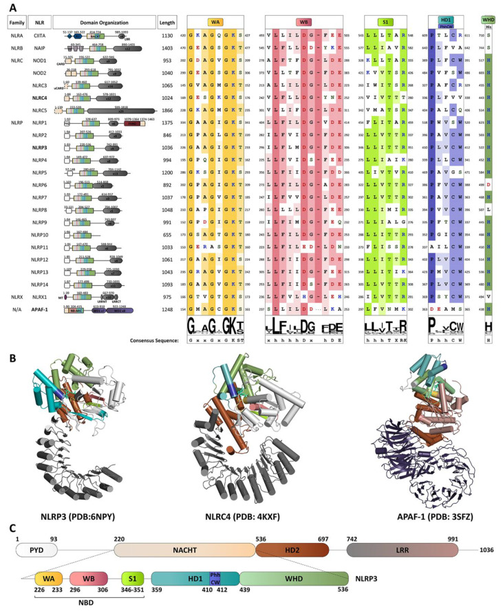
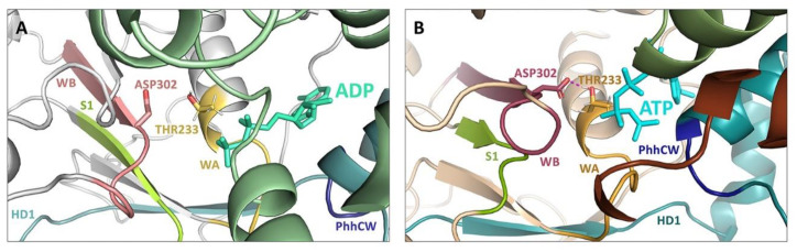
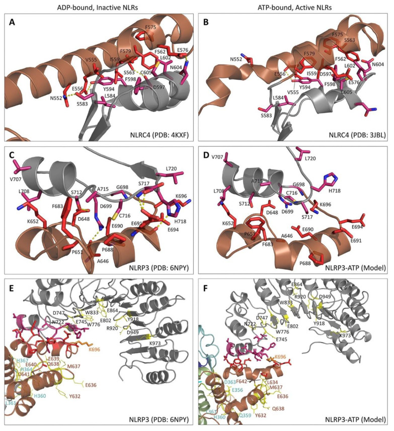
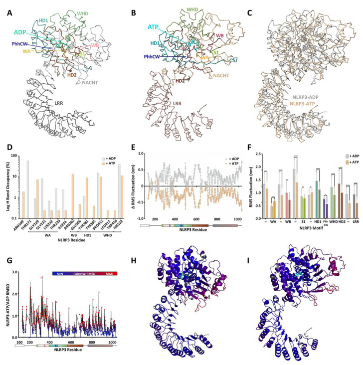
The prototypical model for NOD-like receptor (NLR) inflammasome assembly includes nucleotide-dependent activation of the NLR downstream of pathogen- or danger-associated molecular pattern (PAMP or DAMP) recognition, followed by nucleation of hetero-oligomeric platforms that lie upstream of inflammatory responses associated with innate immunity. As members of the STAND ATPases, the NLRs are generally thought to share a similar model of ATP-dependent activation and effect. However, recent observations have challenged this paradigm to reveal novel and complex biochemical processes to discern NLRs from other STAND proteins. In this review, we highlight past findings that identify the regulatory importance of conserved ATP-binding and hydrolysis motifs within the nucleotide-binding NACHT domain of NLRs and explore recent breakthroughs that generate connections between NLR protein structure and function. Indeed, newly deposited NLR structures for NLRC4 and NLRP3 have provided unique perspectives on the ATP-dependency of inflammasome activation. Novel molecular dynamic simulations of NLRP3 examined the active site of ADP- and ATP-bound models. The findings support distinctions in nucleotide-binding domain topology with occupancy of ATP or ADP that are in turn disseminated on to the global protein structure. Ultimately, studies continue to reveal how the ATP-binding and hydrolysis properties of NACHT domains in different NLRs integrate with signaling modules and binding partners to control innate immune responses at the molecular level.

Keywords: ATPase; NACHT domain; NLR; NLRP; NOD-like receptor; inflammasome; molecular dynamic simulation; nucleotide.


Conflict of interest statement

J.A.M. is cofounder and has an equity position in Arch Biopartners Inc. All other authors declare no conflicts of interest. The funders had no role in the design of the study; in the collection, analyses, or interpretation of data; in the writing of the manuscript, or in the decision to publish the results.

Figures

Figure 1
Figure 1
Characterization of NLRP3 and the NLR family. (A) Multiple sequence alignment of conserved features critical for catalytic activity in the 22 NLR proteins and APAF-1 NBD motifs. The NLR family members are subdivided based on the presence of one of four N-terminal effector domains: NLRAs with an acidic domain (AD), NLRBs with a Baculovirus IAP repeat (BIR), NLRCs with a caspase recruitment domain (CARD), or NLRPs with a pyrin domain (PYD). The naming system approved by the HUGO Gene Nomenclature Committee (HGNC) is used [10]. Domain identities and configuration for NLR proteins are displayed to scale with boundaries numbered as per UniProtKB accession numbers: CIITA, P33076; NAIP, Q13075; NOD1, Q9Y239; NOD2, Q9HC29; NLRC3, Q7RTR2; NLRC4, Q9NPP4; NLRC5, Q86WI3; NLRX1, Q86UT6; NLRP1, Q9C000; NLRP2, Q9NX02; NLRP3, Q96P20; NLRP4, Q96MN2; NLRP5, P59047; NLRP6, P59044; NLRP7, Q8WX94; NLRP8, Q86W28; NLRP9, Q7RTR0; NLRP10, Q86W26; NLRP11, P59045; NLRP12, P59046; NLRP13, Q86W25; and NLRP14, Q86W24. Multiple sequence alignments were created with Jalview [16] and MUSCLE [17] with default options and used to generate sequence conservation logos shown below each motif [18]. These indicate sequence conservation amongst NLRs and APAF-1, with the height of the stack indicating overall sequence conservation at that position in the alignment, while the height of letters within the stack indicates the relative frequency of each amino acid at that position. Amino acid class in the alignment is denoted by coloured font; where hydrophobic residues (A,I,L,M,F,W,V,C,G,P) are coloured in yellow; polar residues in green; polar (S,T,Y,N,Q) in green; acidic (D,E) in red; and basic (K,R,H) in blue. Conservation within each motif is denoted by background shading, where no color indicates <30% conservation, and increasing colour intensity indicates conservation of residue type from 30–60%, 60–80% or >80%. (B) Cartoon representation of NLR proteins NLRP3 and NLRC4 as well as APAF1, a close structural homolog. Domains and motifs are coloured as in (A). (C) Schematic representation of human NLRP3 showing important domains and motifs. The labelled domain boundaries were defined as per UniProtKB accession Q96P20-1. Important motifs within the NACHT domain were identified based on previous definitions. PYD, pyrin domain; NBD, nucleotide-binding domain; HD1, Helical domain 1; NACHT, ATPase domain named after its discovery in NAIP, CIITA, HET-E and TP1 apoptosis regulator proteins; WHD, Winged helix domain; HD2, Helical domain 2; WA, Walker A motif; WB, Walker B motif; S1, Sensor 1 motif; HD1: Helical domain 1; xVP, NLR xVP motif; LRR, Leucine rich repeat; AD, Acidic domain; PST, Proline-serine-threonine-rich domain; CARD, CAspase recruitment domain; uCARD, Untypical CARD domain; FIIND, domain with function to find; MT, Mitochondrial-targeting sequence; LRRNT, LRR N-terminal domain; LRRCT, LRR C-terminal domain; NB-ARC, Nucleotide-binding adaptor shared by APAF-1, R proteins, and CED-4; and WD, WD40 repeats. (For interpretation of the references to colour in this figure legend, the reader is referred to the Web version of this article.).
Figure 2
Figure 2
Molecular dynamic simulations of NLRP3 support distinctions in nucleotide-binding domain structure with occupancy of ATP or ADP ligands. (A) Ribbon diagrams in two orientations of the NLRP3-ADP structure (PDB: 6NPY). Domains are colour-coded and labelled following visual rendering with PyMOL v2.4 (pymol.org). The bound ADP ligand is shown in stick rendering. (B) Ribbon diagrams in two orientations of a representative ATP-bound structure provided with a 10 ns molecular dynamics simulation run with ATP occupancy of the NLRP3 structure (provided by PDB: 6NPY). Domains are colour-coded and labelled, and the bound ATP is shown with stick rendering. The detailed interactions for specific main chain and side chain residues are shown for NLRP3 and ADP (C) determined using the 6NPY structure, or NLRP3 and ATP (D) determined by molecular dynamic simulation. Insets: magnified ligand pocket surface views of the nucleotide-binding domain showing the motifs involved in coordination of the ADP (C) or ATP (D) ligands.
Figure 3
Figure 3
Molecular dynamics simulations of NLRP3 indicate key residues of the ATP-hydrolysing Walker B motif interact with ATP but not ADP. A critical hydrogen bond between the NLRP3 Walker A (WA) and Walker B (WB) positions the two motifs suitably for hydrolysis of the bound ATP (Thr233 and Asp302, respectively). In (A) the 3.8 Å cryo-EM structure of NLRP3 (PDB: 6NPY) is ADP-bound, and the residues are at a distance of 4.8 Å, exceeding the limit for hydrogen bond activity. However, as shown by a representative snapshot in (B) a 10 ns molecular dynamics simulation of NLRP3 with ATP yielded an average distance of 1.75 Å between the side chain Thr hydroxyl group in WA and Asp carboxyl group in WB, consistent with the formation of a hydrogen bond.
Figure 4
Figure 4
HD2-LRR interactions within NLRC4 and NLRP3 proteins. The detailed interaction interfaces are highlighted for the helical domain 2 (HD2, brown) and the N-terminal region of the leucine rich repeat (LRR) domain (gray). The side chains from the HD2 and LRR are coloured in pink and red respectively. Dashed yellow lines indicate hydrogen bonds. (A) ADP-bound NLRC4 at 3.2 Å resolution [15], (B) ATP-bound NLRC4 at 4.7 Å resolution [89], (C) ADP-bound NLRP3 at 3.8 Å resolution [73], and (D) representative ATP-bound NLRP3. Overview of NEK7 interfaces in NLRP3-ADP (E) or NLRP3-ATP (F). NEK7-binding residues are shown in yellow, or orange if overlap occurs with the HD2-LRR interface. In (E,F), labels are colour coded with respect to motifs.
Figure 5
Figure 5
Binding of ATP or ADP ligands to NLRP3 suggests holistic structural differences. Backbone images are provided for the NLRP3-ADP (A) and NLRP3-ATP (B) structures. Important domains and motifs are colour-coded and labelled following visual rendering with PyMOL v2.4 (pymol.org), and the bound nucleotide ligand is shown in stick rendering. In (C), the backbone structures of NLRP3-ADP and NLRP3-ATP were aligned by sequence. (D) Hydrogen bond occupancy between NLRP3 and ADP or ATP observed over the course of 10 ns molecular simulation. The plot indicates how often hydrogen bonds between the nucleotides and the protein were observed during the simulation. (E) Root-mean-square (RMS) fluctuations of NLRP3 residue positions were calculated during the 10 ns molecular simulations with ADP or ATP. The average RMS fluctuations (nm) were plotted along the length of the NLRP3 primary sequence. Note: the PYD was not present in the NLRP3 protein construct used to generate the structure of the 6NPY deposition. (F) The average RMS fluctuations were calculated for each of the important NLRP3-NACHT motifs involved in nucleotide-binding and hydrolysis. The unstructured linker sequences connecting the domains/motifs are indicated by (—). Significantly different between ADP-and ATP-bound forms of NLRP3-NACHT: *, p < 0.05, **, p < 0.01, ***, p < 0.001, and ****, p < 0.0001; two-way ANOVA with Holm-Sidak post hoc test. In (G), the NLRP3-ATP and NLRP3-ADP structures were compared across the 10 ns molecular simulations, and RMSD values were calculated for all the NLRP3 residues and plotted along the length of the NLRP3 primary sequence. The RMSD values were used to colour the NLRP3-ADP (H) and NLRP3-ATP (I) ribbon diagrams. The structures are coloured with heat-mapping, where blue to red, where red designates those residues which fluctuated most between the ADP and ATP bound structures (highest RMSD values).
Figure 6
Figure 6
Small molecule inhibitors targeting the ATPase Activity of NLRP3. Chemical structures of NLRP3 Inhibitors which target the ATPase activity of NLRP3 are provided. The molecular structures were generated in ChemDraw v19, and the Michael acceptor moiety is coloured in red for electrophilic inhibitors. PubChem Compound Identification Numbers: MCC950, 9910393; parthenolide, 7251185; Bay11-7082, 5353431; CY-09, 75070350; MNS, 672296; OLT1177, 12714644; Bot-4-one, 16129399; and INF39, 69150705.

References

    1. Vajjhala P.R., Ve T., Bentham A., Stacey K.J., Kobe B. The molecular mechanisms of signaling by cooperative assembly formation in innate immunity pathways. Mol. Immunol. 2017;86:23–37. doi: 10.1016/j.molimm.2017.02.012. - DOI - PubMed
    1. Franz K.M., Kagan J.C. Innate immune receptors as competitive determinants of cell fate. Mol. Cell. 2017;66:750–760. doi: 10.1016/j.molcel.2017.05.009. - DOI - PMC - PubMed
    1. Martinon F., Mayor A., Tschopp J. The inflammasomes: Guardians of the body. Ann. Rev. Immunol. 2009;27:229–265. doi: 10.1146/annurev.immunol.021908.132715. - DOI - PubMed
    1. Latz E., Xiao T.S., Stutz A. Activation and regulation of the inflammasomes. Nat. Rev. Immunol. 2013;13:397–411. doi: 10.1038/nri3452. - DOI - PMC - PubMed
    1. Sharma D., Kanneganti T.D. The cell biology of inflammasomes: Mechanisms of inflammasome activation and regulation. J. Cell Biol. 2016;213:617–629. doi: 10.1083/jcb.201602089. - DOI - PMC - PubMed

MeSH terms

Substances