. 2020 Oct 7;21(19):7391.
doi: 10.3390/ijms21197391.

An Injectable Hyaluronan-Methylcellulose (HAMC) Hydrogel Combined with Wharton's Jelly-Derived Mesenchymal Stromal Cells (WJ-MSCs) Promotes Degenerative Disc Repair

Affiliations

An Injectable Hyaluronan-Methylcellulose (HAMC) Hydrogel Combined with Wharton's Jelly-Derived Mesenchymal Stromal Cells (WJ-MSCs) Promotes Degenerative Disc Repair

Un Yong Choi et al. Int J Mol Sci. .

Abstract

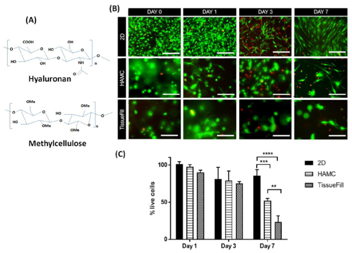
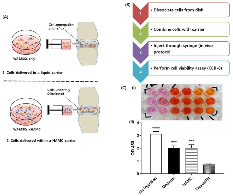
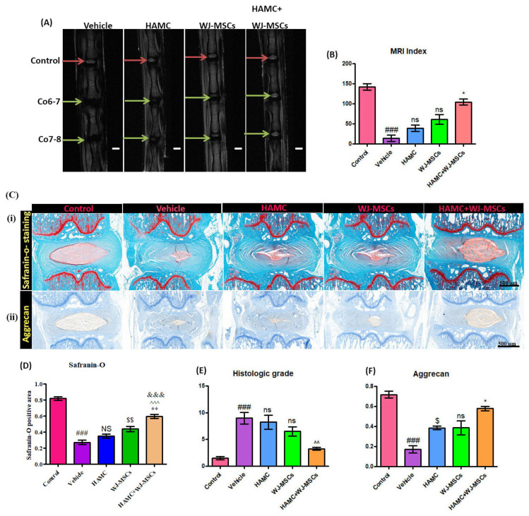
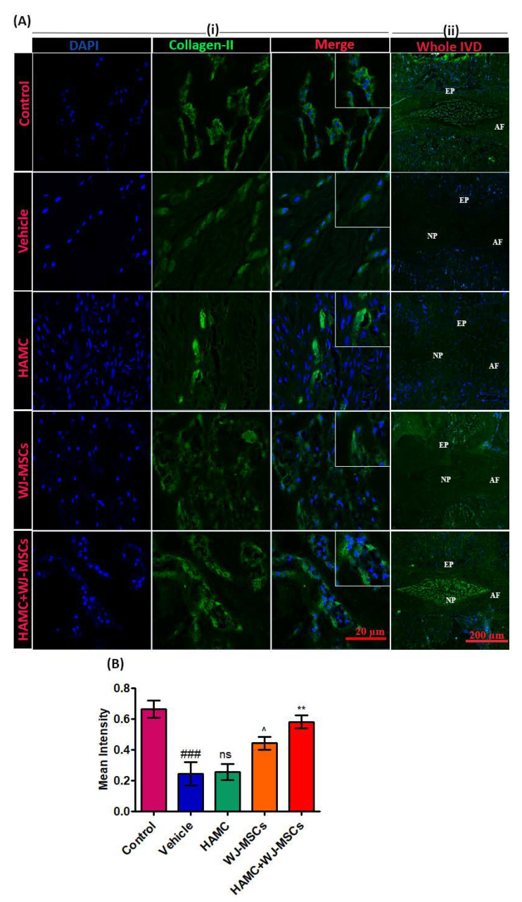
Intervertebral disc (IVD) degeneration is one of the predominant causes of chronic low back pain (LBP), which is a leading cause of disability worldwide. Despite substantial progress in cell therapy for the treatment of IVD degeneration, significant challenges remain for clinical application. Here, we investigated the effectiveness of hyaluronan-methylcellulose (HAMC) hydrogels loaded with Wharton's Jelly-derived mesenchymal stromal cell (WJ-MSCs) in vitro and in a rat coccygeal IVD degeneration model. Following induction of injury-induced IVD degeneration, female Sprague-Dawley rats were randomized into four groups to undergo a single intradiscal injection of the following: (1) phosphate buffered saline (PBS) vehicle, (2) HAMC, (3) WJ-MSCs (2 × 10⁴ cells), and (4) WJ-MSCs-loaded HAMC (WJ-MSCs/HAMC) (n = 10/each group). Coccygeal discs were removed following sacrifice 6 weeks after implantation for radiologic and histologic analysis. We confirmed previous findings that encapsulation in HAMC increases the viability of WJ-MSCs for disc repair. The HAMC gel maintained significant cell viability in vitro. In addition, combined implantation of WJ-MSCs and HAMC significantly promoted degenerative disc repair compared to WJ-MSCs alone, presumably by improving nucleus pulposus cells viability and decreasing extracellular matrix degradation. Our results suggest that WJ-MSCs-loaded HAMC promotes IVD repair more effectively than cell injection alone and supports the potential clinical use of HAMC for cell delivery to arrest IVD degeneration or to promote IVD regeneration.

Keywords: Wharton jelly; extracellular matrix; hyaluronic acid; intervertebral disc degeneration; mesenchymal stromal cell; methylcellulose; regeneration.


Conflict of interest statement

The authors declare no conflict of interest.

Figures

Figure 1
Figure 1
HAMC supports 3D WJ-MSCs survival in vitro. Cells were cultured in 3D HAMC and compared to 2D plastic culture or 3D TissueFill. Cell viability was significantly higher for cells cultured in HAMC compared to TissueFill. (A) Molecular structure of hyaluronan (HA) and methyl cellulose (MC). (B) Representative immunofluorescence images for live (green) and dead (red) assay in 2D, HAMC and TissueFill at day 0, day 1, day 3 and day 7 following WJ-MSCs culture. (C) Quantitative bar graph illustrating the percentage of live cells in 2D, HAMC and TissueFill at day 0, day 1, day 3 and day 7 following WJ-MSC culture. After 7 days of culture HAMC had significantly higher viability than TissueFill. Mean + SD, n = 3 biological replicates. Two-way ANOVA with Tukey’s post hoc, ** p < 0.01, *** p < 0.001, **** p < 0.0001. Scale bar = 50 µm.
Figure 2
Figure 2
Encapsulation in HAMC maintains post-injection viability compared to TissueFill. WJ-MSCs were encapsulated in different vehicles and injected through a fine-gauge needle to assess post-injection viability. (A) Schematic illustrating the comparison of WJ-MSC stability and viability between HAMC-encapsulated, and non-encapsulated WJ-MSCs post-injection. (B) Stepwise post-injection procedure. (C) (i) Representative CCK-8 assay, (ii) quantitative result after CCK-8 assay, compareing the cell viability of WJ-MSCs in medium alone, TissueFill or HAMC. Cells injected in medium or HAMC retained significantly higher viability compared to TissueFill. Mean + SD, n = 3 biological replicates. One-way ANOVA with Tukey’s post hoc, *** p < 0.001, **** p < 0.0001 compared to TissueFill group.
Figure 3
Figure 3
WJ-MSCs-loaded HAMC injection promotes repair of the degenerated disc (A) Representative T2-weighted MRI of the coccygeal discs: control, vehicle-treated, HAMC-treated, WJ-MSCs only treated, and HAM + CWJ-MSC-treated disc (coronal view) (red arrow = control, green arrow = needle-punctured disc). (B) MRI index for discs of each group. Mean ± SEM, (n = 3), one-way ANOVA followed by Tukey’s post-test. ## p < 0.05 (vehicle vs. control), * p < 0.05 (HAMC + WJ-MSCs vs. WJ-MSCs); ns = non-significant. MRI performed at 6 weeks after implantation showed the best restoration of disc anatomy and water content of the disc in HAMC/WJ-MSC-implanted discs. (C) (i) and (D) Representative images of safranin-O staining and quantitative safranin-O positive area, revealed the highest preservation of disc structure in HAMC/WJ-MSCs-injected disc (×200). Mean ± SEM, (n = 3), one-way ANOVA followed by Tukey’s post-test. ### p < 0.001(vehicle vs. Sham), $$ p < 0.01 (WJ-MSCs vs. HAMC), ** p < 0.01 (HAMC + WJ-MSCs vs. WJ-MSCs), ^^^ p < 0.001 (HAMC + WJ-MSCs vs. HAMC), and &&& p < 0.001 (HAMC + WJ-MSCs vs. vehicle). (E) The quantitative histological score and (C) (ii) and (F) immunoreactivity for aggrecan showed the highest expression in HAMC/WJ-MSCs-implanted discs. Mean ± SEM, (n = 3), one-way ANOVA followed by Tukey’s post-test. ### p < 0.001(vehicle vs. Sham), $ p < 0.05 (HAMC vs. vehicle), * p < 0.05 (HAMC + WJ-MSCs vs. WJ-MSCs).
Figure 4
Figure 4
Combined injection of HAMC and WJ-MSC significantly restores collagen type II in degenerated disc. (A) Representative collagen II immunofluorescent staining. (i) Collagen expression at region of interest (ROI) and (ii) full power images demarcating entire disc (nucleus pulposus—NP, annulus fibrosus—AF and end plate—EP), and collagen II expression in whole NP area. (B) Quantitative immunofluorescence intensity for collagen II expression. Mean ± SEM, (n = 3), one-way ANOVA followed by Tukey’s post-test. ### p < 0.001 (vehicle vs. control), ^ p < 0.05 (WJ-MSCs vs. HAMC), ** p < 0.01 (HAMC + WJ-MSCs vs. WJ-MSCs), ns = non-significant. Combined injection of HAMC and WJ-MSCs significantly preserved the extracellular matrix (ECM) content, collagen type II compared to the vehicle, HAMC, or WJ-MSCs only injected discs.
Figure 5
Figure 5
Combined injection of HAMC and WJ-MSC substantially reduced matrix metalloproteinase-13 (MMP-13) expression in degenerated disc. (A) Representative MMP-13 protein levels as determined by immunofluorescence staining. (i) MMP-13 expression (green) at region of interest (ROI), DAPI (blue) and merge signal (ii) full power images (far right panel) demarcating entire disc (nucleus pulposus—NP, annulus fibrosus—AF and end plate—EP), and MMP-13 expression in whole NP area. (B) Quantitative immunofluorescence intensity for MMP-13 expression. Immunopositivity was counted in at least three random low power fields (×40) and calculated as mean intensity. Mean ± SEM, (n = 3), one-way ANOVA followed by Tukey’s post-test. ### p < 0.001 (vehicle vs. control), ** p < 0.01 (HAMC + WJ-MSCs vs. WJ-MSCs). Ns = non-significant. We found that combined injection of HAMC and WJ-MSC significantly diminished the expression of matrix-degrading enzyme, MMP-13.
Figure 6
Figure 6
Combined injection of HAMC and WJ-MSC inhibits the mRNA expression of pro-inflammatory cytokine and matrix-degrading enzymes. (A) iNOS mRNA expression, (B) MMP-13 mRNA expression, (C) Adamts4 mRNA expression, and (D) Cox-2 mRNA expression. In the HAMC/WJ-MSCs-injected discs, mRNA expression of pro-inflammatory cytokine (inducible nitric oxide synthase; iNOS) and matrix-degrading enzymes, including MMP-13, A disintegrin and metalloproteinase with thrombospondin motifs 4 (Adamts4) and Cycloxygenase-2 (Cox-2) were found to be significantly downregulated compared to the vehicle-injected disc. Data are presented as mean ± SEM, one-way ANOVA followed by Tukey’s post-hoc test. (n = 3). For time, glyceraldehyde 3-phosphate dehydrogenase (GAPDH) was used as internal control. # p < 0.05, ## p < 0.01, ### p < 0.001 (vehicle vs.control), $ p < 0.05, $$$ p < 0.001 (HAMC vs. vehicle), ^ p < 0.05, ^^^ p < 0.001 (WJ-MSCs vs. vehicle only), * p < 0.05, ** p < 0.01, *** p < 0.001 (HAMC + WJ-MSCs vs. vehicle only).

References

    1. Kumar H., Ha D.-H., Lee E.-J., Park J.H., Shim J.H., Ahn T.-K., Kim K.-T., Ropper A.E., Sohn S., Kim C.-H. Safety and tolerability of intradiscal implantation of combined autologous adipose-derived mesenchymal stem cells and hyaluronic acid in patients with chronic discogenic low back pain: 1-year follow-up of a phase I study. Stem Cell Res. Ther. 2017;8:1–14. doi: 10.1186/s13287-017-0710-3. - DOI - PMC - PubMed
    1. Takeoka Y., Yurube T., Nishida K. Gene therapy approach for intervertebral disc degeneration: An update. Neurospine. 2020;17:3. doi: 10.14245/ns.2040042.021. - DOI - PMC - PubMed
    1. Han I., Ropper A.E., Konya D., Kabatas S., Toktas Z., Aljuboori Z., Zeng X., Chi J.H., Zafonte R., Teng Y.D. Biological approaches to treating intervertebral disk degeneration: Devising stem cell therapies. Cell Transplant. 2015;24:2197–2208. doi: 10.3727/096368915X688650. - DOI - PubMed
    1. Ahn J., Park E.-M., Kim B.J., Kim J.-S., Choi B., Lee S.-H., Han I. Transplantation of human Wharton’s jelly-derived mesenchymal stem cells highly expressing TGFβ receptors in a rabbit model of disc degeneration. Stem Cell Res. Ther. 2015;6:190. doi: 10.1186/s13287-015-0183-1. - DOI - PMC - PubMed
    1. Meisel H.-J., Agarwal N., Hsieh P.C., Skelly A., Park J.-B., Brodke D., Wang J.C., Yoon S.T., Buser Z. Cell therapy for treatment of intervertebral disc degeneration: A systematic review. Glob. Spine J. 2019;9:39S–52S. doi: 10.1177/2192568219829024. - DOI - PMC - PubMed

MeSH terms

LinkOut - more resources