

General Aspects of Metal Ions as Signaling Agents in Health and Disease
- PMID: 33036384
- PMCID: PMC7600656
- DOI: 10.3390/biom10101417
General Aspects of Metal Ions as Signaling Agents in Health and Disease
Abstract
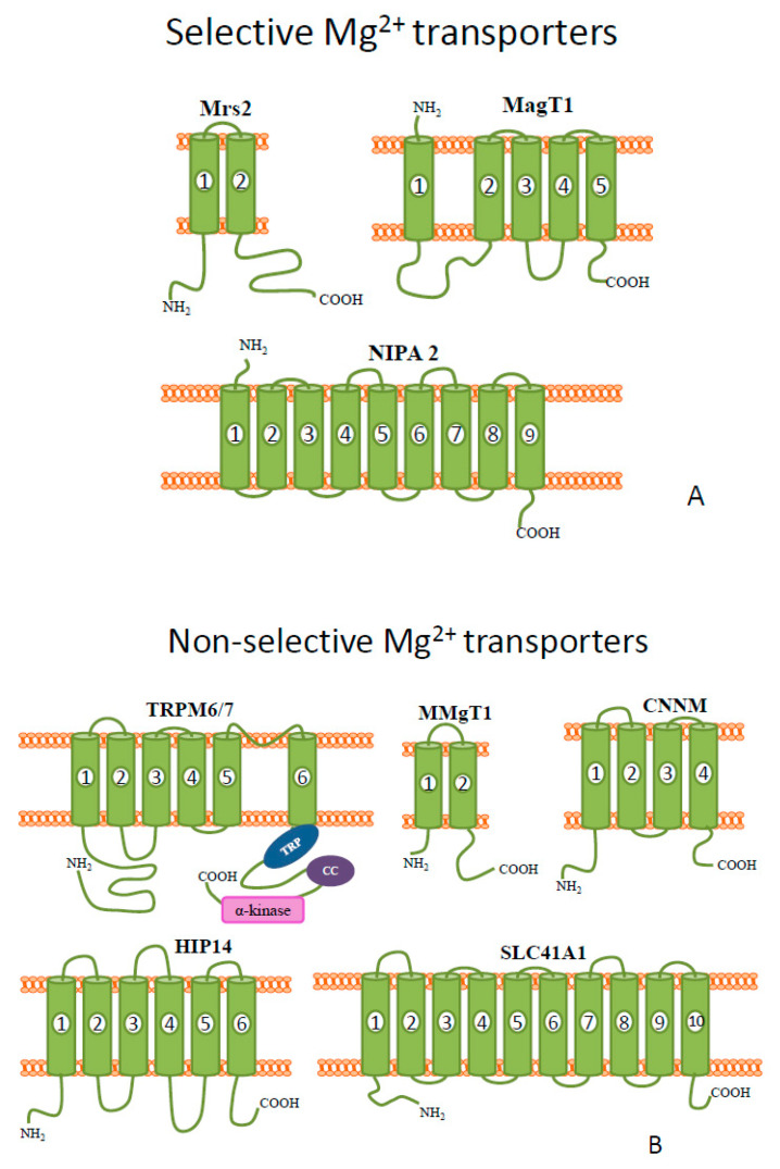
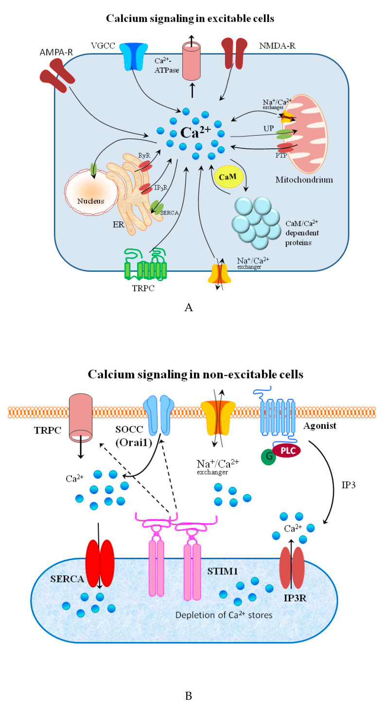
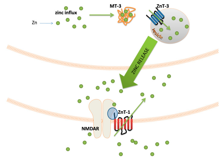
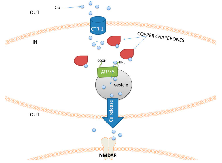
This review focuses on the current knowledge on the involvement of metal ions in signaling processes within the cell, in both physiological and pathological conditions. The first section is devoted to the recent discoveries on magnesium and calcium-dependent signal transduction-the most recognized signaling agents among metals. The following sections then describe signaling pathways where zinc, copper, and iron play a key role. There are many systems in which changes in intra- and extra-cellular zinc and copper concentrations have been linked to important downstream events, especially in nervous signal transduction. Iron signaling is mostly related with its homeostasis. However, it is also involved in a recently discovered type of programmed cell death, ferroptosis. The important differences in metal ion signaling, and its disease-leading alterations, are also discussed.
Keywords: cell signaling; ferroptosis; metal homeostasis.
Conflict of interest statement
The authors declare no conflict of interest.
Figures
Similar articles
-
Neurotoxicity Linked to Dysfunctional Metal Ion Homeostasis and Xenobiotic Metal Exposure: Redox Signaling and Oxidative Stress.Antioxid Redox Signal. 2018 Jun 20;28(18):1669-1703. doi: 10.1089/ars.2017.7272. Epub 2018 Mar 28. Antioxid Redox Signal. 2018. PMID: 29402131 Free PMC article. Review.
-
Intracellular metal ion-based chemistry for programmed cell death.Chem Soc Rev. 2025 Feb 3;54(3):1552-1582. doi: 10.1039/d4cs00930d. Chem Soc Rev. 2025. PMID: 39744985 Review.
-
Metalloimmunology: The metal ion-controlled immunity.Adv Immunol. 2020;145:187-241. doi: 10.1016/bs.ai.2019.11.007. Epub 2019 Dec 9. Adv Immunol. 2020. PMID: 32081198 Review.
-
The mechanistic role of chemically diverse metal ions in the induction of autophagy.Pharmacol Res. 2017 May;119:118-127. doi: 10.1016/j.phrs.2017.01.009. Epub 2017 Jan 10. Pharmacol Res. 2017. PMID: 28087444 Review.
-
The role of metal ions in stroke: Current evidence and future perspectives.Ageing Res Rev. 2024 Nov;101:102498. doi: 10.1016/j.arr.2024.102498. Epub 2024 Sep 5. Ageing Res Rev. 2024. PMID: 39243890 Review.
Cited by
-
Multifaceted Sulfonamide-Derived Thiosemicarbazones: Combining Metal Chelation and Carbonic Anhydrases Inhibition in Anticancer Therapy.Int J Mol Sci. 2025 Jan 30;26(3):1225. doi: 10.3390/ijms26031225. Int J Mol Sci. 2025. PMID: 39940992 Free PMC article.
-
Nanoencapsulation of basil essential oil alleviates the oxidative stress, genotoxicity and DNA damage in rats exposed to biosynthesized iron nanoparticles.Heliyon. 2021 Jul 10;7(7):e07537. doi: 10.1016/j.heliyon.2021.e07537. eCollection 2021 Jul. Heliyon. 2021. PMID: 34345731 Free PMC article.
-
Impact of Heavy Metal Pollution in the Environment on the Metabolic Profile of Medicinal Plants and Their Therapeutic Potential.Plants (Basel). 2024 Mar 21;13(6):913. doi: 10.3390/plants13060913. Plants (Basel). 2024. PMID: 38592933 Free PMC article. Review.
-
Metal coordination polymer nanoparticles for cancer therapy.Essays Biochem. 2025 Apr 10;69(2):EBC20253012. doi: 10.1042/EBC20253012. Essays Biochem. 2025. PMID: 40209056 Free PMC article. Review.
-
A Comprehensive Review of Computation-Based Metal-Binding Prediction Approaches at the Residue Level.Biomed Res Int. 2022 Mar 31;2022:8965712. doi: 10.1155/2022/8965712. eCollection 2022. Biomed Res Int. 2022. PMID: 35402609 Free PMC article. Review.
References
-
- Penner R., Neher E. The role of calcium in stimulus-secretion coupling in excitable and non-excitable cells. J. Exp. Biol. 1988;139:329–345. - PubMed
Publication types
MeSH terms
Substances
Grants and funding
LinkOut - more resources
Full Text Sources
