. 2020 Oct 7;25(19):4579.
doi: 10.3390/molecules25194579.

Verbascoside-Rich Abeliophyllum distichum Nakai Leaf Extracts Prevent LPS-Induced Preterm Birth Through Inhibiting the Expression of Proinflammatory Cytokines from Macrophages and the Cell Death of Trophoblasts Induced by TNF-α

Affiliations

Verbascoside-Rich Abeliophyllum distichum Nakai Leaf Extracts Prevent LPS-Induced Preterm Birth Through Inhibiting the Expression of Proinflammatory Cytokines from Macrophages and the Cell Death of Trophoblasts Induced by TNF-α

Ho Won Kim et al. Molecules. .

Abstract

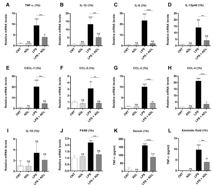
Background: Preterm birth is a known leading cause of neonatal mortality and morbidity. The underlying causes of pregnancy-associated complications are numerous, but infection and inflammation are the essential high-risk factors. However, there are no safe and effective preventive drugs that can be applied to pregnant women. Objective: The objectives of the study were to investigate a natural product, Abeliophyllum distichum leaf (ADL) extract, to examine the possibility of preventing preterm birth caused by inflammation. Methods: We used a mouse preterm birth model by intraperitoneally injecting lipopolysaccharides (LPS). ELISA, Western blot, real-time PCR and immunofluorescence staining analyses were performed to confirm the anti-inflammatory efficacy and related mechanisms of the ADL extracts. Cytotoxicity and cell death were measured using Cell Counting Kit-8 (CCK-8) analysis and flow cytometer. Results: A daily administration of ADL extract significantly reduced preterm birth, fetal loss, and fetal growth restriction after an intraperitoneal injection of LPS in mice. The ADL extract prevented the LPS-induced expression of TNF-α in maternal serum and amniotic fluid and attenuated the LPS-induced upregulation of placental proinflammatory genes, including IL-1β, IL-6, IL-12p40, and TNF-α and the chemokine gene CXCL-1, CCL-2, CCL3, and CCL-4. LPS-treated THP-1 cell-conditioned medium accelerated trophoblast cell death, and TNF-α played an essential role in this effect. The ADL extract reduced LPS-treated THP-1 cell-conditioned medium-induced trophoblast cell death by inhibiting MAPKs and the NF-κB pathway in macrophages. ADL extract prevented exogenous TNF-α-induced increased trophoblast cell death and decreased cell viability. Conclusions: We have demonstrated that the inhibition of LPS-induced inflammation by ADL extract can prevent preterm birth, fetal loss, and fetal growth restriction.

Keywords: Abeliophyllum distichum Nakai; TNF-α; inflammation; macrophage; preterm birth; trophoblast.


Conflict of interest statement

The authors have declared no competing financial interests.

Figures

Figure 1
Figure 1
HPLC chromatogram of A. distichum ethanolic extract and five standard phenolic compounds. (A) The standard phenolic compounds are 1: chlorogenic acid, 2: caffeic acid, 3: rutin, 4: ferulic acid, 5: verbascoside. (B) The extracts were analyzed with a Shimadzu liquid chromatography system (LC-20AD) with a column (C18, 4.6 × 250 mm, 5 μm), and the mobile phase consisted of solvents A and B. Solvent A was 0.1% trifluoroacetic acid in water, and solvent B was 0.1% trifluoroacetic acid in acetonitrile. The gradient was 0 min, 10% B; 0–20 min, 20% B; 20–30 min, 20% B; 30–35 min, 100% B; 35–40 min, 100% B; 40–41 min, 10% B; 41–50 min, 10% B. The run time was 50 min, and the flow rate was 1.0 mL/min.
Figure 2
Figure 2
ADL extract prevents LPS-induced preterm birth and fetal loss. (A) Schematic illustration of the mouse model of LPS-induced preterm birth and fetal loss. In the LPS+ADL groups, pregnant mice were gavaged with ADL extract (100 µg/kg) daily from GD 1 to GD 15. In the LPS alone and LPS+ADL groups, pregnant mice were i.p. injected with LPS (40 μg/kg) on GD 16 and then observed for preterm birth within 72 h of LPS administration. All pregnant mice were sacrificed on GD 19. (B) The ratio of pups associated with different maternal outcomes relative to the total number of pups 72 h after LPS injection. Pregnancy outcomes were classified as normal (>1 live fetus) or preterm (preterm delivery on GD < 19). The number of pregnant mice per group is shown in parentheses. (C) The ratio of pups associated with different fetal outcomes relative to the total number of pups 72 h after LPS injection. Fetal outcomes were classified as (i) viable, in utero, (ii) nonviable, in utero, or (iii) dead, delivered preterm (GD < 19). The number of pups per group is shown in parentheses. The Chi2 test was used for statistical comparisons (* < 0.05, ** < 0.01, and **** < 0.001). (D) Maternal weights were recorded daily from GD 2 to GD 19. The data represent the mean ± standard deviation, n = 4. *** < 0.005 and ns > 0.05 compared with the CNT group. (E) Representative photographs of uterine horns from each group on GD 19 are shown. “*” indicates nonviable fetuses in utero. Scale bar = 10 mm.
Figure 3
Figure 3
ADL extract pretreatment alleviates LPS-induced fetal growth restriction. In the LPS+ADL groups, pregnant mice were gavaged with ADL extract (100 µg/kg) daily from GD 1 to GD 15. In the LPS alone and LPS+ADL groups, pregnant mice were i.p. injected with LPS (40 μg/kg) on GD 16. All mice were sacrificed on GD 19. Representative photographs of (A) fetuses and (D) placentas from each group are shown. A: scale bar = 10 mm; D: scale bar = 4 mm. (B) The fetal weight, (C) crown–rump length, and (E) placental weight of mice from the different groups. All data represent the mean ± standard deviation. ns > 0.05, * < 0.05, ** < 0.01, and *** < 0.005 compared with the CNT group.
Figure 4
Figure 4
ADL extract prevents LPS-induced expression of proinflammatory cytokines and chemokines. In the ADL alone and LPS+ADL groups, pregnant mice were gavaged with ADL extract (100 μg/kg) daily from GD 1 to GD15. In the LPS alone and LPS+ADL groups, pregnant mice were i.p. injected with LPS (40 μg/kg) on GD 16. Mouse placental tissues were collected 1 h after LPS (40 μg/kg) injection. Placental samples were collected from maternal mice of each group, pooled, and used for analysis. Relative mRNA expression levels of (A) TNF-α, (B) IL-1β, (C) IL-6, (D) IL-12p40, (E) CXCL-1, (F) CCL-2, (G) CCL-3, (H) CCL-4, (I) IL-10 and (J) F4/80 in the placenta were measured by real-time RT-PCR and normalized to the level of β-actin. Maternal serum and amniotic fluid were collected 1 h after LPS (40 μg/kg) injection. Amniotic fluid samples were collected from maternal mice from each group, pooled, and used for analysis. The concentration of TNF-α was measured in (K) maternal serum and (L) amniotic fluid using ELISA. All data represent the mean ± standard deviation, n = 3–4. ns > 0.05, * < 0.05, ** < 0.01, *** < 0.005, and **** < 0.001 compared with the CNT group.
Figure 5
Figure 5
ADL extract inhibits LPS-induced TNF-α production by suppressing MAPK/NF-κB signaling in macrophages. Human promonocytic (THP-1) cells differentiated with phorbol myristic acetate (PMA) (100 μM) overnight were cotreated with LPS (100 ng/mL) and ADL extract (20, 100, or 500 µg/mL) for 24 h. Murine bone marrow-derived macrophages (BMDMs) were cotreated with LPS (100 ng/mL) and ADL extract (20, 100, or 200 μg/mL) for 24 h. (A, B) The production of TNF-α in the supernatant was measured by ELISA. THP-1 cells and BMDMs were cotreated with LPS (100 ng/mL) and ADL extract (100 μg/mL) for 30 min. The data represent the mean ± standard deviation, n = 3. ns > 0.05, * < 0.05, ** < 0.01, *** < 0.005, and **** < 0.001 compared with the untreated group. (C, D) Whole-cell lysates were prepared, and phospho-ERK/ERK, phospho-JNK/JNK, phospho-p38/p38, phospho-IκBα/IκBα, and β-actin levels were analyzed by Western blotting. (E) Confocal imaging of p65 and 4′,6-diamidino-2-phenylindole (DAPI) were performed. BMDMs were pretreated with ADL extract (100 μg/mL) for 1 h and stimulated with LPS (100 ng/mL) for 30 min. Cells were stained with an Alexa Fluor 488-labeled NF-κB/p65 antibody (green), and the nuclei were stained with DAPI (blue). Images were captured at 100×.
Figure 6
Figure 6
Effects of conditioned medium from LPS-stimulated and ADL extract-treated THP-1 cells on trophoblasts. Trophoblasts were treated with culture medium (CNT), THP-1 cell-conditioned medium (CM-CNT), or LPS-treated THP-1 cell-conditioned medium (CM-LPS) for 72 h. (A) The Cell Counting Kit-8 (CCK-8) assay was performed to measure the viability of JEG-3 and BeWo cells. The ratio of viable cells to total cells was normalized to that of the CNT group. CM-LPS treatment significantly downregulated trophoblast viability in both cell lines. (B) Trophoblasts were treated with culture media (CNT), CM-CNT, CM-LPS, or conditioned medium from THP-1 cells cotreated with LPS and different concentrations of ADL extract (CM-LPS+ADL) for 72 h. The CCK-8 assay was performed to measure the viability of both cell types. In both cell types, CM-LPS+ADL treatment increased trophoblast viability in a dose-dependent manner. The ratio of viable cells to total cells was normalized to that of the CNT group. (C) Death of JEG-3 and BeWo was analyzed by the lactate dehydrogenase (LDH) assay after treatment with culture media (CNT), CM-CNT, CM-LPS, or conditioned medium from THP-1 cells cotreated with LPS and different concentrations of ADL extract (CM-LPS+ADL) for 72 h. The ratio of LDH release was normalized to that of the lysis control. All data represent the mean ± standard deviation, n = 3. ns > 0.05, ** < 0.01, *** < 0.005, and **** < 0.001 compared with the CNT group.
Figure 7
Figure 7
Inhibition of TNF-α rescues trophoblast death caused by LPS-treated THP-1 cell-conditioned medium. (A) The viability of JEG-3 and BeWo cells was analyzed by the CCK-8 assay after stimulation with THP-1-conditioned medium in the presence and absence of specific neutralizing antibodies against TNF-α. The ratio of viable cells relative to total cells was normalized to that of the CNT group. (B) Death of JEG-3 and BeWo was analyzed by the LDH assay after stimulation with THP-1 cell-conditioned medium in the presence and absence of specific neutralizing antibodies against TNF-α. The ratio of LDH release was normalized to that of the lysis control. Death of these cells was significantly induced by CM-LPS, but the effect is abolished after TNF-α depletion. All data represent the mean ± standard deviation, n = 3. ns > 0.05, ** < 0.01, *** < 0.005, and **** < 0.001 compared with the CNT group.
Figure 8
Figure 8
ADL extract attenuates TNF-α-induced death of trophoblasts. (A) The viability of JEG-3 and BeWo was analyzed by the CCK-8 assay after stimulation with different concentrations of TNF-α for 72 h. (B) The percentage of dead JEG-3 and BeWo cells after stimulation with TNF-α proteins and ADL extract for 72 h using the cell counter. The percentage of trypan blue-stained cells from three independent experiments is shown. (C) The CCK-8 assay was performed to measure the viability of both cell types. Trophoblasts were cotreated with TNF-α and different concentrations of ADL extract for 72 h. The ratio of viable cells relative to total cells was normalized to that of the CNT group. (D) Death of both cell types was analyzed by the LDH assay. Trophoblasts were cotreated with TNF-α and different concentrations of ADL extract for 72 h. The ratio of LDH release was normalized to that of the lysis control. All data represent the mean ± standard deviation, n = 3–4. ns > 0.05, * < 0.05, ** < 0.01, *** < 0.005, and **** < 0.001 compared with the untreated group.

Similar articles

Cited by

References

    1. Born Too Soon: The Global Action Report on Preterm Birth. WTO; Geneve, Switzerland: 2012.
    1. Romero R., Roslansky P., Oyarzun E., Wan M., Emamian M., Novitsky T.J., Gould M.J., Hobbins J.C. Labor and infection: II. Bacterial endotoxin in amniotic fluid and its relationship to the onset of preterm labor. Am. J. Obstet. Gynecol. 1988;158:1044–1049. doi: 10.1016/0002-9378(88)90216-5. - DOI - PubMed
    1. Buhimschi I.A., Buhimschi C.S., Weiner C.P. Protective effect of N-acetylcysteine against fetal death and preterm labor induced by maternal inflammation. Am. J. Obstet. Gynecol. 2003;188:203–208. doi: 10.1067/mob.2003.112. - DOI - PubMed
    1. Chen Y.H., Xu D.X., Wang J.P., Wang H., Wei L.Z., Sun M.F., Wei W. Melatonin protects against lipopolysaccharide-induced intra-uterine fetal death and growth retardation in mice. J. Pineal Res. 2006;40:40–47. doi: 10.1111/j.1600-079X.2005.00274.x. - DOI - PubMed
    1. Rivera D., Olister S., Liu X., Thompson J., Zhang X.J., Pennline K., Azuero R., Clark D., Miller M. Interleukin-10 attenuates experimental fetal growth restriction and demise. FASEB J. 1998;12:189–197. doi: 10.1096/fasebj.12.2.189. - DOI - PubMed

MeSH terms

LinkOut - more resources