. 2020 Oct 9;5(1):235.
doi: 10.1038/s41392-020-00334-0.

SARS-CoV-2 triggers inflammatory responses and cell death through caspase-8 activation

Affiliations

SARS-CoV-2 triggers inflammatory responses and cell death through caspase-8 activation

Shufen Li et al. Signal Transduct Target Ther. .

Abstract

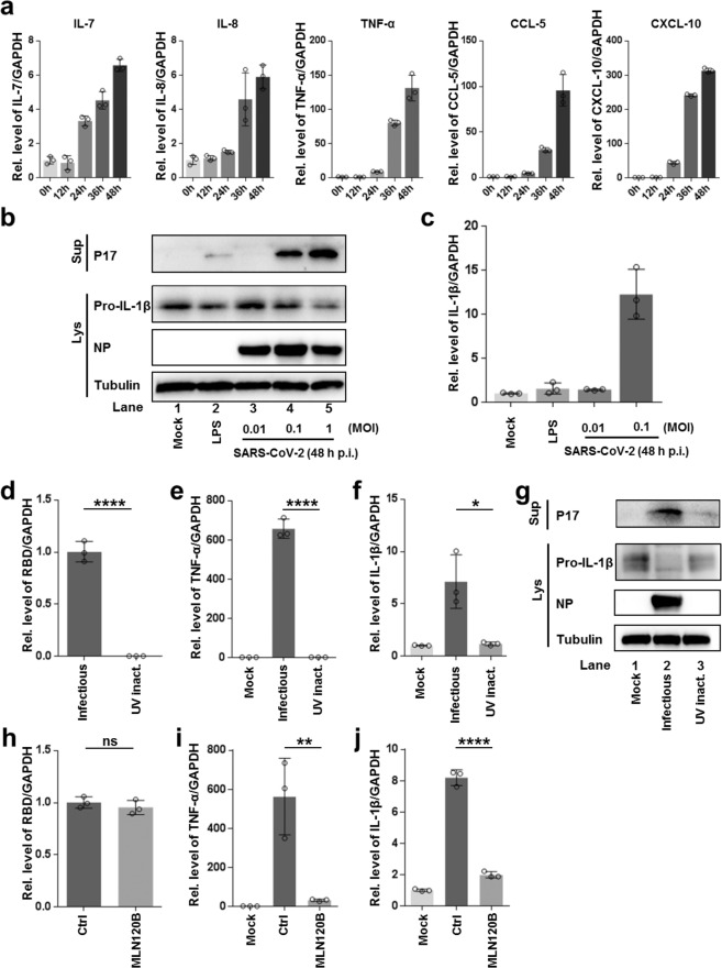
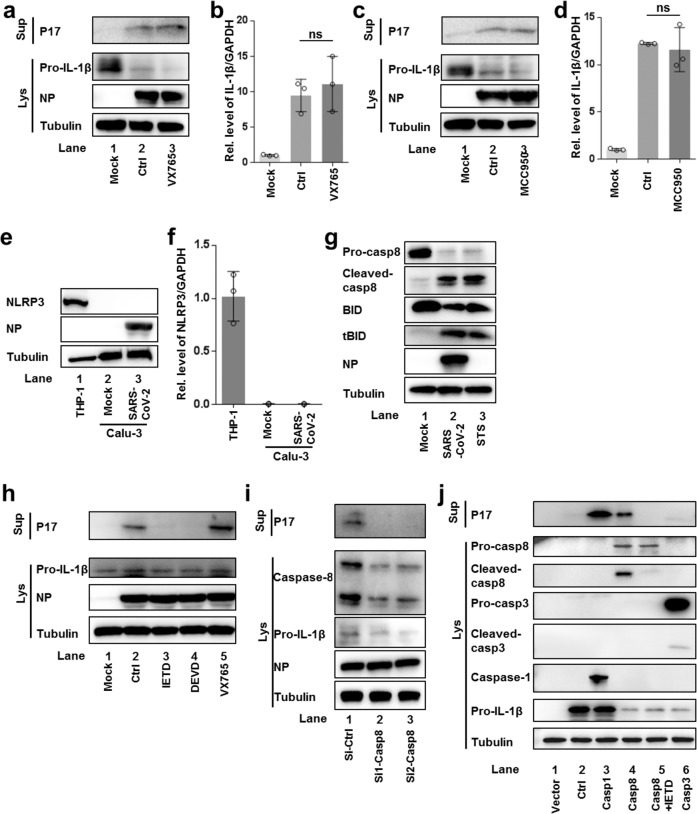
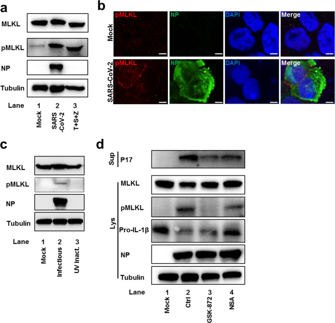
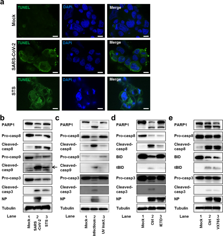
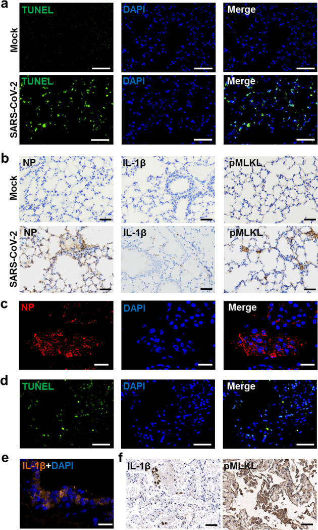
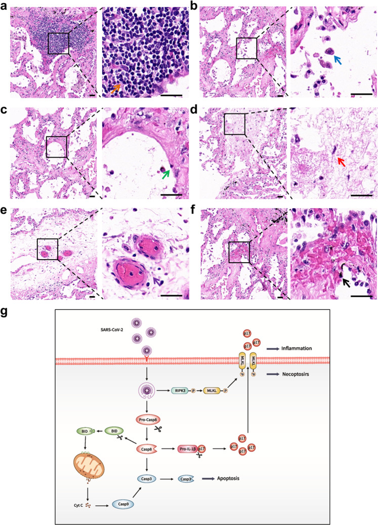
Severe acute respiratory syndrome coronavirus 2 (SARS-CoV-2) infection can lead to respiratory illness and multi-organ failure in critically ill patients. Although the virus-induced lung damage and inflammatory cytokine storm are believed to be directly associated with coronavirus disease 2019 (COVID-19) clinical manifestations, the underlying mechanisms of virus-triggered inflammatory responses are currently unknown. Here we report that SARS-CoV-2 infection activates caspase-8 to trigger cell apoptosis and inflammatory cytokine processing in the lung epithelial cells. The processed inflammatory cytokines are released through the virus-induced necroptosis pathway. Virus-induced apoptosis, necroptosis, and inflammation activation were also observed in the lung sections of SARS-CoV-2-infected HFH4-hACE2 transgenic mouse model, a valid model for studying SARS-CoV-2 pathogenesis. Furthermore, analysis of the postmortem lung sections of fatal COVID-19 patients revealed not only apoptosis and necroptosis but also massive inflammatory cell infiltration, necrotic cell debris, and pulmonary interstitial fibrosis, typical of immune pathogenesis in the lung. The SARS-CoV-2 infection triggered a dual mode of cell death pathways and caspase-8-dependent inflammatory responses may lead to the lung damage in the COVID-19 patients. These discoveries might assist the development of therapeutic strategies to treat COVID-19.


Conflict of interest statement

The authors declare no competing interests.

Figures

Fig. 1
Fig. 1
SARS-CoV-2 infection induces inflammatory responses through activation of the NFκB pathway. a Calu-3 cells were infected with SARS-CoV-2 (MOI = 0.1) with indicated time; intracellular mRNA levels of IL-7, IL-8, TNF-α, CCL5, and CXCL10 were measured with quantitative RT-PCR (qRT-PCR). b, c Calu-3 cells were treated with LPS (5 μg/ml) or infected with SARS-CoV-2 with indicated MOIs for 48 h. Mature IL-1β (P17) levels in the supernatants and intracellular levels of pro-IL-1β were determined by western blotting (b). Intracellular mRNA levels of IL-1β were measured with qRT-PCR (c). dg Calu-3 cells were infected with SARS-CoV-2 (MOI = 0.1) or inoculated with UV-inactivated virus (equal amount) for 48 h. Intracellular mRNA levels of viral RBD (d),TNF-α (e), and IL-1β (f) were determined with qRT-PCR. P17 levels in the supernatants and intracellular levels of pro-IL-1β were determined by western blotting (g). hj Calu-3 cells were treated with MLN120B (10 μM) followed by SARS-CoV-2 (MOI = 0.1) infection for 48 h. Intracellular mRNA levels of RBD (h), TNF-α (i), and IL-1β (j) were determined with qRT-PCR. Experiments were performed in triplicates. Data shown are means ± SD. Comparison of mean values (df, hj) between two groups was analyzed by Student’s t-test. *p < 0.05; **p < 0.01; ****p < 0.0001; NS, no significance
Fig. 2
Fig. 2
SARS-CoV-2 infection induces caspase-8 activation to mediate pro-IL-1β processing. a, b Calu-3 cells pretreated with VX-765 (20 μM) were infected with SARS-CoV-2 (MOI = 0.1) for 48 h. P17 levels in the supernatants and intracellular levels of pro-IL-1β were determined by western blotting (a). Intracellular mRNA levels of IL-1β were measured by qRT-PCR (b). c, d Calu-3 cells pretreated with MCC950 (100 μM) were infected with SARS-CoV-2 (MOI = 0.1) for 48 h. P17 levels in the supernatants and intracellular levels of pro-IL-1β were determined by western blotting (c). Intracellular mRNA levels of IL-1β were measured by qRT-PCR (d). e, f Calu-3 cells were mock-treated or infected with SARS-CoV-2 (MOI = 0.1); NLRP3 levels were determined by western blotting (e) or qRT-PCR (f) with THP-1 cells as a control. g Calu-3 cells infected with SARS-CoV-2 (MOI = 0.1) for 48 h were subjected to western blotting using the indicated antibodies. Cells treated with staurosporine (STS, 1 μM) for 24 h were used as a positive control. h Calu-3 cells were treated with Z-IETD-FMK (IETD, 50 μM), Z-DEVD-FMK (DEVD, 50 μM), or VX-765 (50 μM) followed by SARS-CoV-2 (MOI = 0.03) infection. P17 levels in the supernatants and intracellular levels of pro-IL-1β were determined by western blotting. i Calu-3 cells were transfected with siRNAs against caspase-8 for 48 h followed by SARS-CoV-2 (MOI = 0.1) infection. Cells and supernatants were collected at 48 h post infection. P17 levels in the supernatants and intracellular levels of pro-IL-1β were determined by western blotting. j Vero cells were co-transfected with plasmid expressing caspase-1, caspase-8, or caspase-3 and plasmid expressing IL-1β. Transfected cells were treated with Z-IETD-FMK (IETD, 50 μM) or left untreated. Twenty-four hours post transfection, the supernatants and cells were collected for western blotting analysis. Experiments were performed in triplicates. Data shown are means ± SD. Comparison of mean values (b, d) between two groups were analyzed by Student’s t-test. NS, no significance
Fig. 3
Fig. 3
SARS-CoV-2 activates the necroptosis pathway to mediate IL-1β secretion. a Calu-3 cells were mock-treated or infected with SARS-CoV-2 (MOI = 0.1) for 48 h and subjected to western blotting using the indicated antibodies. Cells treated with 100 ng/ml of TNF-α, 50 nM of SM-164, and 100 μM of Z-VAD-FMK (T + S + Z) for 48 h were used as a positive control. b Mock-treated or SARS-CoV-2 (MOI = 0.1)-infected Calu-3 cells were fixed at 36 h post infection, followed by staining with antibodies against pMLKL (red) and viral antigen NP (green). Nuclei were stained by DAPI (blue). Scale bar: 5 μm. c Calu-3 cells were infected with SARS-CoV-2 (MOI = 0.1) or inoculated with UV-inactivated virus (equal amount) for 48 h and subjected to western blotting using the indicated antibodies. d Calu-3 cells pretreated with GSK-872 (5 μM) or NSA (2.5 μM) were infected with SARS-CoV-2 (MOI = 0.1) for 48 h. P17 levels in the supernatants and intracellular levels of MLKL, pMLKL, and pro-IL-1β were determined by western blotting
Fig. 4
Fig. 4
SARS-CoV-2 infection triggers apoptosis through caspase-8 activation. a Mock-treated, SARS-CoV-2 (MOI = 0.1)-infected, or STS (1 μM)-treated Calu-3 cells were fixed at 36 h post infection, followed by labeling with TUNEL (green) and staining with DAPI (blue). Scale bar: 10 μm. b Mock-treated or SARS-CoV-2 (MOI = 0.1)-infected Calu-3 cells were collected at 48 h post infection and subjected to western blotting using the indicated antibodies. Cells treated with staurosporine (STS, 1 μM) for 24 h were used as a positive control. The arrow indicates the specific band of cleaved caspase-9. c Calu-3 cells infected with SARS-CoV-2 (MOI = 0.1) or inoculated with UV-inactivated virus (equal amount) were collected at 48 h post infection and subjected to western blotting using the indicated antibodies. d Calu-3 cells pretreated with Z-IETD-FMK (IETD, 50 μM) were infected with SARS-CoV-2 (MOI = 0.1) for 48 h and subjected to western blotting using the indicated antibodies. e Calu-3 cells pretreated with VX-765 (50 μM) were infected with SARS-CoV-2 (MOI = 0.1) for 48 h and subjected to western blotting using the indicated antibodies
Fig. 5
Fig. 5
SARS-CoV-2 induces cell death and inflammatory responses in the HFH4-hACE2 mouse model and human postmortem lung. a, b Paraffin-embedded lung sections from mock-treated or SARS-CoV-2-infected HFH4-hACE2 mice (3 × 104 TCID50) at 3 days post infection were labeled with TUNEL (green) and stained with DAPI (blue) (a), or subjected to immunohistochemistry with the indicated antibodies (b). cf Human postmortem lung sections were prepared from a fatal COVID-19 patient. Sections were stained with viral antigen SARS-CoV-2-NP (Red) and DAPI (blue) (c), or labeled with TUNEL (green) and stained with DAPI (blue) (d), or stained with IL-1β (orange) and DAPI (blue) (e), or subjected to immunohistochemistry with indicated antibodies (f). a, b, d, f Scale bar: 50 μm. c, e Scale bar: 20 μm
Fig. 6
Fig. 6
Pathological changes in human postmortem lung and a proposed model of SARS-CoV-2 infection induces inflammatory responses and cell death. af Sections prepared from human postmortem lung were stained with hematoxylin and eosin to observe pathological changes. Lymphocyte aggregation (a, yellow arrow), macrophage infiltration (b, blue arrow), necrotic cell debris (c, green arrow), early fibrosis formation (d, red arrow), extravasated blood (e, purple arrow), and hemorrhage (f, black arrow) were observed in the lung sections. Right panels are enlarged pictures of the boxed regions. Scale bar, 25 μm. g A proposed model of SARS-CoV-2 infection induces inflammatory responses and cell death. SARS-CoV-2 infection induces the cell death through the activation of caspase-8. The activation of caspase-8 promotes pro-IL-1β cleavage, leading to the secretion of mature IL-1β (P17) through necroptosis pathway

References

    1. Coronaviridae Study Group of the International Committee on Taxonomy of Viruses. The species severe acute respiratory syndrome-related coronavirus: classifying 2019-nCoV and naming it SARS-CoV-2. Nat. Microbiol. 5, 536–544 (2020). - PMC - PubMed
    1. Moore JB, June CH. Cytokine release syndrome in severe COVID-19. Science. 2020;368:473–474. doi: 10.1126/science.abb8925. - DOI - PubMed
    1. Chen G, et al. Clinical and immunological features of severe and moderate coronavirus disease 2019. J. Clin. Invest. 2019;130:2620–2629. doi: 10.1172/JCI137244. - DOI - PMC - PubMed
    1. Ruan Q, Yang K, Wang W, Jiang L, Song J. Clinical predictors of mortality due to COVID-19 based on an analysis of data of 150 patients from Wuhan, China. Intens. Care Med. 2020;46:846–848. doi: 10.1007/s00134-020-05991-x. - DOI - PMC - PubMed
    1. Jose RJ, Manuel A. COVID-19 cytokine storm: the interplay between inflammation and coagulation. Lancet Resp. Med. 2020;8:e46–e47. doi: 10.1016/S2213-2600(20)30216-2. - DOI - PMC - PubMed

Publication types

MeSH terms