. 2020 Oct 9;11(1):5090.
doi: 10.1038/s41467-020-18906-7.

The reductive glycine pathway allows autotrophic growth of Desulfovibrio desulfuricans

Affiliations

The reductive glycine pathway allows autotrophic growth of Desulfovibrio desulfuricans

Irene Sánchez-Andrea et al. Nat Commun. .

Abstract

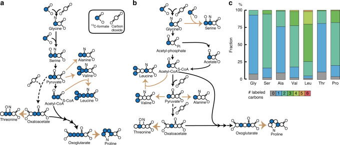
Six CO2 fixation pathways are known to operate in photoautotrophic and chemoautotrophic microorganisms. Here, we describe chemolithoautotrophic growth of the sulphate-reducing bacterium Desulfovibrio desulfuricans (strain G11) with hydrogen and sulphate as energy substrates. Genomic, transcriptomic, proteomic and metabolomic analyses reveal that D. desulfuricans assimilates CO2 via the reductive glycine pathway, a seventh CO2 fixation pathway. In this pathway, CO2 is first reduced to formate, which is reduced and condensed with a second CO2 to generate glycine. Glycine is further reduced in D. desulfuricans by glycine reductase to acetyl-P, and then to acetyl-CoA, which is condensed with another CO2 to form pyruvate. Ammonia is involved in the operation of the pathway, which is reflected in the dependence of the autotrophic growth rate on the ammonia concentration. Our study demonstrates microbial autotrophic growth fully supported by this highly ATP-efficient CO2 fixation pathway.


Conflict of interest statement

The authors declare no competing interests.

Figures

Fig. 1
Fig. 1. Comparison of growth of D. desulfuricans under autotrophic and heterotrophic conditions.
Autotrophic growth (H2/CO2/sulphate) after some adaptation transfers (a) is similar to heterotrophic growth (acetate/H2/CO2/sulphate (b). Increasing ammonium concentrations do have an effect on the autotrophic growth (c), but not on heterotrophic growth (d). All growth experiments were performed at 30 °C and 175 rpm in triplicates in 250 ml glass bottles containing 100 ml anoxic minimal medium. Error bars represent the standard deviation. Source data are provided with this paper, and raw data can be found in Supplementary Data 1.
Fig. 2
Fig. 2. Volcano plot for protein up-regulation of autotrophic vs. heterotrophic growth conditions.
Autotrophic (H2/CO2/sulphate) and heterotrophic (acetate/H2/CO2/sulphate) cultivations were performed in four biological replicates to perform proteomic analysis. Growth conditions were 30 °C and 175 rpm in 250 ml glass bottles containing 100 ml anoxic minimal medium. ACK (acetate kinase); ACS (acetyl-CoA synthetase); FDHa, FDHb, and FDHc (formate dehydrogenase: alpha, beta and gamma subunits); FTL (formate–tetrahydrofolate ligase); METCD (methenyltetrahydrofolate cyclohydrolase); GCS_Pa and GCS_Pb (glycine dehydrogenase/decarboxylating, subunit 1 and 2, glycine cleavage system P-protein); GCS_H (glycine cleavage system H-protein); GCS_T (aminomethyltransferase, glycine cleavage system T-protein); GDH (glutamate dehydrogenase); GR_A (glycine reductase complex, component A); GR_Ba (glycine reductase complex; component B, subunit beta); GR_Ca and GR_Cb (glycine reductase complex, component C, subunit alpha and beta); GR_TRX (thioredoxin domain); GR_TRXR (thioredoxin-disulphide reductase); GS (glutamate-ammonia ligase or glutamine synthetase); METCD (methenyltetrahydrofolate cyclohydrolase); PFOR (pyruvate: ferredoxin oxidoreductase) and SHMT (serine hydroxymethyltransferase). Source data are provided with this paper, and complete proteomics data can be found in Supplementary Data 5.
Fig. 3
Fig. 3. Comparative omics for genes and proteins involved in the reductive glycine pathway.
Plots per enzyme represent the log10-fold change in autotrophic condition (H2/CO2/sulphate) versus heterotrophic condition (acetate/H2/CO2/sulphate) and versus heterotrophic growth on lactate as sole energy source (lactate/CO2/sulphate), for both proteome and transcriptome analysis. Long-dashed lines are the transporters involved, short dashes incidate the alternative variant route via serine. Cultures were performed in four biological replicates. Growth conditions were 30 °C and 175 rpm in 250 ml glass bottles containing 100 ml anoxic minimal medium. Abbreviations are described in the legend of Fig. 2, with the addition of: ATP/ADP (adenosine tri/diphosphate); CoA (co-enzyme A); cytred/ox (reduced/oxidised cytochrome); FDHap (formate dehydrogenase, accessory protein); FT (formate transporter); GR_Bb (glycine reductase complex; component B, subunit alpha); NAD(P)H (nicotinamide adenine dinucleotide (phosphate)); SDA (serine dehydratase-like); THF: tetrahydrofolate; ND: not detected. Source data are provided with this paper, and complete transcriptomics and proteomics data can be found in Supplementary Data 4 and 5, respectively.
Fig. 4
Fig. 4. Confirmation of the reductive glycine pathway via 13C-labelled metabolomics.
13C-labelled carbon signature expected in the serine route (a), glycine route (b) and obtained pattern of D. desulfuricans grown in autotrophic conditions (c) with addition of 13C-formate. Autotrophic cultures (H2/CO2/sulphate) were supplemented with 75 mM sodium 13C-formate (99% 13C, Sigma-Aldrich) and grown in 30 mL bottles in 10 mL anoxic medium, cultivations were performed in biological triplicates. The dotted arrows indicate anaplerotic production of oxaloacetate via pyruvate carboxylase. Gly (glycine); Ser (serine); Ala (alanine); Val (valine); Leu (leucine); Thr (threonine); Pro (proline). Source data are provided with this paper, and data can be found in Supplementary Data 6.

References

    1. Smith, D. W. Ecological actions of sulfate-reducing bacteria. In The Sulfate-reducing Bacteria: Contemporary Perspectives (eds Odom, J.M. & Singleton, R.) 161–188 (Springer-Verlag, 1993).
    1. Postgate JR, Campbell LL. Classification of Desulfovibrio species, the nonsporulating sulfate-reducing bacteria. Bacteriol. Rev. 1966;30:732–738. doi: 10.1128/MMBR.30.4.732-738.1966. - DOI - PMC - PubMed
    1. Badziong W, Thauer RK, Zeikus JG. Isolation and characterization of Desulfovibrio growing on hydrogen plus sulfate as the sole energy source. Arch. Microbiol. 1978;116:41–49. doi: 10.1007/BF00408732. - DOI - PubMed
    1. Jansen K, Thauer RK, Widdel F, Fuchs G. Carbon assimilation pathways in sulfate reducing bacteria. Formate, carbon dioxide, carbon monoxide, and acetate assimilation by Desulfovibrio baarsii. Arch. Microbiol. 1984;138:257–262. doi: 10.1007/BF00402132. - DOI
    1. McInerney MJ, Bryant MP, Pfennig N. Anaerobic bacterium that degrades fatty acids in syntrophic association with methanogens. Arch. Microbiol. 1979;122:129–135. doi: 10.1007/BF00411351. - DOI - PubMed

Publication types

MeSH terms