. 2020 Nov;17(11):1119-1125.
doi: 10.1038/s41423-020-00550-2. Epub 2020 Oct 9.

A compromised specific humoral immune response against the SARS-CoV-2 receptor-binding domain is related to viral persistence and periodic shedding in the gastrointestinal tract

Affiliations

A compromised specific humoral immune response against the SARS-CoV-2 receptor-binding domain is related to viral persistence and periodic shedding in the gastrointestinal tract

Fengyu Hu et al. Cell Mol Immunol. 2020 Nov.

Abstract

Severe acute respiratory syndrome coronavirus 2 (SARS-CoV-2) has been redetected after discharge in some coronavirus disease 2019 (COVID-19) patients. The reason for the recurrent positivity of the test and the potential public health concern due to this occurrence are still unknown. Here, we analyzed the viral data and clinical manifestations of 289 domestic Chinese COVID-19 patients and found that 21 individuals (7.3%) were readmitted for hospitalization after detection of SARS-CoV-2 after discharge. First, we experimentally confirmed that the virus was involved in the initial infection and was not a secondary infection. In positive retests, the virus was usually found in anal samples (15 of 21, 71.4%). Through analysis of the intracellular viral subgenomic messenger RNA (sgmRNA), we verified that positive retest patients had active viral replication in their gastrointestinal tracts (3 of 16 patients, 18.7%) but not in their respiratory tracts. Then, we found that viral persistence was not associated with high viral titers, delayed viral clearance, old age, or more severe clinical symptoms during the first hospitalization. In contrast, viral rebound was associated with significantly lower levels of and slower generation of viral receptor-binding domain (RBD)-specific IgA and IgG antibodies. Our study demonstrated that the positive retest patients failed to create a robust protective humoral immune response, which might result in SARS-CoV-2 persistence in the gastrointestinal tract and possibly in active viral shedding. Further exploration of the mechanism underlying the rebound in SARS-CoV-2 in this population will be crucial for preventing virus spread and developing effective vaccines.

Keywords: SARS-CoV-2; gastrointestinal infection; protective antibody; virus recurrence.


Conflict of interest statement

The authors declare no competing interests.

Figures

Fig. 1
Fig. 1
Existence of active replicating SARS-CoV-2 in anal samples from positive retest patients. a Frequency of viral RNA detection in anal samples between the negative retest and positive retest groups. The percentage (%) and number (n) are labeled. The total case number (n) is shown under the pie chart. p Values (chi-square test) are indicated. b Viral detection in positive retest patients during the second admission. Throat and anal samples are shown. Neg. negative samples, Pos. positive samples. c sgmRNA reads in samples from positive retest patients. The read numbers were normalized to reads per million (RPM) to minimize sequencing size variation. Patient numbers are shown. The positive controls were two intracellular nucleic acid samples extracted from cells with actively replicating SARS-CoV-2 (dilution factor PC1: 1 × 10−4, PC2: 1 × 10−5). Red triangle, throat sample from Patient 08 during the first admission; red circle, anal sample from Patient 08 during the second admission; pink triangle, throat sample from Patient 12 during the second admission
Fig. 2
Fig. 2
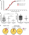
Clinical features of SARS-CoV-2 RNA-positive patients. a Kinetics of SARS-CoV-2 RNA clearance. The cumulative viral clearance (percentage, %) is shown for the negative retest (black circle) and positive retest groups (red square). p Values (log-rank (Mantel–Cox) test) are indicated. b Maximum viral concentration distribution. Each data point represents the maximum viral concentration (Ct value) in each patient during the entire admission. Patient numbers are labeled below each group. c Age distribution. An unpaired t test with Welch’s correction was used. p Values with a significant difference are shown. d Clinical symptom severity. Percentage (%) and number (n) are indicated
Fig. 3
Fig. 3
Features of anti-RBD-specific IgM, IgA, and IgG. a Concentrations (cut-off index, COI) of anti-RBD-specific IgM (upper), IgA (middle), and IgG (bottom) antibodies at different time points. Times (weeks after symptom onset, W) are as marked. First, serum detection for each patient within 1 week after symptom onset was grouped separately as “1st detect.” Patient numbers at each timepoint are labeled separately for the positive retest group (red filled circle, positive retest.) and the negative retest group (black open circle, negative retest.). An unpaired t test with Welch’s correction was used. p Value: *p < 0.05, **p < 0.01, ***p < 0.001. b The speed of anti-RBD-specific antibody generation. Cumulative patient numbers (%) with anti-RBD IgM > 5 COI (upper), IgA > 5 COI (middle), and IgG > 10 (bottom) are shown. Positive retest group, red filled circle; negative retest group, black filled square. p Values (calculated by the log-rank (Mantel–Cox) test) are shown
Fig. 4
Fig. 4
Kinetics of viral RNA, anti-RBD antibodies, and neutralizing capacity. a Profiles of three representative negative retest COVID-19 patients with high levels of anti-RBD antibodies. Fifty-eight of 60 negative retest patients were in this group (see Supplementary Fig. 4). b Profiles of nine representative positive retest COVID-19 patients with low levels of anti-RBD antibodies. Seventeen of 19 positive retest patients were included in this group. c Profiles of one negative retest patient with low anti-RBD antibody titers and nonprotective neutralizing activity. Two of 60 negative retest patients were in this group. d Profiles of two positive retest patients with high levels of antibodies and neutralizing activity. Two of 19 positive retest patients were in this group. Black lines, IgM, IgA, and IgG; red line, viral load; green line, serum microneutralization. Patient ID numbers are shown on the top. Viral RNA below the detection limit was set at 1.44 log10. Folds of serum dilution were used as microneutralization titers

References

    1. Hao, X. et al. Reconstruction of the full transmission dynamics of COVID-19 in Wuhan. Nature. 10.1038/s41586-020-2554-8 (2020). - PubMed
    1. He X, et al. Temporal dynamics in viral shedding and transmissibility of COVID-19. Nat. Med. 2020;26:672–675. - PubMed
    1. Team TNCPERE. Vital surveillances: the epidemiological characteristics of an outbreak of 2019 novel coronavirus diseases (COVID-19)—China, 2020. China CDC Wkly. 2020;2:113–122. - PMC - PubMed
    1. Wu, Z. & McGoogan, J. M. Characteristics of and important lessons from the Coronavirus Disease 2019 (COVID-19) Outbreak in China: summary of a Report of 72314 Cases From the Chinese Center for Disease Control and Prevention. J. Am. Med. Assoc.10.1001/jama.2020.2648 (2020). - PubMed
    1. Lan, L. et al. Positive RT-PCR test results in patients recovered from COVID-19. J. Am. Med. Assoc.10.1001/jama.2020.2783 (2020). - PMC - PubMed

Publication types

MeSH terms