. 2021 Jun;42(6):932-941.
doi: 10.1038/s41401-020-00538-8. Epub 2020 Oct 9.

Engineered cardiac tissues: a novel in vitro model to investigate the pathophysiology of mouse diabetic cardiomyopathy

Affiliations

Engineered cardiac tissues: a novel in vitro model to investigate the pathophysiology of mouse diabetic cardiomyopathy

Xiang Wang et al. Acta Pharmacol Sin. 2021 Jun.

Abstract

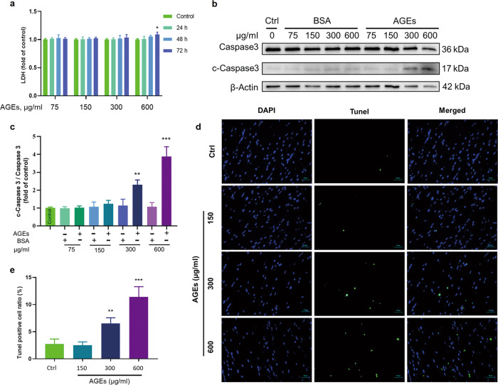
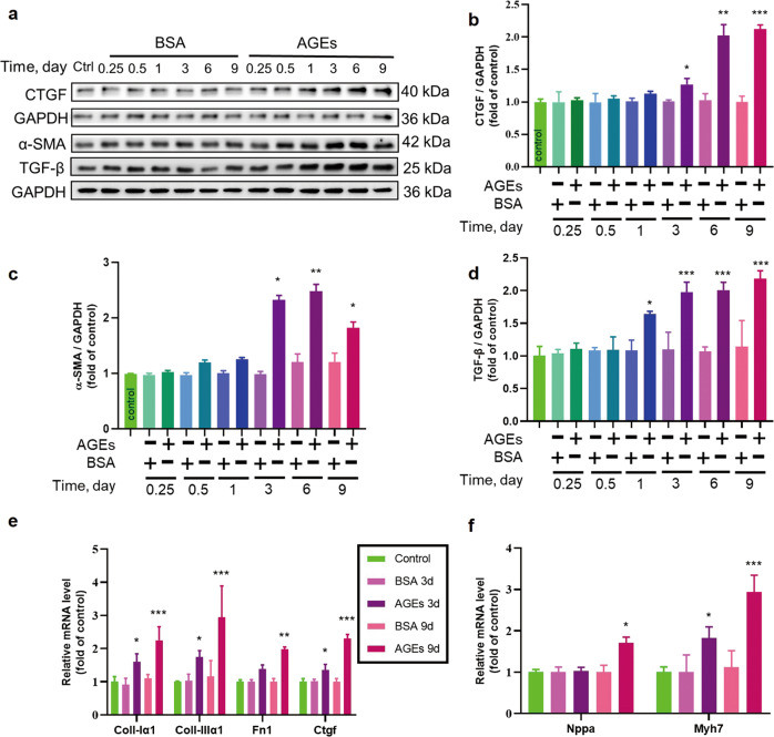
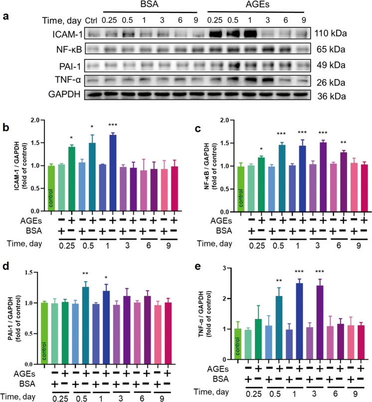
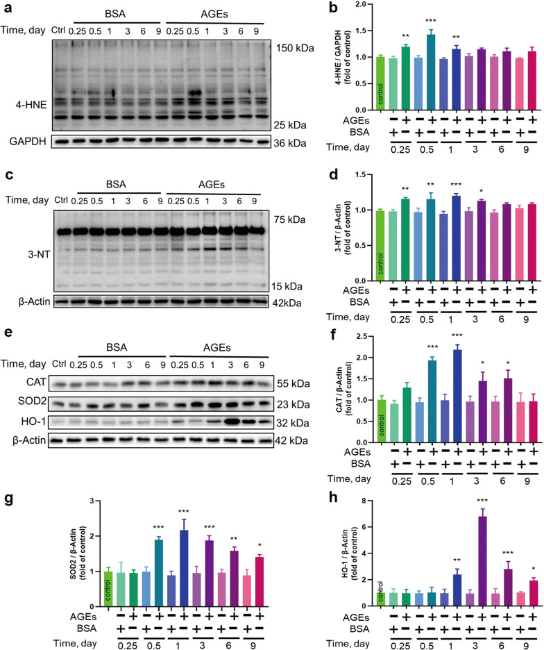
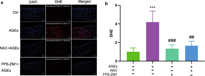
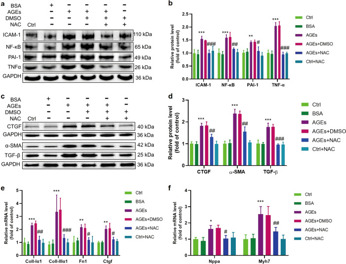
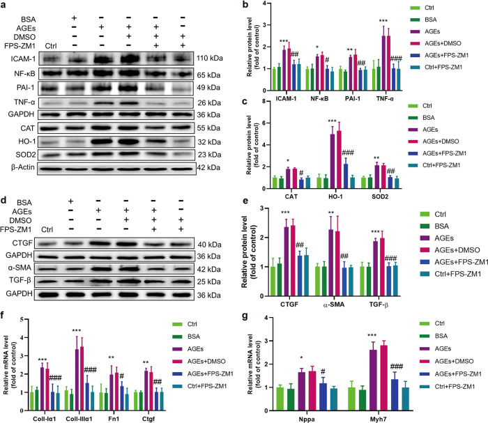
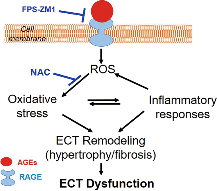
Rodent diabetic models, used to understand the pathophysiology of diabetic cardiomyopathy (DCM), remain several limitations. Engineered cardiac tissues (ECTs) have emerged as robust 3D in vitro models to investigate structure-function relationships as well as cardiac injury and repair. Advanced glycation end-products (AGEs), produced through glycation of proteins or lipids in response to hyperglycemia, are important pathogenic factor for the development of DCM. In the current study, we developed a murine-based ECT model to investigate cardiac injury produced by AGEs. We treated ECTs composed of neonatal murine cardiac cells with AGEs and observed AGE-related functional, cellular, and molecular alterations: (1) AGEs (150 µg/mL) did not cause acute cytotoxicity, which displayed as necrosis detected by medium LDH release or apoptosis detected by cleaved caspase 3 and TUNEL staining, but negatively impacted ECT function on treatment day 9; (2) AGEs treatment significantly increased the markers of fibrosis (TGF-β, α-SMA, Ctgf, Collagen I-α1, Collagen III-α1, and Fn1) and hypertrophy (Nppa and Myh7); (3) AGEs treatment significantly increased ECT oxidative stress markers (3-NT, 4-HNE, HO-1, CAT, and SOD2) and inflammation response markers (PAI-1, TNF-α, NF-κB, and ICAM-1); and (4) AGE-induced pathogenic responses were all attenuated by pre-application of AGE receptor antagonist FPS-ZM1 (20 µM) or the antioxidant glutathione precursor N-acetylcysteine (5 mM). Therefore, AGEs-treated murine ECTs recapitulate the key features of DCM's functional, cellular and molecular pathogenesis, and may serve as a robust in vitro model to investigate cellular structure-function relationships, signaling pathways relevant to DCM and pharmaceutical intervention strategies.

Keywords: FPS-ZM1; N-acetylcysteine; advanced glycation end-products; cardiac fibrosis and hypertrophy; cardiomyopathic in vitro model; diabetic cardiomyopathy; engineered cardiac tissue; inflammation response; oxidative stress.


Conflict of interest statement

The authors declare no competing interests.

Figures

Fig. 1
Fig. 1. Determination of the noncytotoxic dose of AGEs.
a LDH assays of the medium from ECTs at different time points treated with different concentrations of AGEs revealed significant necrosis occurred only after AGEs treatment at 600 µg/mL for 72 h. n = 3 per group per time point. b, c Western blotting of c-Caspase 3/Caspase 3 in ECTs treated with different concentrations of AGEs. n = 3 per group, treatment for 72 h. d, e Representative ECT immunofluorescent staining for the terminal transferase dUTP nick end labeling assay (TUNEL, green) and of nuclei (DAPI, blue) following treatment with different concentrations of AGEs for 12 days (×40 magnification). n = 3 per group. *P < 0.05, **P < 0.01, ***P < 0.001 versus the respective control groups. Data are summarized as the normalized mean ± SD.
Fig. 2
Fig. 2. The effect of long-term AGEs exposure on ECT remodeling.
ad The fibrosis markers CTGF, α-SMA, and TGF-β were assessed by Western blotting. n = 3 per group. e The fibrosis markers Collagen I-α1, Collagen III-α1, Fn1, and Ctgf were assessed by qRT-PCR. n = 3 per group. f The cardiomyocyte hypertrophy markers Nppa and Myh7 were assessed by qRT-PCR. n = 3 per group. *P < 0.05, **P < 0.01, ***P < 0.001 versus the respective control groups. Data are summarized as the normalized mean ± SD.
Fig. 3
Fig. 3. AGEs induced the expression of markers of inflammation.
ae The inflammatory markers ICAM-1, NF-κB, PAI-1, and TNF-α were assessed by Western blotting. n = 3 per group per time point, *P < 0.05, **P < 0.01, ***P < 0.001 versus the respective control groups. Each panel shows the normalized mean ± SD.
Fig. 4
Fig. 4. AGEs induced the expression of markers of oxidative stress.
ah The oxidative stress markers 4-HNE, 3-NT, CAT, SOD2, and HO-1 were assessed by Western blotting. n = 3 per group per time point, *P < 0.05, **P < 0.01, ***P < 0.001 versus the respective control groups. Each panel shows the normalized mean ± SD.
Fig. 5
Fig. 5. NAC and FPS-ZM1 could attenuate AGE-induced production of ROS as assessed by DHE staining.
a, b DHE staining of ROS (×10 magnification). AGEs treatment was performed for 3 h before DHE staining. DMSO, FPS-ZM1 (20 µM), or NAC (5 mM) was added to ECTs 2 h before control or AGEs treatment. n = 5 per group, ***P < 0.001 vs control. ##P < 0.01, ###P < 0.001 versus AGEs group. Each panel shows the normalized mean ± SD.
Fig. 6
Fig. 6. AGE-induced inflammatory responses and remodeling could be attenuated by depleting ROS with NAC.
a, b The inflammatory markers PAI-1, TNF-α, NF-κB, and ICAM-1 were assessed by Western blotting; n = 3 per group. c, d The fibrosis markers CTGF, α-SMA, and TGF-β were assessed by Western blotting; n = 3 per group. e The fibrosis markers collagen I-α1, collagen III-α1, Fn1, and Ctgf were assessed by qRT-PCR; n = 4 per group. f The cardiomyocyte hypertrophy markers Nppa and Myh7 were assessed by qRT-PCR; n = 4 per group. NAC, N-acetylcysteine; *P < 0.05, **P < 0.01, ***P < 0.001 vs Control. #P < 0.05, ##P < 0.01, ###P < 0.001 versus AGEs+DMSO group. Each panel shows the normalized mean ± SD.
Fig. 7
Fig. 7. AGE-induced expression of markers of oxidative stress, inflammation, and remodeling could be attenuated by blocking RAGE.
a, b The inflammatory markers ICAM-1, NF-κB, PAI-1, and TNF-α were assessed by Western blotting; n = 3 per group. a, c The oxidative stress markers CAT, SOD2, and HO-1 were assessed by Western blotting; n = 3 per group. d, e The fibrosis markers CTGF, α-SMA, and TGF-β were assessed by Western blotting; n = 3 per group. f The fibrosis markers Collagen I-α1, Collagen III-α1, Fn1, and Ctgf were assessed by qRT-PCR; n = 4 per group. g The cardiomyocyte hypertrophy markers Nppa and Myh7 were assessed by qRT-PCR; n = 4 per group. FPS-ZM1, inhibitor of RAGE; *P < 0.05, **P < 0.01, ***P < 0.001 vs control. #P < 0.05, ##P < 0.01, ###P < 0.001 versus AGEs+DMSO groups. Each panel shows the normalized mean ± SD.
Fig. 8
Fig. 8. Illustration of AGE-induced ECT remodeling and dysfunction and the underlying mechanisms.
AGEs (pathogenic factors of diabetic cardiomyopathy) interact with RAGE to induce ROS generation. Excessive oxidative stress can lead to ECT remodeling (cell hypertrophy and accumulation of fibrotic products) directly or indirectly via the inflammatory response. These alterations result in ECT functional abnormalities. ECT engineered cardiac tissue, FPS-ZM1 RAGE antagonist, NAC N-acetylcysteine.

References

    1. Saeedi P, Petersohn I, Salpea P, Malanda B, Karuranga S, Unwin N, et al. Global and regional diabetes prevalence estimates for 2019 and projections for 2030 and 2045: Results from the International Diabetes Federation Diabetes Atlas, 9th edition. Diabetes Res Clin Pr. 2019;157:107843. - PubMed
    1. Boudina S, Abel ED. Diabetic cardiomyopathy, causes and effects. Rev Endocr Metab Disord. 2010;11:31–9. - PMC - PubMed
    1. Rubler SDJ, YuceogluYZ, Kumral T, Branwood AW, Grishman A. New type of cardiomyopathy associated with diabetic glomerulosclerosis. Am J Cardiol. 1972;30:596–602. - PubMed
    1. Fuentes-Antras J, Picatoste B, Gomez-Hernandez A, Egido J, Tunon J, Lorenzo O. Updating experimental models of diabetic cardiomyopathy. J Diabetes Res. 2015;2015:656795. - PMC - PubMed
    1. Yu HT, Zhen J, Xu JX, Cai L, Leng JY, Ji HL, et al. Zinc protects against cadmium-induced toxicity in neonatal murine engineered cardiac tissues via metallothionein-dependent and independent mechanisms. Acta Pharmacol Sin. 2020;41:638–49. - PMC - PubMed

MeSH terms