. 2020 Oct 20;119(8):1538-1549.
doi: 10.1016/j.bpj.2020.08.038. Epub 2020 Sep 11.

ATP-Driven Nonequilibrium Activation of Kinase Clients by the Molecular Chaperone Hsp90

Affiliations

ATP-Driven Nonequilibrium Activation of Kinase Clients by the Molecular Chaperone Hsp90

Huafeng Xu. Biophys J. .

Abstract

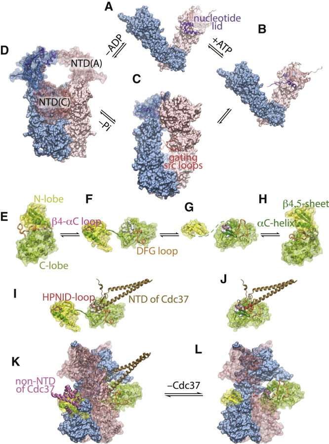
The molecular chaperone 90-kDa heat-shock protein (Hsp90) assists the late-stage folding and activation of diverse types of protein substrates (called clients), including many kinases. Previous studies have established that the Hsp90 homodimer undergoes an ATP-driven cycle through open and closed conformations. Here, I propose a model of client activation by Hsp90 that predicts that this cycle enables Hsp90 to use ATP energy to drive a client out of thermodynamic equilibrium toward its active conformation. My model assumes that an Hsp90-bound client can transition between a deactivating conformation and an activating conformation. It suggests that the cochaperone Cdc37 aids Hsp90 to activate kinase clients by differentiating between these two intermediate conformations. My model makes experimentally testable predictions, including how modulating the stepwise kinetics of the Hsp90 cycle-for example, by various cochaperones-affects the activation of different clients. My model may inform client-specific and cell-type-specific therapeutic intervention of Hsp90-mediated protein activation.


Figures

Figure 1
Figure 1
Structural basis for my model of nonequilibrium activation of client kinases by Hsp90. The underlined texts below are Protein Data Bank IDs and chain IDs. The Hsp90 homodimer goes through four states in A to D. (A) Hsp90open: nucleotide free and open; 2IOQ. (B) Hsp90openATP: ATP bound and open; 2IOQ but with the NTD of one chain replaced with the ATP-bound NTD from the structure 2CG9. ATP binding induces a structural change in the nucleotide lid in NTD. (C) Hsp90closedATP: ATP bound and closed; 2CG9. The hole between the two MDs in the closed Hsp90 dimer is gated by a flexible, often disordered src loop (the src loops from 2CG9, 5FWM, 2IOP, and 4IYN are superimposed). (D) Hsp90compactADP: ADP-bound and compact; 2IOP. The ADP-bound NTD may have a number of orientations with respect to MD; e.g., the NTD of chain A may move to the position occupied by the NTD of chain C in the crystal structure, resulting in a compact conformation consistent with electron microscopy images (12) but clashing with the bound kinase client. In (B)–(D), the nucleotides are shown in spheres. The kinase client may transition through four states in (E) to (H). (E) The inactive conformation: i; 1HCL:A. (F) The deactivating conformation: d; 5FWM:K. (G) The hypothetical maturing conformation: m. (H) The active conformation: a; 1FIN:A. The deactivating structure is of the client kinase Cdk4 in the Hsp90-Cdc37-Cdk4 complex; the inactive and active structures are of Cdk2. (I) Complex between Cdc37 and Cdk4 in the d-state (5FWM:K + E) is shown. (J) Steric clash prevents Cdc37 from binding to Cdk4 in the m-state. Three residues (in spheres) in the DFG loop—in addition to perhaps the β4-αC loop—would collide with Cdc37, whose clashing residues are highlighted in red. (K) The ternary complex of Hsp90-Cdc37-Cdk4 (5FWM), in which the client kinase is in the d-state, is shown. (L) The hypothetical complex of the closed Hsp90 dimer with the client kinase in the m-state is shown. To see this figure in color, go online.
Figure 2
Figure 2
The reactions in Hsp90-mediated nonequilibrium activation of v-Src and the reactive fluxes at the steady state. Each circle represents a molecular species; the radius of the circle is linear in the logarithm of its steady-state concentration. The reactions are labeled with the reaction numbers in Table 1. Each half arrow indicates either a forward or a reverse reaction; its length is linear in the logarithm of the value of the corresponding rate constant (concentration in micromolars and time in seconds; see Table 1). The top four circles represent the free client molecule in its four different conformations; the others represent complexes between the client and Hsp90. The molecular species are divided by the vertical dashed lines according to the client conformation and into the colored horizontal bands according to the conformational and nucleotide state of Hsp90. The ones that lie in the orange vertical band are ternary complexes of Hsp90-client-Cdc37. The Hsp90 ATP cycles are highlighted in red. The intermediate molecular species in the Cdc37-assisted activation pathway are labeled. The directions of the steady-state reactive fluxes are indicated by the green arrows. At the steady state, the concentrations are unchanging; thus, the fluxes for the reactions id and ma are zero and not shown. Hsp90 elevates the m-state concentration above and suppresses the d-state concentration below their respective equilibrium values, resulting in a steady-state flux from m to d in the dm reaction. The reaction conditions are [Hsp90] = [Cdc37] = 1.3 μM, [v-Src] = 0.32 μM, [ATP] = 20 μM, and [ADP] = [Pi] = 1 μM. To see this figure in color, go online.
Figure 3
Figure 3
Activation of v-Src by Hsp90. (A) Normalized v-Src activity at the steady state at different Hsp90 and Cdc37 concentrations is shown. The experimental data (4) are shown as points, and the results calculated from my model are shown as solid lines. (B) The time course of v-Src activation by Hsp90 in the presence of Cdc37 and the concomitant ATP consumption are given, assuming that two ATP molecules are hydrolyzed per Hsp90 cycle. To see this figure in color, go online.
Figure 4
Figure 4
Cdc37 expands Hsp90’s clientele by recruiting kinases in the d-state. (A) Kinase activation by Hsp90 is shown at different ratios of its binding affinity for the d-state to that for the m-state of the kinase. Here, the KD ratio is varied while keeping the conformational equilibrium constants of the apo and the Hsp90-bound client unchanged (reactions 6 and 14 in Table 1), which is satisfied by setting Kd,closure/Km,closure = KD,m⋅Hsp90/KD,d⋅Hsp90, where Ks,closure is the equilibrium constant of the reaction s·Hsp90openATPs·Hsp90closedATP for s = d, m. In the presence of Cdc37, kinases with low KD ratios are activated by Hsp90. The reaction conditions differ from those in Fig. 2 only in [Cdc37] and in that [ATP] = 250 μM. (B) The fraction of v-Src trapped in its d- and m-states by the closed Hsp90 dimer with the nonhydrolyzable ATP analog AMPPNP at different Cdc37 concentrations is shown. The reaction conditions are [Hsp90] = 15 μM, [v-Src] = 0.32 μM, and [AMPPNP] = 250 μM. To see this figure in color, go online.
Figure 5
Figure 5
Changing the kinetic rates of the steps in the ATP cycle affects client activation by Hsp90. Normalized activity (black line and left y axis) and ATP cycle time (red dashed line and right y axis) are calculated for different rates of (A) Hsp90 transition from the open to the closed conformation, (B) ATP hydrolysis after Hsp90 adopts the closed conformation, and (C) ADP release and the return of Hsp90 to the open conformation. The proportions of time spent in the four Hsp90 states—open and nucleotide free, open and ATP bound, closed and ATP bound, and compact and ADP bound—are shown as colored bands. Note the logarithmic scale of the times: for the same apparent thickness, a higher band represents a much larger occupancy than a lower band; in particular, the nucleotide-free open state has a higher occupancy than it seems. (D) Modulation of client activation by the cochaperone Aha1 is shown. Aha1 can augment or diminish client activation depending on whether the client itself accelerates (orange) or slows down (blue) the rate that the Hsp90 dimer closes. Here, [ATP] = 250 μM, [Cdc37] = 0 μM, [Hsp90] = 1.3 μM, [client] = 0.32 μM, and [ADP] = [Pi] = 1 μM. To see this figure in color, go online.

References

    1. Taipale M., Jarosz D.F., Lindquist S. HSP90 at the hub of protein homeostasis: emerging mechanistic insights. Nat. Rev. Mol. Cell Biol. 2010;11:515–528. - PubMed
    1. Schopf F.H., Biebl M.M., Buchner J. The HSP90 chaperone machinery. Nat. Rev. Mol. Cell Biol. 2017;18:345–360. - PubMed
    1. Xu Y., Lindquist S. Heat-shock protein hsp90 governs the activity of pp60v-src kinase. Proc. Natl. Acad. Sci. USA. 1993;90:7074–7078. - PMC - PubMed
    1. Boczek E.E., Reefschläger L.G., Buchner J. Conformational processing of oncogenic v-Src kinase by the molecular chaperone Hsp90. Proc. Natl. Acad. Sci. USA. 2015;112:E3189–E3198. - PMC - PubMed
    1. Jakob U., Lilie H., Buchner J. Transient interaction of Hsp90 with early unfolding intermediates of citrate synthase. Implications for heat shock in vivo. J. Biol. Chem. 1995;270:7288–7294. - PubMed

MeSH terms

LinkOut - more resources