. 2020 Oct 26;55(2):224-236.e6.
doi: 10.1016/j.devcel.2020.09.016. Epub 2020 Oct 9.

Super-Resolution Microscopy and FIB-SEM Imaging Reveal Parental Centriole-Derived, Hybrid Cilium in Mammalian Multiciliated Cells

Affiliations

Super-Resolution Microscopy and FIB-SEM Imaging Reveal Parental Centriole-Derived, Hybrid Cilium in Mammalian Multiciliated Cells

Zhen Liu et al. Dev Cell. .

Abstract

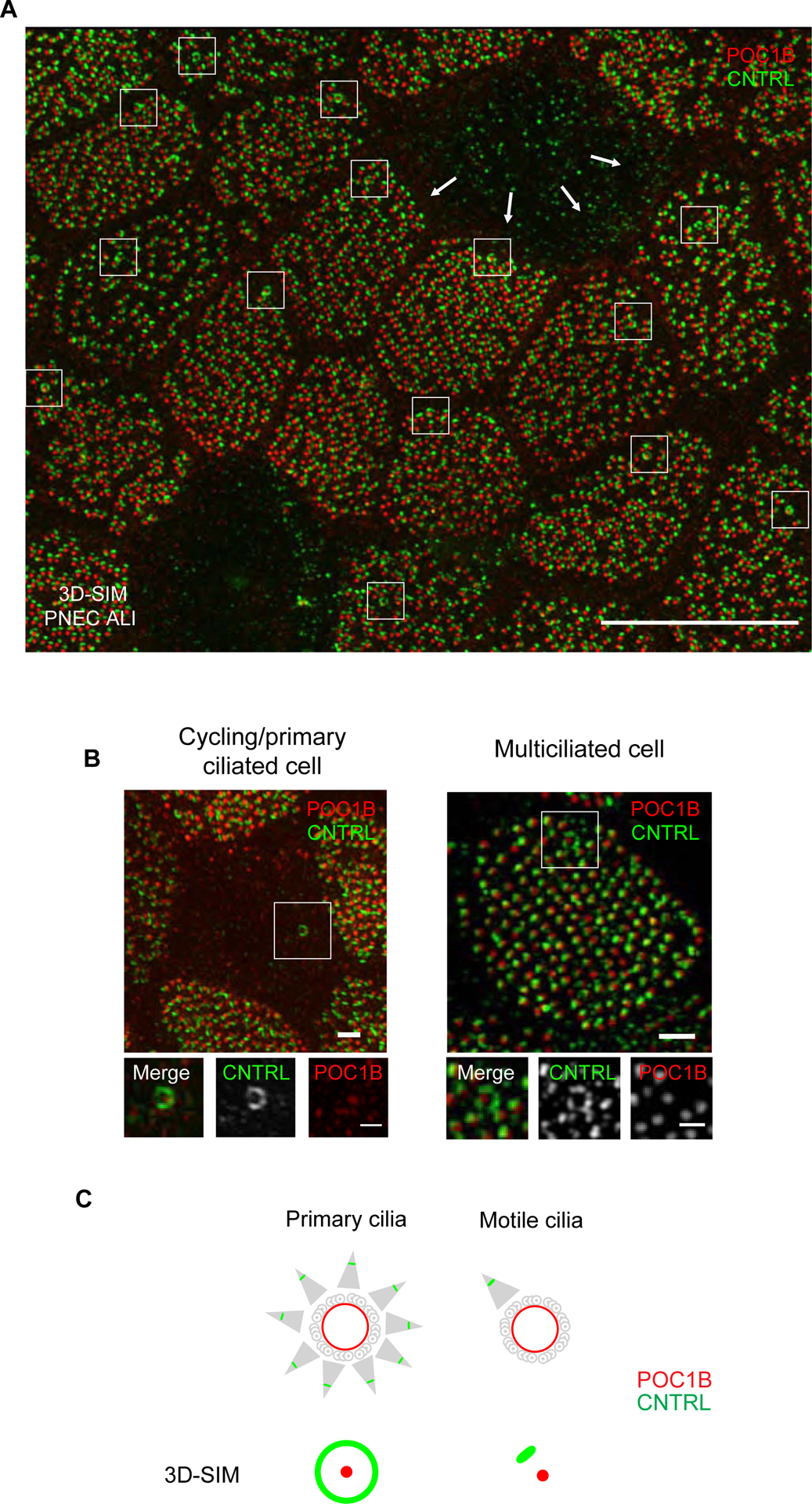
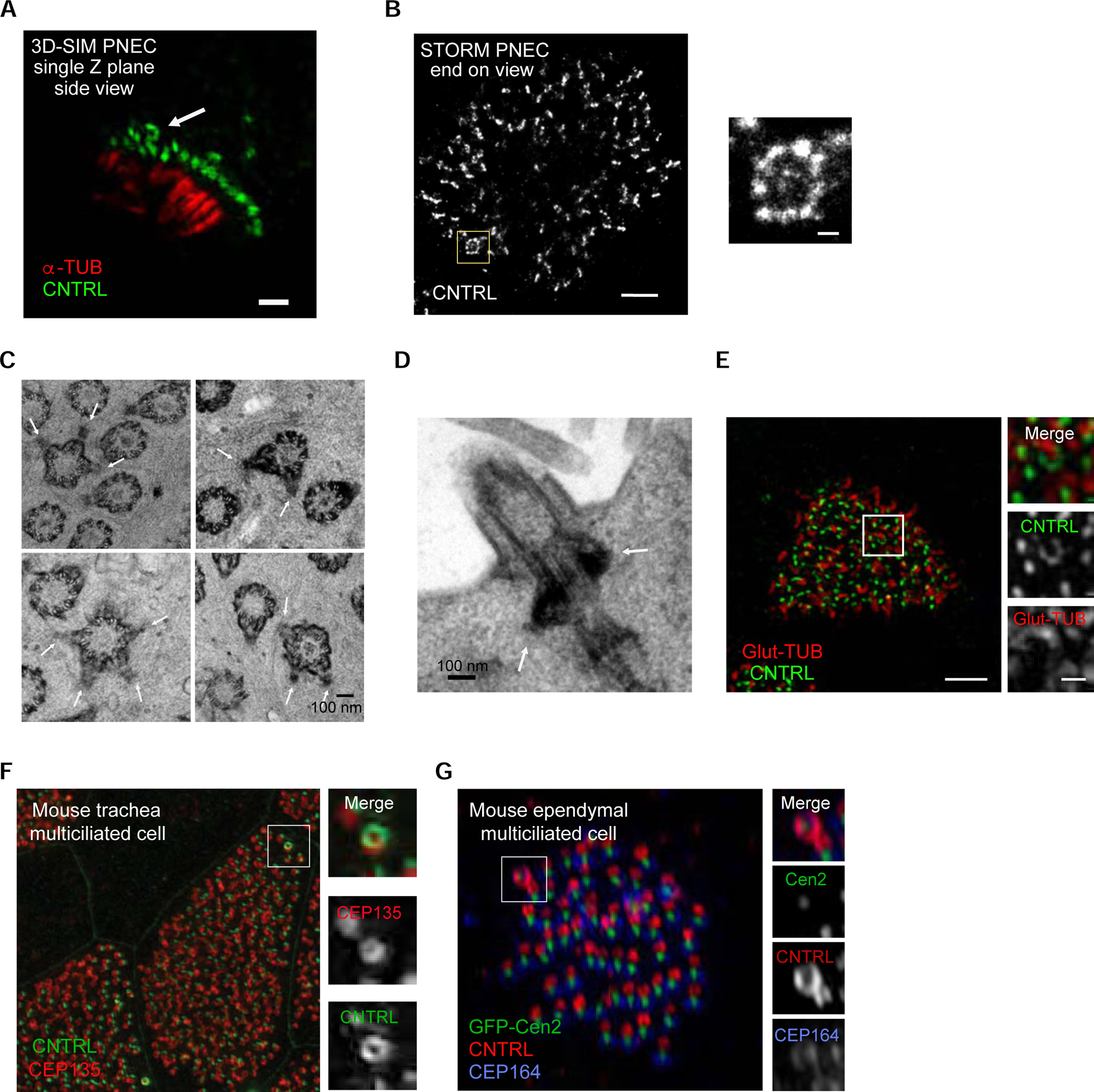
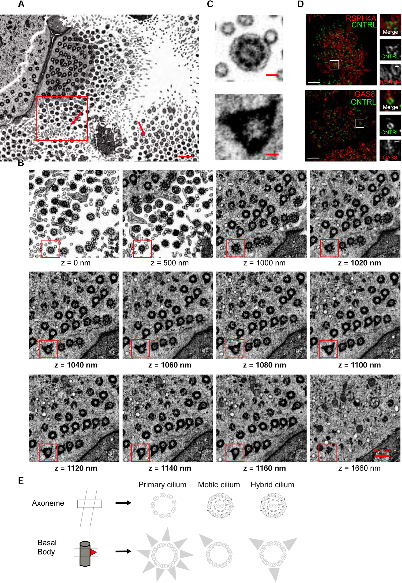
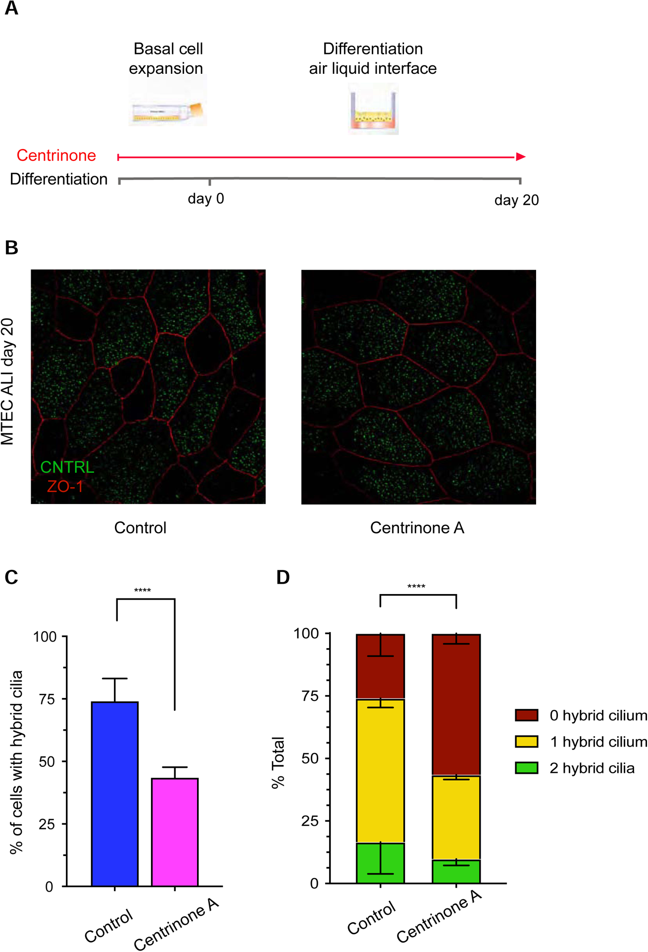
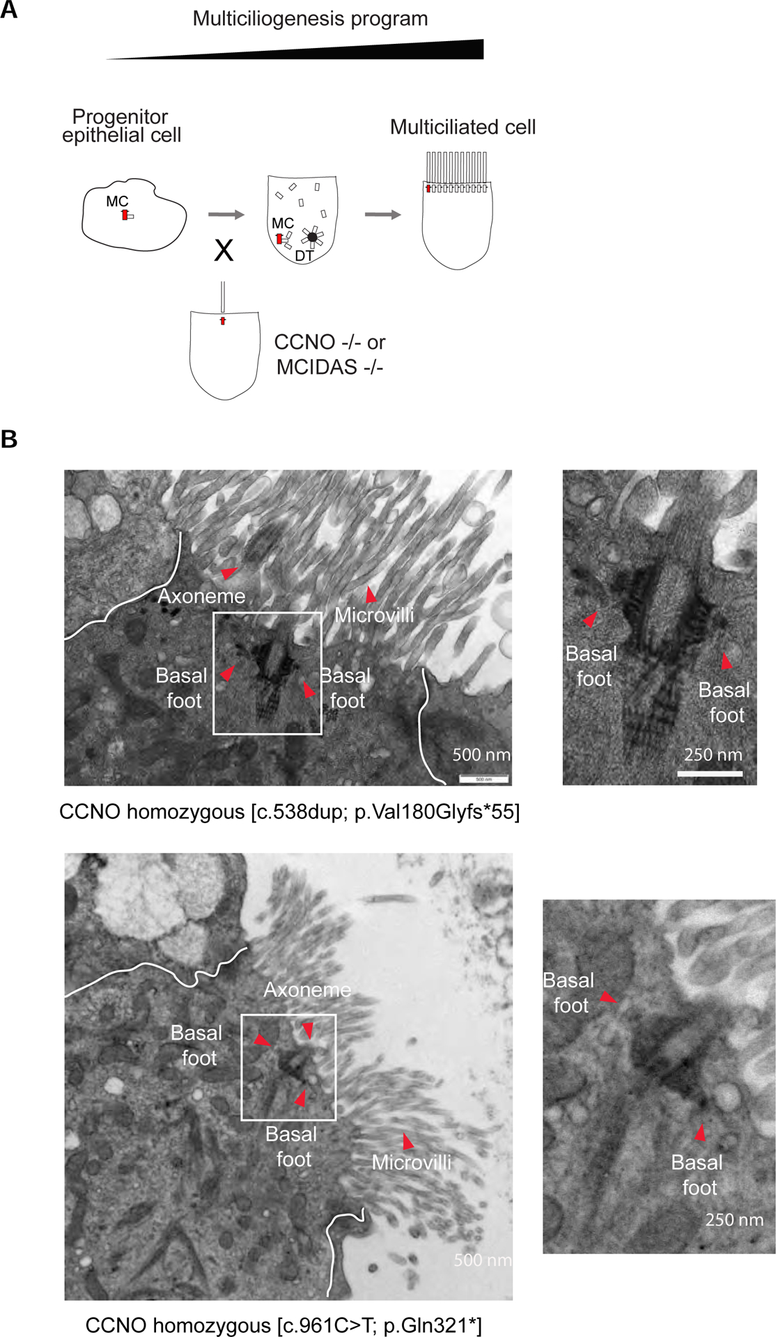
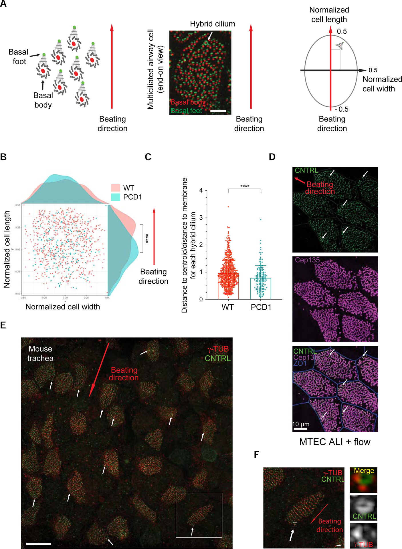
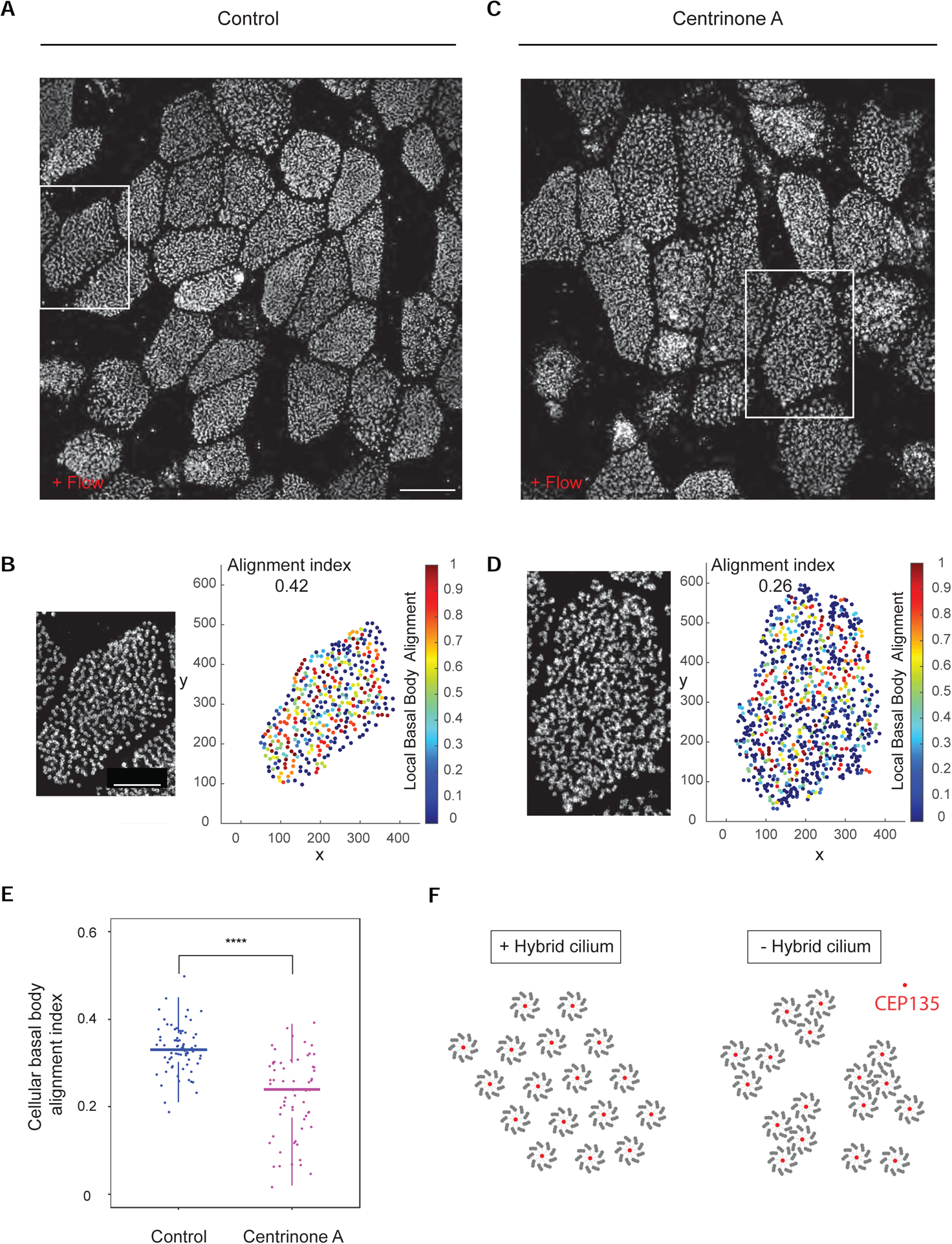
Motile cilia are cellular beating machines that play a critical role in mucociliary clearance, cerebrospinal fluid movement, and fertility. In the airways, hundreds of motile cilia present on the surface of a multiciliated epithelia cell beat coordinately to protect the epithelium from bacteria, viruses, and harmful particulates. During multiciliated cell differentiation, motile cilia are templated from basal bodies, each extending a basal foot-an appendage linking motile cilia together to ensure coordinated beating. Here, we demonstrate that among the many motile cilia of a multiciliated cell, a hybrid cilium with structural features of both primary and motile cilia is harbored. The hybrid cilium is conserved in mammalian multiciliated cells, originates from parental centrioles, and its cellular position is biased and dependent on ciliary beating. Furthermore, we show that the hybrid cilium emerges independently of other motile cilia and functions in regulating basal body alignment.

Keywords: FIB-SEM; airway; appendages; basal bodies; basal foot; centrosome; cilia; electron microscopy; primary ciliary dyskinesia; quantitative imaging; super-resolution imaging.


Conflict of interest statement

Declarations of Interests The authors declare no competing interests.

Figures

Figure 1.
Figure 1.. Super-resolution reveals a basal body with multiple basal feet in airway multiciliated cells
(A) 3D-SIM volume maximum intensity projection of large field of view of nasal primary airway multiciliated cell (PNEC) grown in air liquid interphase labeled with anti-CNTRL (green) and anti-POC1B (red) antibodies. Note the ring-like pattern of CNTRL localization encircling the basal body labeled by POC1B (boxed areas). Scale bar represents 10 μm. ALI, air liquid interface. (B) 3D-SIM volume maximum intensity projection of an airway cycling/primary ciliated cell (left) or nasal airway multiciliated cell (right) grown in ALI labeled with anti-CNTRL (green) and anti-POC1B (red) antibodies. Scale bars represent 1 μm and 500 nm (boxed areas). (C) Cartoon representation of basal body-basal foot structure by TEM in cells with primary or motile cilia (upper panel) and by 2 colour 3D-SIM imaging of basal body and basal foot (lower panel). By 3D-SIM microscopy the basal body protein POC1B appears as a dot while the basal foot protein CNTRL appears as a dot in motile cilia, but as a ring in primary cilia. Red represents localization of POC1B and green represents localization of CNTRL. See also Figure S1.
Figure 2.
Figure 2.. The basal body extrudes a cilium and is conserved in different mammalian primary cells, tissues and species.
(A) 3D-SIM single-plane image of a human nasal airway multiciliated cell freshly isolated from a healthy individual, labeled with anti-CNTRL (green) and anti-alpha-tubulin (red) antibodies, showing the presence of the basal body with multiple basal feet. Scale bar represents 1 μm. (B) Left: STORM micrograph of airway multiciliated cell labeled with anti-CNTRL antibody, showing a distinct ring-like distribution of CNTRL (boxed area). Right: High-magnification view of boxed area. Scale bars represent 1 μm (left) and 100 nm (right). (C) Collage of representative TEM micrographs showing basal bodies harbouring multiple basal feet in ALI-cultured human airway multiciliated cells (see white arrows). Scale bar represents 100 nm. (D) TEM micrograph showing axoneme emanating from basal bodies harbouring multiple basal feet (in white arrows) in human airway multiciliated cell. Scale bar represents 1 μm. (E) Left: 3DSIM volume maximum intensity projection of an ALI-cultured human airway multiciliated cell labeled with anti-CNTRL (green) and anti-Glut-TUB (red) antibodies. Note the axoneme emanating from ring-like structure labeled with CNTRL (boxed area). Right: High-magnification view of boxed area with individual channels. Scale bars represent 2 μm (left) and 500 nm (right). (F) 3D-SIM volume maximum intensity projection of mouse tracheal multiciliated cell (ALI D20), labeled with anti-CNTRL (green) and anti-CEP135 (red) antibodies. Right: High-magnification view of boxed area with individual channels. Scale bar represents 2 μm. (G) 3D-SIM volume maximum intensity projection of adult mouse ependymal multiciliated cells (P16), labeled with GFP-Centrin2 (basal body), anti-CNTRL (red) and anti-CEP164 (blue, distal appendage protein) antibodies. Right: High-magnification view of boxed area labeled in left. Scale bar represents 1 μm and 200 nm.
Figure 3.
Figure 3.. The cilium has hybrid features between primary and motile cilia
(A) Representative section from FIB-SEM tomogram of human primary nasal multiciliated cells. Arrows indicate basal bodies with multiple basal feet. Scale bar represents 1 μm. (B) High-magnification view of boxed area in (A) at different z positions of the tomogram from z=0 nm to z=1660 nm. Note the hybrid cilium axoneme and central pair (z=0 nm), transition fibers (z=1000–1040 nm), multiple basal feet (z=1160–1160 nm) and the absence of the endocytic pocket (z=1660 nm). Scale bar represents 500 nm. (C) A high-magnification view of boxed area in (B) highlighting the basal body with a central pair and multiple basal feet. Scale bar represents 100 nm. (D) 2D projection micrographs of 3D-SIM volume of human airway multiciliated cells (left), and high-magnification views of boxed areas (right), labeled with anti-CNTRL (green), anti-RSPH4A (red, top) and anti-GAS8 (red, bottom) antibodies. Scale bars represent 2 μm. (E) Cartoon representation of hybrid cilium structure relative to the primary cilium and motile cilium. The axoneme extends from the basal body (grey), each with a basal foot (red triangle). The basal body of the primary cilium presents multiple basal feet, while the one of the motile cilium has one basal foot. In regard to the axoneme, the motile cilium presents outer dynein arms, inner dynein arms, nexin-dynein regulatory complexes, radial spokes and central pair complexes (black) , which are critical for in plane ciliary beating. The hybrid cilium has features of both the primary and motile cilium, namely multiple basal feet and protein complexes critical for ciliary beating.
Figure 4.
Figure 4.. Hybrid cilium originates from parental centrioles
(A) Cartoon representation of centrinone A treatment in mouse tracheal multiciliated cells. (B) 2D projection micrograph of 3D-SIM volume of representative example of mouse tracheal multiciliated cells at ALI D20 treated with DMSO control or centrinone A labeled with anti-CNTRL (green) and anti-ZO-1 (red) antibodies. Arrowheads indicate CNTRL rings. Scale bar represents 5 μm. (C) Bar graphs representing percentage of cells with hybrid cilium in DMSO control (blue) and centrinone A-treated (pink) cells treated during basal cell expansion and throughout differentiation; n>800 over three independent biological replicates. Data are represented as mean ± SD. Statistical analysis was done using Cochran-Mantel-Haenszel test. **** p<0.0001. (D) Bar graph representing percentage of cells with none (red), one (yellow) or two (green) hybrid cilia in ALI D20 mouse tracheal multiciliated cells treated with DMSO control (left) or centrinone A (right); n>800 over three independent biological replicates. Data are represented as mean ± SD. Statistical analysis was done using Chi-square test. See also Figures S2 and S3.
Figure 5.
Figure 5.. Hybrid cilium formation is independent from other motile cilia in airway multiciliated cells
(A) Cartoon depicting a simplified version of the multiciliogenesis cellular program. Note CCNO and MCIDAS loss of function mutations lead to oligocilia phenotype. MC, mother centriole. DT, deuterostome. (B) Left: TEM micrographs of a human airway multiciliated cells isolated from nasal epithelium of PCD patients with CCNO loss of function early termination mutations. Boxed areas represent basal bodies with multiple basal feet. Red arrowheads indicate axoneme, basal feet and microvilli. Right: High-magnification views of boxed area. Scale bars represent 500 nm (left) and 250 nm (right).
Figure 6.
Figure 6.. Hybrid cilium position is biased in the direction of beating and dependent on flow
(A) Left: Cartoon depiction of the strategy for analysis of the position of the hybrid cilium relative to cilia beating direction. Middle: representative 3DSIM image used for data analysis. Scale bar represents 5μm. Right: MATLAB-based analysis strategy to assess rotational polarity in multiciliated cells. The position is calculated relative to coordinates (−0.5
Figure 7.
Figure 7.. Hybrid cilium is critical for establishing basal body alignment
(A) 3D-SIM volume maximum intensity projection of mouse tracheal multiciliated cell (ALI D20), treated with DMSO (control) from basal cells expansion throughout ALI differentiation labeled with anti-CEP135 antibodies. Scale bar represents 10 μm. (B) Left: High-magnification view of boxed area in (A). Right: relative basal body local and global alignment measurements. (C) 3D-SIM volume maximum intensity projection of mouse tracheal multiciliated cell (ALI D20), treated centrinone A (right) from basal cells expansion throughout ALI differentiation labeled with anti-CEP135 antibodies. Scale bar represents 10 μm. (D) Left: High-magnification view of boxed area in (C). Right: relative basal body local and global alignment measurements. (E) Quantification of cellular basal body alignment index in cells treated with DMSO or centrinone A throughout multiciliated cells differentiation (n=70 over 3 independent biological replicates, total 63 cells. DMSO=0.33±0.05; centrinone A=0.23±0.09). Statistical analysis was done using unpaired t-test. Data are represented as mean ± SD. **** p<0.0001. (F) A model showing function of the hybrid cilia. When hybrid cilium is present, basal bodies are well aligned. However, when hybrid cilium is removed by centrinone A, basal body alignment in the cell is disrupted and basal bodies tend to group into clusters. Red present basal body protein CEP135, surrounded by 9 axonemal microtubule triplets.

Comment in

References

    1. Al Jord A, Lemaitre AI, Delgehyr N, Faucourt M, Spassky N, and Meunier A (2014). Centriole amplification by mother and daughter centrioles differs in multiciliated cells. Nature 516, 104–107. - PubMed
    1. Boisvieux-Ulrich E, Laine MC, and Sandoz D (1985). The orientation of ciliary basal bodies in quail oviduct is related to the ciliary beating cycle commencement. Biol Cell 55, 147–150. - PubMed
    1. Boon M, Wallmeier J, Ma L, Loges NT, Jaspers M, Olbrich H, Dougherty GW, Raidt J, Werner C, Amirav I, et al. (2014). MCIDAS mutations result in a mucociliary clearance disorder with reduced generation of multiple motile cilia. Nature communications 5, 4418. - PubMed
    1. Chong WM, Wang WJ, Lo CH, Chiu TY, Chang TJ, Liu YP, Tanos B, Mazo G, Tsou MB, Jane WN, et al. (2020). Super-resolution microscopy reveals coupling between mammalian centriole subdistal appendages and distal appendages. Elife 9. - PMC - PubMed
    1. Clare DK, Magescas J, Piolot T, Dumoux M, Vesque C, Pichard E, Dang T, Duvauchelle B, Poirier F, and Delacour D (2014). Basal foot MTOC organizes pillar MTs required for coordination of beating cilia. Nat Commun 5, 4888. - PMC - PubMed

Publication types