. 2020 Dec;98(6):1502-1518.
doi: 10.1016/j.kint.2020.09.015. Epub 2020 Oct 8.

SARS-CoV-2 receptor networks in diabetic and COVID-19-associated kidney disease

Affiliations

SARS-CoV-2 receptor networks in diabetic and COVID-19-associated kidney disease

Rajasree Menon et al. Kidney Int. 2020 Dec.

Abstract

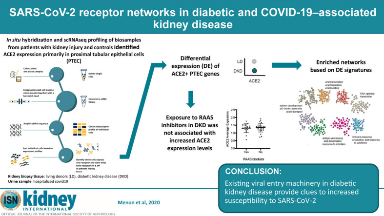
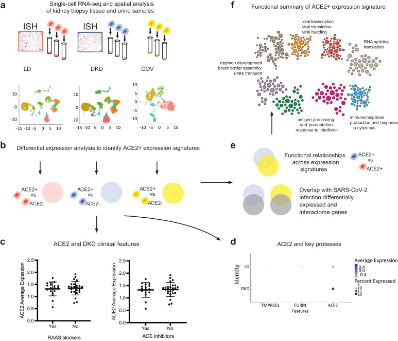
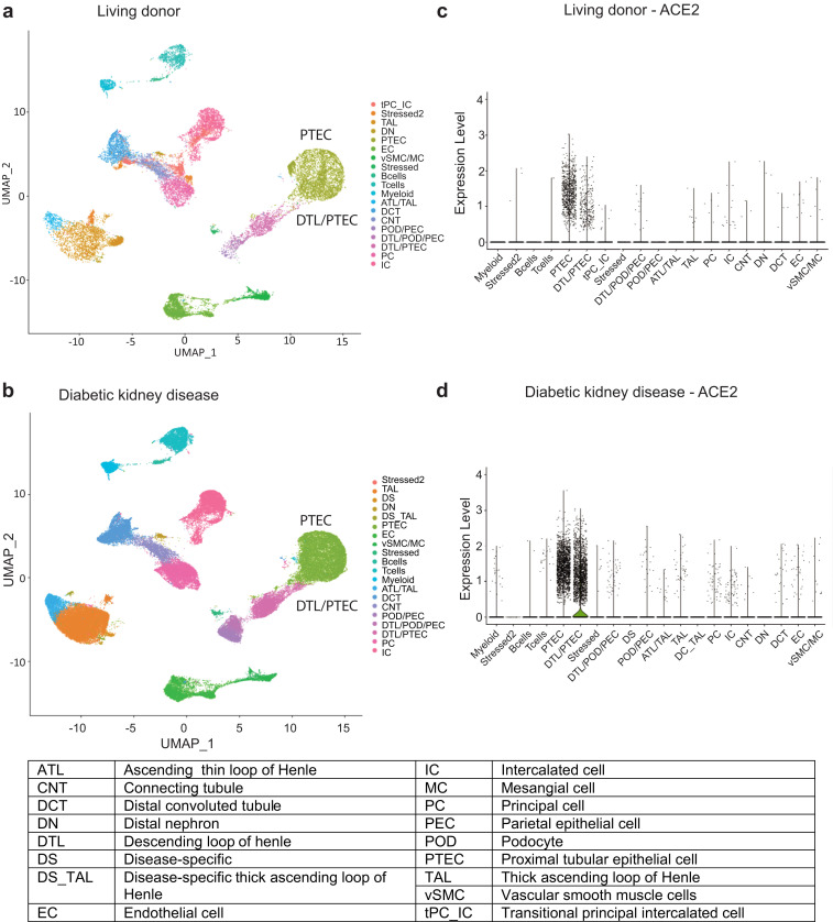
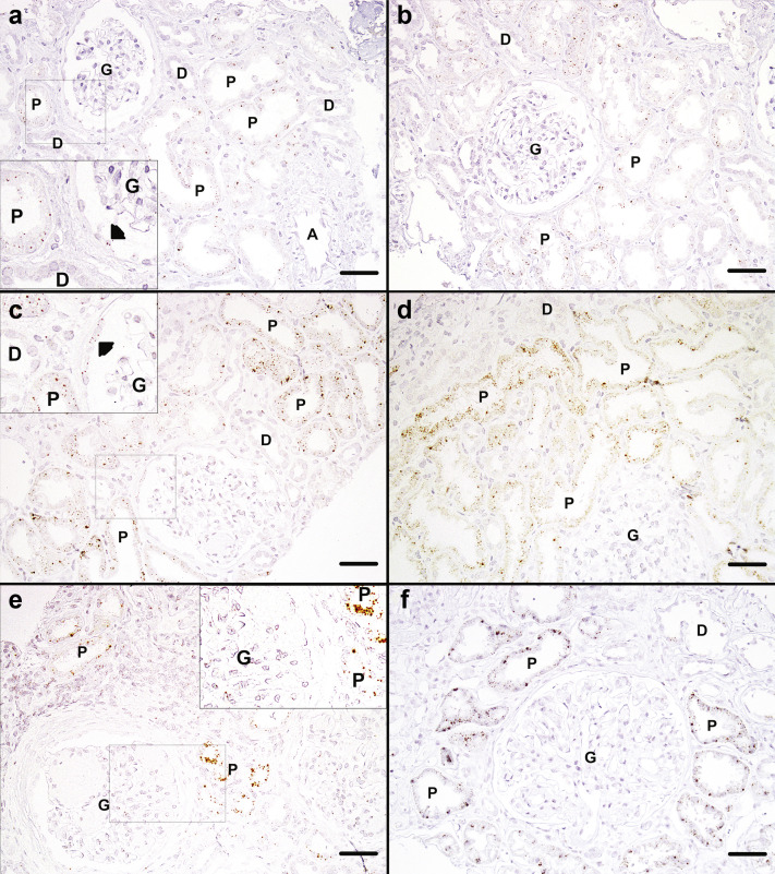
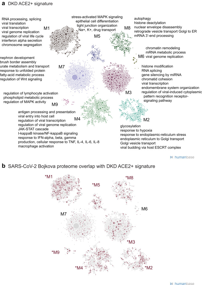
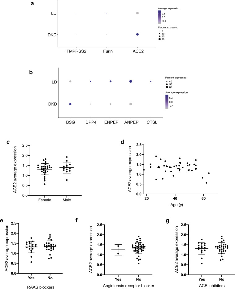
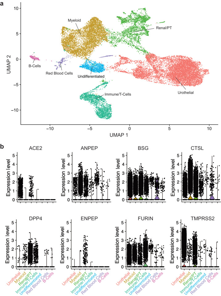
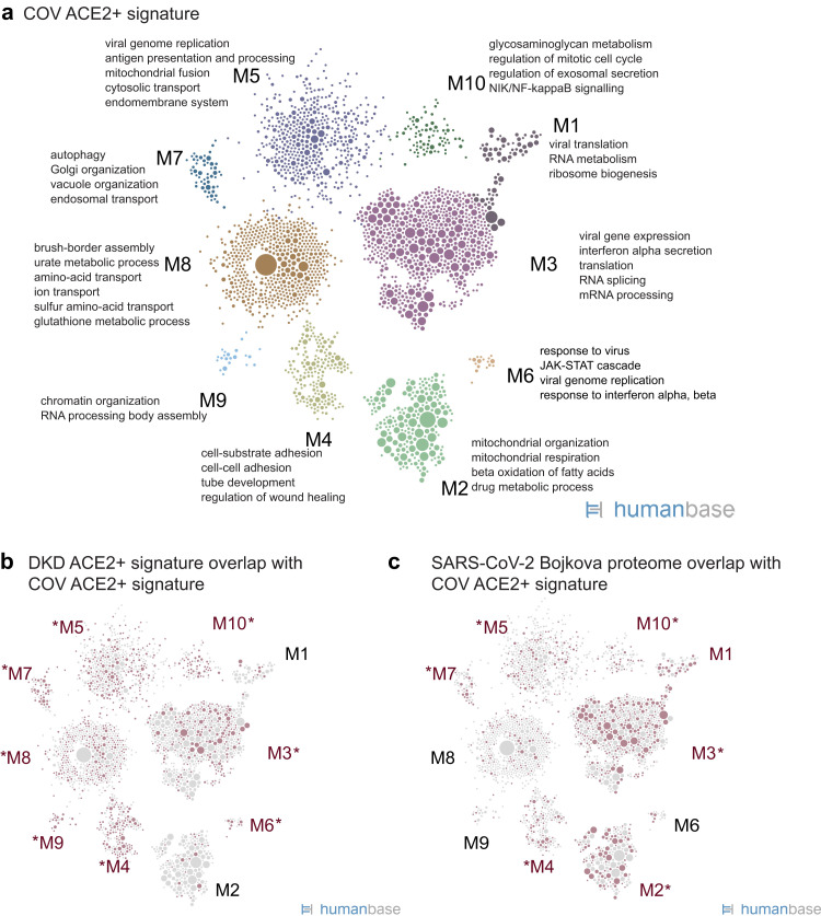
COVID-19 morbidity and mortality are increased via unknown mechanisms in patients with diabetes and kidney disease. SARS-CoV-2 uses angiotensin-converting enzyme 2 (ACE2) for entry into host cells. Because ACE2 is a susceptibility factor for infection, we investigated how diabetic kidney disease and medications alter ACE2 receptor expression in kidneys. Single cell RNA profiling of kidney biopsies from healthy living donors and patients with diabetic kidney disease revealed ACE2 expression primarily in proximal tubular epithelial cells. This cell-specific localization was confirmed by in situ hybridization. ACE2 expression levels were unaltered by exposures to renin-angiotensin-aldosterone system inhibitors in diabetic kidney disease. Bayesian integrative analysis of a large compendium of public -omics datasets identified molecular network modules induced in ACE2-expressing proximal tubular epithelial cells in diabetic kidney disease (searchable at hb.flatironinstitute.org/covid-kidney) that were linked to viral entry, immune activation, endomembrane reorganization, and RNA processing. The diabetic kidney disease ACE2-positive proximal tubular epithelial cell module overlapped with expression patterns seen in SARS-CoV-2-infected cells. Similar cellular programs were seen in ACE2-positive proximal tubular epithelial cells obtained from urine samples of 13 hospitalized patients with COVID-19, suggesting a consistent ACE2-coregulated proximal tubular epithelial cell expression program that may interact with the SARS-CoV-2 infection processes. Thus SARS-CoV-2 receptor networks can seed further research into risk stratification and therapeutic strategies for COVID-19-related kidney damage.

Keywords: ACE inhibitors; COVID-19; SARS-CoV-2; acute kidney injury; diabetic nephropathy; molecular networks; proximal tubules; scRNAseq.


Figures

None
Graphical abstract
Figure 1
Figure 1
Study overview. To understand how the kidney may be affected by coronavirus disease 2019 (COVID-19) we performed spatial, systems, and clinical association analyses of angiotensin-converting enzyme 2 (ACE2) and other severe acute respiratory syndrome coronavirus 2 (SARS-CoV-2) host factors in kidney biopsies from living donors (LD) and patients with diabetic kidney disease (DKD) and kidney cells isolated from the urine of hospitalized COVID-19 patients (COV). (a) Biopsy samples from DKD and LD were processed for in situ hybridization (ISH) and single-cell RNA sequencing (scRNAseq) profiling. scRNAseq of LD, DKD, and urine cell pellets from COV samples were analyzed to determine cell type expression specificity of ACE2 in healthy and disease states. (b) For each scRNAseq dataset, ACE2+ differential expression signatures were identified. (c) Association of ACE2 expression levels in DKD with clinical characteristics were evaluated, including exposure to renin-angiotensin-aldosterone system (RAAS) blockers and ACE inhibitors. (d) Expression of ACE2 and key proteases between LD and DKD proximal tubule epithelial cells (PTECs) were compared. (e) ACE2 expression signatures across datasets identified aspects induced in PTECs expressing DKD samples compared to LD. These gene sets significantly overlapped those reported to be affected by direct SARS-CoV-2 infection. (f) The biological processes in ACE2+ expression signatures were characterized by projecting these signature genes onto PTEC-specific functional networks at HumanBase (https://hb.flatironinstitute.org/covid-kidney). These networks represent genes and their interactions in biological processes and pathways active in PTECs.
Figure 2
Figure 2
Unsupervised clustering of cells from living donors (LD) and patients with diabetic kidney disease (DKD). UMAP plots showing the distributions in unsupervised clustering of (a) 25,163 LD kidney cells into 19 clusters and (b) 85,872 cells from DKD into 21 clusters; 22% of the total cells in both DKD and LD were cubilin-positive proximal tubular epithelial cells (PTECs). The violin plots for (c) LD and (d) DKD show cell type specificity of ACE2 expression. DTL, distal limb of loop of Henle.
Figure 3
Figure 3
In situ detection of angiotensin-converting enzyme 2 (ACE2) in deidentified living donor (LD) and diabetic kidney disease tissue.In situ hybridization for ACE2 mRNA in (a,b) 2 LD reference control biopsies, (c,d) 2 renal biopsies from patients with mild diabetic nephropathy, and (e,f) 2 cases of advanced diabetic nephropathy. Small brown dots representing ACE2 mRNA transcripts are seen scattered at low density in proximal tubules (PTs) of control biopsies (a,b) and at higher density in mild (c,d) and advanced (e,f) diabetic nephropathy. Small brown dots can also be seen in parietal cells in control biopsies (a) and mild diabetic nephropathy (c). No ACE2 transcript signal is seen in distal tubules (D), glomeruli (G), or arterioles (A). Original magnification ×200. Bars = 50 μm. Arrowheads indicate parietal cells. To optimize viewing of this image, please see the online version of this article at www.kidney-international.org/.
Figure 4
Figure 4
Functional summary of diabetic kidney disease (DKD) angiotensin-converting enzyme 2–positive (ACE2+) expression signature. (a) ACE2+ coexpression signatures were used for community clustering in a positive proximal tubular epithelial cell–specific functional network to identify enriched processes and pathways in DKD biopsy samples. (b) Highlight of ACE2+ signature genes (red circles) shared with the set of proteins that increase expression in response to severe acute respiratory syndrome coronavirus 2 (SARS-CoV-2) infection (Bojkova proteome36; Supplementary Table S6). Red text indicates modules with the strongest overlap (P < 0.05), highlighting modules and processes in the ACE2+ signature relevant to SARS-CoV-2 infection.
Figure 5
Figure 5
Severe acute respiratory syndrome coronavirus 2–related host gene expression in diabetic kidney disease (DKD) biopsy samples and relationships to clinical phenotypes. (a,b) Expression of angiotensin-converting enzyme 2 (ACE2) and related proteases in proximal tubular epithelial cells (PTECs). Dot plots showing the expression of ACE2 and proteases in PTECs from DKD and LD tissue according to single-cell analysis. Color indicates expression level, and the size of the dot indicates the percentage of cells expressing the gene. Proteases were grouped into 2 sets based on the overall expression pattern in proximal cells (a, expression in <30% of the cells; b, expression in ≥30% of the cells). (c–g) Clinical associations of ACE2 expression in DKD. Relationships between ACE2 expression in PTECs and (c) biological sex, (d) age, (e) exposure to any renin-angiotensin-aldosterone-system (RAAS) blockers, (f) exposure to angiotensin receptor blockers, and (g) exposure to ACE inhibitors were examined. No significant correlations between clinical factors and ACE2 expression were found in the DKD cohort.
Figure 6
Figure 6
Single-cell analysis of cells isolated from the urine of patients hospitalized with coronavirus disease 2019. (a) UMAP plot showing the 7 clusters identified from 25,791 cells. (b) Violin plots of severe acute respiratory syndrome coronavirus 2–associated proteases ACE2, ANPEP, BSG, CTSL, DPP4, ENPEP, FURIN, and TMPRSS2 in the 7 identified cell types.
Figure 7
Figure 7
Functional summary of angiotensin-converting enzyme 2–positive (ACE2+) expression signature in kidney cells isolated from the urine of hospitalized corona virus disease 2019 (COVID-19) patients (COV). (a) Community clustering and enriched processes of the ACE2+ expression signature in proximal tubule epithelial cells (PTECs) isolated from COV. (b) Highlight of genes (red circles) from the DKD ACE2+ signature (Figure 4a; Supplementary Table S2) that overlap the ACE2+ signature in PTEC from COVID-19 patients. Red text indicates modules with the strongest gene overlap (P < 0.0105), thus indicating ACE2+-associated processes shared between PTECs in diabetic kidneys and PTECs in the context of COVID-19. (c) Highlight of ACE2+ signature genes (red circles) shared with the set of proteins that increase expression in response to SARS-CoV-2 infection (Bojkova proteome36; Supplementary Table S6).

Update of

References

    1. Argenziano M.G., Bruce S.L., Slater C.L., et al. Characterization and clinical course of 1000 patients with coronavirus disease 2019 in New York: retrospective case series. BMJ. 2020;369:m1996. - PMC - PubMed
    1. Apicella M., Campopiano M.C., Mantuano M., et al. COVID-19 in people with diabetes: understanding the reasons for worse outcomes. Lancet Diabetes Endocrinol. 2020;8:782–792. - PMC - PubMed
    1. Muniyappa R., Gubbi S. COVID-19 pandemic, corona viruses, and diabetes mellitus. Am J Physiol Endocrinol Metab. 2020;318:E736–E741. - PMC - PubMed
    1. Shahid Z., Kalayanamitra R., McClafferty B., et al. COVID-19 and older adults: what we know. J Am Geriatr Soc. 2020;68:926–929. - PMC - PubMed
    1. Emami A., Javanmardi F., Pirbonyeh N., et al. Prevalence of underlying diseases in hospitalized patients with COVID-19: a systematic review and meta-analysis. Arch Acad Emerg Med. 2020;8 e35–e35. - PMC - PubMed

Publication types

MeSH terms

Substances