

Quantitative MRI demonstrates abnormalities of the third ventricle subventricular zone in neurofibromatosis type-1 and sporadic paediatric optic pathway glioma
- PMID: 33038669
- PMCID: PMC7554210
- DOI: 10.1016/j.nicl.2020.102447
Quantitative MRI demonstrates abnormalities of the third ventricle subventricular zone in neurofibromatosis type-1 and sporadic paediatric optic pathway glioma
Abstract
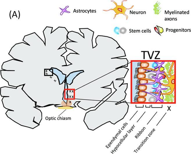
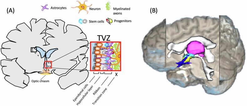
Background: The subventricular zone of the third ventricle (TVZ) is a germinal stem cell niche, identified as the possible location of optic pathway glioma (OPG) cell origin. Paediatric OPGs are predominantly diagnosed as low-grade astrocytomas, which are either sporadic or are associated with neurofibromatosis type-1 (NF1). These tumours often cause a significant impairment to visual acuity (VA). Infiltrative/invasive tumour activity is associated with increased apparent diffusion coefficient (ADC) and cerebral blood flow (CBF). This study aimed to determine whether TVZ imaging features differed between sporadic-OPG, NF1-OPG and controls, and whether the ADC and CBF profile at the germinal stem cell niche (the TVZ) correlated with the primary outcome of VA.
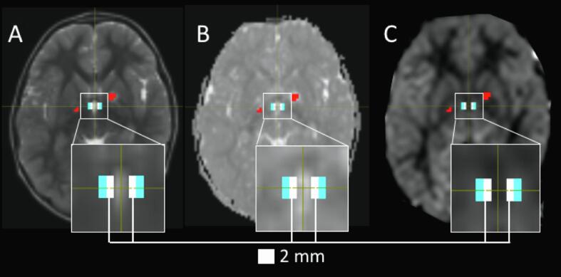
Methods: ADC and CBF MRI data were acquired from 30 paediatric OPG patients (median age 6 years; range 8 months-17 years), along with VA measurements, during clinical surveillance of their tumour. Values for mean ADC and maximum CBF were measured at the TVZ, and normalized to normal-appearing grey matter. These values were compared between the two OPG groups and the healthy control subjects, and multivariate linear regression was used to test the linear association between these values and patient's VA.
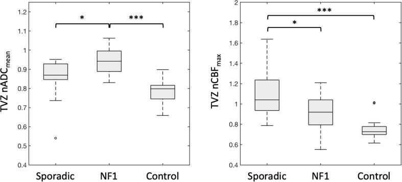
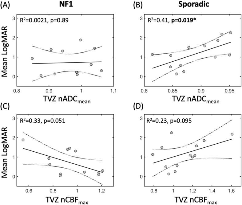
Results: In the TVZ, normalized mean ADC was higher in NF1-associated OPG patients (N = 15), compared to both sporadic OPG patients (N = 15; p = 0.010) and healthy controls (N = 14; p < 0.001). In the same region, normalized maximum CBF was higher in sporadic OPG patients compared to both NF1-OPG patients (p = 0.016) and healthy controls (p < 0.001). In sporadic OPG patients only, normalized mean ADC in the TVZ was significantly correlated with visual acuity (R2 = 0.41, p = 0.019). No significant correlations were found between TVZ CBF and ADC values and visual acuity in the NF1-associated OPG patients.
Conclusion: Quantitative MRI detects TVZ abnormalities in both sporadic and NF1-OPG patients, and identifies TVZ features that differentiate the two. TVZ features may be useful MRI markers of interest in future predictive studies involving sporadic OPG.
Keywords: Neurofibromatosis type-1; Optic pathway glioma; Paediatric; Quantitative magnetic resonance imaging; Third ventricle subventricular zone.
Copyright © 2020 The Authors. Published by Elsevier Inc. All rights reserved.
Conflict of interest statement
The authors declare that they have no known competing financial interests or personal relationships that could have appeared to influence the work reported in this paper.
Figures
Similar articles
-
Visual outcomes after chemotherapy for optic pathway glioma in children with and without neurofibromatosis type 1: results of the International Society of Paediatric Oncology (SIOP) Low-Grade Glioma 2004 trial UK cohort.Br J Ophthalmol. 2018 Oct;102(10):1367-1371. doi: 10.1136/bjophthalmol-2017-311305. Epub 2018 Jan 17. Br J Ophthalmol. 2018. PMID: 29343527 Clinical Trial.
-
Optical coherence tomography of the macular ganglion cell layer in children with neurofibromatosis type 1 is a useful tool in the assessment for optic pathway gliomas.PLoS One. 2024 Jul 11;19(7):e0305548. doi: 10.1371/journal.pone.0305548. eCollection 2024. PLoS One. 2024. PMID: 38990917 Free PMC article.
-
Delineation of the visual pathway in paediatric optic pathway glioma patients using probabilistic tractography, and correlations with visual acuity.Neuroimage Clin. 2017 Oct 11;17:541-548. doi: 10.1016/j.nicl.2017.10.010. eCollection 2018. Neuroimage Clin. 2017. PMID: 29527480 Free PMC article.
-
Optic Pathway Glioma and Cerebral Focal Abnormal Signal Intensity in Patients with Neurofibromatosis Type 1: Characteristics, Treatment Choices and Follow-up in 134 Affected Individuals and a Brief Review of the Literature.Anticancer Res. 2016 Aug;36(8):4095-121. Anticancer Res. 2016. PMID: 27466519 Review.
-
Insights into optic pathway glioma vision loss from mouse models of neurofibromatosis type 1.J Neurosci Res. 2019 Jan;97(1):45-56. doi: 10.1002/jnr.24250. Epub 2018 Apr 28. J Neurosci Res. 2019. PMID: 29704429 Free PMC article. Review.
Cited by
-
Neuroimaging of pediatric tumors of the sellar region-A review in light of the 2021 WHO classification of tumors of the central nervous system.Front Pediatr. 2023 Jun 21;11:1162654. doi: 10.3389/fped.2023.1162654. eCollection 2023. Front Pediatr. 2023. PMID: 37416813 Free PMC article. Review.
References
-
- Alsop D.C., Detre J.A., Golay X., Günther M., Hendrikse J., Hernandez-Garcia L., Lu H., MacIntosh B.J., Parkes L.M., Smits M., van Osch M.J.P., Wang D.J.J., Wong E.C., Zaharchuk G. Recommended implementation of arterial spin-labeled perfusion MRI for clinical applications: a consensus of the ISMRM perfusion study group and the European consortium for ASL in dementia. Magn. Reson. Med. 2015;73:102–116. doi: 10.1002/mrm.25197. - DOI - PMC - PubMed
-
- Alvarez-Buylla, A., García-Verdugo, J.M., 2002. Neurogenesis in adult subventricular zone. The Journal of neuroscience 22, 629–634. https://doi.org/0270-6474/02/220629-06. - PMC - PubMed
-
- Aronen H.J., Gazit I.E., Louis D.N., Buchbinder B.R., Pardo F.S., Weisskoff R.M., Harsh G.R., Cosgrove G.R., Halpern E.F., Hochberg F.H. Cerebral blood volume maps of gliomas: comparison with tumor grade and histologic findings. Radiology. 1994;191:41–51. doi: 10.1148/radiology.191.1.8134596. - DOI - PubMed
Publication types
MeSH terms
Grants and funding
LinkOut - more resources
Full Text Sources
Research Materials
Miscellaneous
