Review
. 2020 Nov;30(4):505-516.
doi: 10.1016/j.nic.2020.07.003. Epub 2020 Sep 17.

Diverse Applications of Artificial Intelligence in Neuroradiology

Affiliations
Review

Diverse Applications of Artificial Intelligence in Neuroradiology

Michael Tran Duong et al. Neuroimaging Clin N Am. 2020 Nov.

Abstract

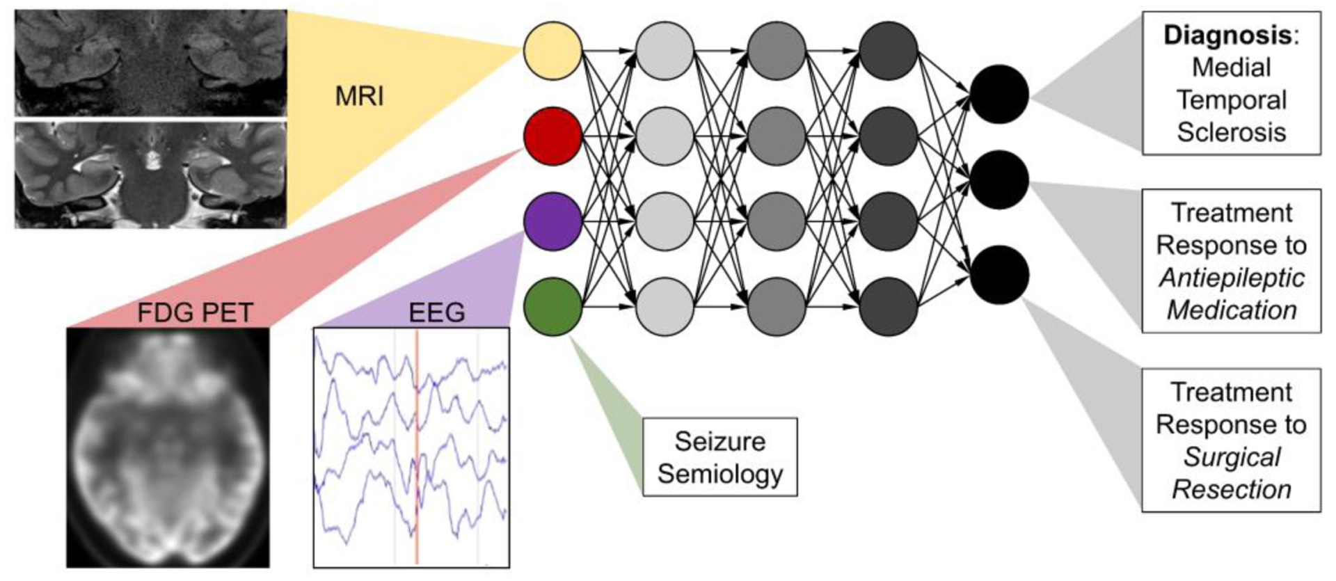
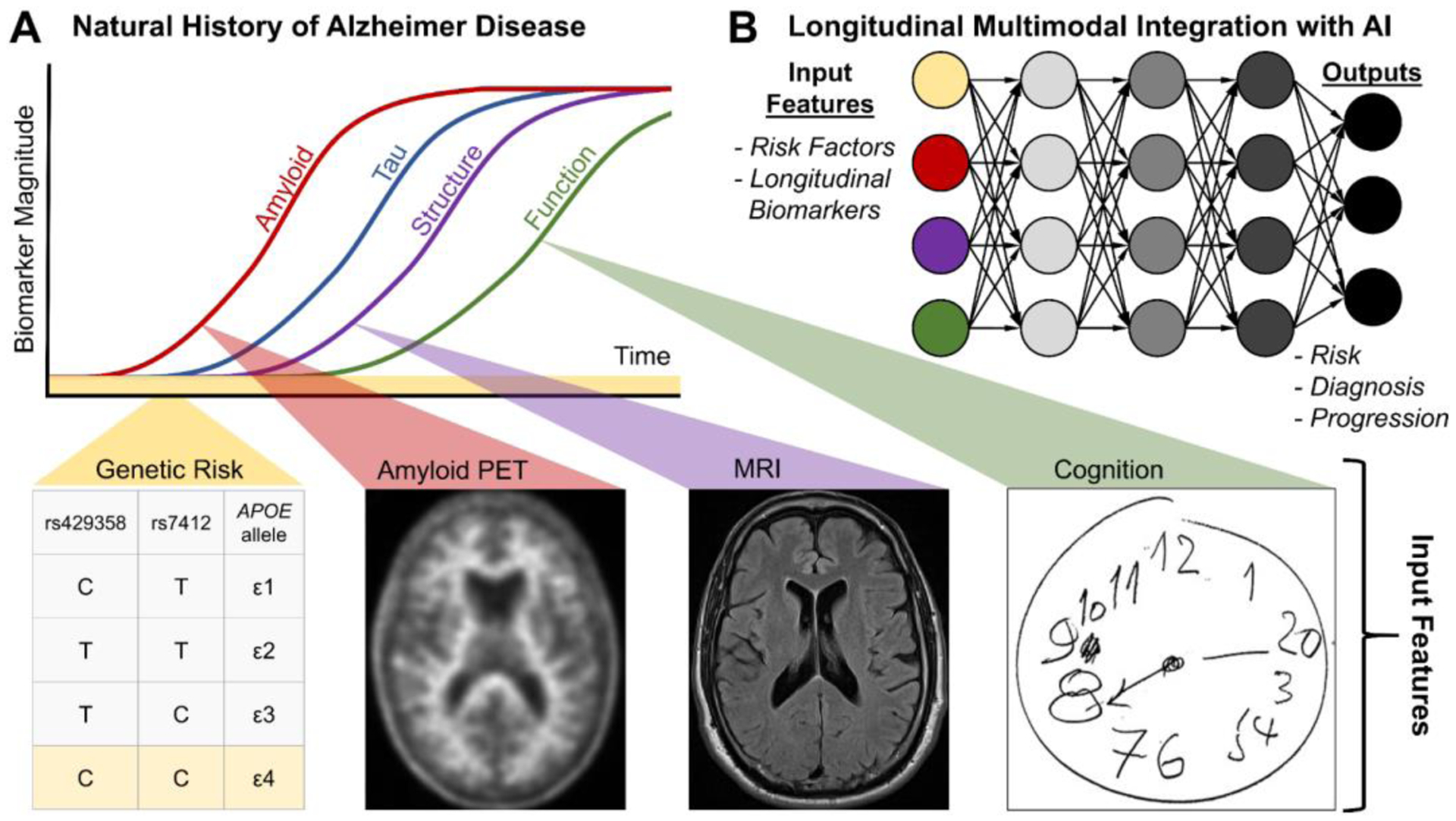
Recent advances in artificial intelligence (AI) and deep learning (DL) hold promise to augment neuroimaging diagnosis for patients with brain tumors and stroke. Here, the authors review the diverse landscape of emerging neuroimaging applications of AI, including workflow optimization, lesion segmentation, and precision education. Given the many modalities used in diagnosing neurologic diseases, AI may be deployed to integrate across modalities (MR imaging, computed tomography, PET, electroencephalography, clinical and laboratory findings), facilitate crosstalk among specialists, and potentially improve diagnosis in patients with trauma, multiple sclerosis, epilepsy, and neurodegeneration. Together, there are myriad applications of AI for neuroradiology."

Keywords: Artificial intelligence; Deep learning; Epilepsy; Multiple sclerosis; Neural network; Neurodegeneration; Neuroradiology; Trauma.


Conflict of interest statement

Disclosure S. Mohan has research grants from Galileo CDS and Novocure, USA. M.T. Duong and A.M. Rauschecker have nothing to disclose.

Figures

Figure 1.
Figure 1.
Cumulative frequency of PubMed indexed articles on AI and imaging/radiology (grey line) and AI and neuroimaging/neuroradiology (blue dashed line). Boolean search query for radiology was “AI/ML/DL/NN and clinical/medical and imaging/radiology/body” and for neuroradiology was “AI/ML/DL/NN and clinical/medical and neuroimaging/neuroradiology/brain.” PubMed last accessed November 27, 2019.
Figure 2.
Figure 2.
The Adaptive Radiology Interpretation and Education System (ARIES) distinguishes between ependymoma and astrocytoma in the spine network. (A) Features based on signal, spatial and clinical information are selected by the trainee in blue. Unselected features are grey and the most differentiating unanswered features are highlighted in orange. Differential diagnoses by (B) imaging features only vs (C) a combination of clinical and imaging features are derived from manually selected features (A). Probabilities are calculated by a naïve Bayes network.
Figure 3.
Figure 3.
Feedback loop framework for longitudinal AI-augmented precision education.
Figure 4.
Figure 4.
Model of multimodal integration of AI in diagnosis and assessment of disease progression in multiple sclerosis.
Figure 5.
Figure 5.
Model of multimodal integration in diagnosis and management of epilepsy. Bilateral mesial temporal sclerosis demonstrates increased signal and volume loss in hippocampal MRI, temporal lobe hypometabolism on 18F-FDG-PET and ictal events on EEG.
Figure 6.
Figure 6.
Model of multimodal integration of AI for diagnosis and management in Alzheimer disease. (A) Natural history of Alzheimer disease (adapted from). (B) AI-enabled multimodal integration of input features for longitudinal (re)assessment of risk, diagnosis and progression.

References

    1. World Health Organization. Global health estimates 2015: Disease burden by cause, age, sex, by country and by region, 2000–2015. Geneva: 2016. http://www.who.int/healthinfo/global_burden_disease/estimates/en/index1....
    1. Hassabis D, Kumaran D, Summerfield C, et al. Neuroscience-Inspired Artificial Intelligence. Neuron 2017; 95:245–258. doi:10.1016/j.neuron.2017.06.011 - DOI - PubMed
    1. Kriegeskorte N. Deep Neural Networks: A New Framework for Modeling Biological Vision and Brain Information Processing. Annu Rev Vision Sci 2015;1:417–446. doi:10.1146/annurev-vision-082114-035447 - DOI - PubMed
    1. Erickson BJ, Korfiatis P, Akkus Z, et al. Machine learning for medical imaging. Radiographics 2017; 37: 505–15. doi:10.1148/rg.2017160130 - DOI - PMC - PubMed
    1. Kohli M, Prevedello LM, Filice RW, et al. Implementing machine learning in radiology practice and research. AJR Am J Roentgenol 2017;208:754–60. doi:10.2214/AJR.16.17224 - DOI - PubMed

MeSH terms