Review
. 2021 Apr:199:101919.
doi: 10.1016/j.pneurobio.2020.101919. Epub 2020 Oct 8.

The intracellular renin-angiotensin system: Friend or foe. Some light from the dopaminergic neurons

Affiliations
Review

The intracellular renin-angiotensin system: Friend or foe. Some light from the dopaminergic neurons

Jose L Labandeira-Garcia et al. Prog Neurobiol. 2021 Apr.

Abstract

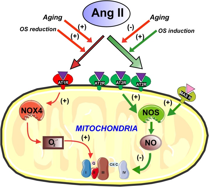
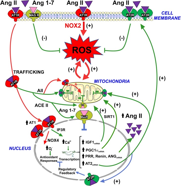
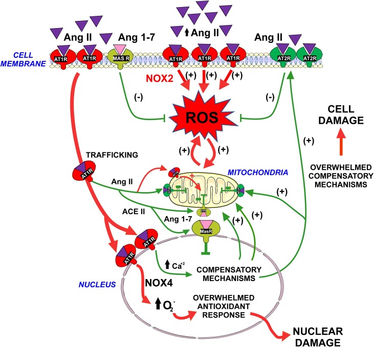
The renin-angiotensin system (RAS) is one of the oldest hormone systems in vertebrate phylogeny. RAS was initially related to regulation of blood pressure and sodium and water homeostasis. However, local or paracrine RAS were later identified in many tissues, including brain, and play a major role in their physiology and pathophysiology. In addition, a major component, ACE2, is the entry receptor for SARS-CoV-2. Overactivation of tissue RAS leads several oxidative stress and inflammatory processes involved in aging-related degenerative changes. In addition, a third level of RAS, the intracellular or intracrine RAS (iRAS), with still unclear functions, has been observed. The possible interaction between the intracellular and extracellular RAS, and particularly the possible deleterious or beneficial effects of the iRAS activation are controversial. The dopaminergic system is particularly interesting to investigate the RAS as important functional interactions between dopamine and RAS have been observed in the brain and several peripheral tissues. Our recent observations in mitochondria and nucleus of dopaminergic neurons may clarify the role of the iRAS. This may be important for the developing of new therapeutic strategies, since the effects on both extracellular and intracellular RAS must be taken into account, and perhaps better understanding of COVID-19 cell mechanisms.

Keywords: ACE/angiotensin receptors/renin angiotensin system; Angiotensin; COVID-19; Cell biology/structural biology; Cell signaling/signal transduction; Intracrine; Mitochondria; Nucleus; Oxidant stress; Oxidative stress.


Conflict of interest statement

The authors report no declarations of interest.

Figures

Fig. 1
Fig. 1
Modulation of oxidative phosphorylation by mitochondrial angiotensin receptors. Mitochondrial AT1 receptors, via Nox4, induce superoxide production and increase respiration. AT2 receptors are more numerous than AT1 in mitochondria. AT2 and Mas receptors induce, via nitric oxide, a reduction in mitochondrial respiration and regulate oxidative phosphorylation without significant change in mitochondrial membrane potential, which suggests that mitochondrial bioenergetic properties are not altered. Treatment of cells with oxidative stress (OS) inducers leads to a compensatory increase in levels of mitochondrial AT2 receptors, while treatment of cells with antioxidants increases AT1 receptor levels. Mitochondrial AT2 and Mas receptors may modulate respiration and offset the effects of low levels of OS during the normal cell functioning. Aging leads to a decrease in AT2 and Mas receptors and an increase in AT1 mitochondrial receptors, which may contribute to mitochondrial dysfunction and cell death. Images were produced using Servier Medical Art (http://www.servier.com).
Fig. 2
Fig. 2
Modulation by nuclear angiotensin receptors of the pro-oxidative effects of activation of the plasma membrane AT1-Nox2 axis . Activation of surface AT1 leads to generation of intracellular superoxide/H2O2 and oxidative stress (red arrows). However, activation of AT1 also induces internalization of the Ang II-AT1 receptor complex to the nucleus (red arrows). Nuclear AT1 receptor activation leads to an increase in NOX4/superoxide/H2O2 and IP3/Ca2+ levels that, via regulation of gene expression, trigger several mechanisms that may protect cells against oxidative stress (green arrows): (i) an increase in the levels of protective AT2 receptors that traffic to mitochondria and cell membrane leading to a compensatory upregulation of the RAS protective arm (i.e. AII/AT2); (ii) an increase in the synthesis of intracellular angiotensinogen/AngII to act on intracellular AT2 receptors and, via Ang 1-7, intracellular Mas receptors; (iii) upregulation of mRNA expression for PGC-1α and IGF-1, which, possibly interacting with SIRT1, enhance mitochondrial protection and reduce oxidative damage. Nuclear AT2 and Mas receptors modulate the effects of nuclear AT1 receptors by increasing nuclear levels of NO (blue arrows). Abbreviations: ANG, angiotensinogen; Ang II, angiotensin II; Ang 1-7, angiotensin 1-7; AT1, angiotensin type 1; AT2, angiotensin type 2; IGF-1, insulin-like growth factor 1; IP3R, inositol 1,4,5-trisphosphate receptor; MAS, Mas receptors; PGC-1α, peroxisome proliferator-activated receptor gamma coactivator 1-alpha; PRR, prorenin/renin receptors; ROS, reactive oxygen species; SIRT1, sirtuin 1. Images were produced using Servier Medical Art (http://www.servier.com).
Fig. 3
Fig. 3
Physiologic compensatory mechanisms may be overwhelmed. Intracellular RAS may buffer the pro-oxidative effects of activation of plasma membrane AT1 receptors by extracellular Ang II. Internalization of the Ang II-AT1 complex to the nucleus, and activation of nuclear AT1 receptors by intracellular Ang II triggers a number of mechanisms that may protect cells against oxidative stress during the normal cell function (see Fig. 1, Fig. 2). However, excessive Ang II /AT1 activation or an increase in the AT1/AT2 ratio, as a result of disease-related mechanisms or aging, may overwhelm the compensatory mechanisms, leading to cell damage and progression of disease. Abbreviations: Ang II, angiotensin II; Ang 1-7, angiotensin 1-7; AT1, angiotensin type 1; AT2, angiotensin type 2; MAS, Mas receptors; ROS, reactive oxygen species. Images were produced using Servier Medical Art (http://www.servier.com).

References

    1. Abadir P.M., Foster D.B., Crow M., Cooke C.A., Rucker J.J., Jain A., Smith B.J., Burks T.N., Cohn R.D., Fedarko N.S., Carey R.M., O’Rourke B., Walston J.D. Identification and characterization of a functional mitochondrial angiotensin system. Proc. Natl. Acad. Sci. U. S. A. 2011;108:14849–14854. - PMC - PubMed
    1. Abadir P.M., Walston J.D., Carey R.M. Subcellular characteristics of functional intracellular renin-angiotensin systems. Peptides. 2012;38:437–445. - PMC - PubMed
    1. AbdAlla S., Lother H., Abdel-tawab A.M., Quitterer U. The angiotensin II AT2 receptor is an AT1 receptor antagonist. J. Biol. Chem. 2001;276:39721–39726. - PubMed
    1. Albiston A.L., McDowall S.G., Matsacos D., Sim P., Clune E., Mustafa T., Lee J., Mendelsohn F.A., Simpson R.J., Connolly L.M., Chai S.Y. Evidence that the angiotensin IV (AT(4)) receptor is the enzyme insulin-regulated aminopeptidase. J. Biol. Chem. 2001;276:48623–48626. - PubMed
    1. Alzayadneh E.M., Chappell M.C. Angiotensin-(1-7) abolishes AGE-induced cellular hypertrophy and myofibroblast transformation via inhibition of ERK1/2. Cell. Signal. 2014;26:3027–3035. - PMC - PubMed

Publication types