Review
. 2020 Dec:201:108294.
doi: 10.1016/j.exer.2020.108294. Epub 2020 Oct 8.

Inflammatory basis for dry eye disease flares

Affiliations
Review

Inflammatory basis for dry eye disease flares

Victor L Perez et al. Exp Eye Res. 2020 Dec.

Abstract

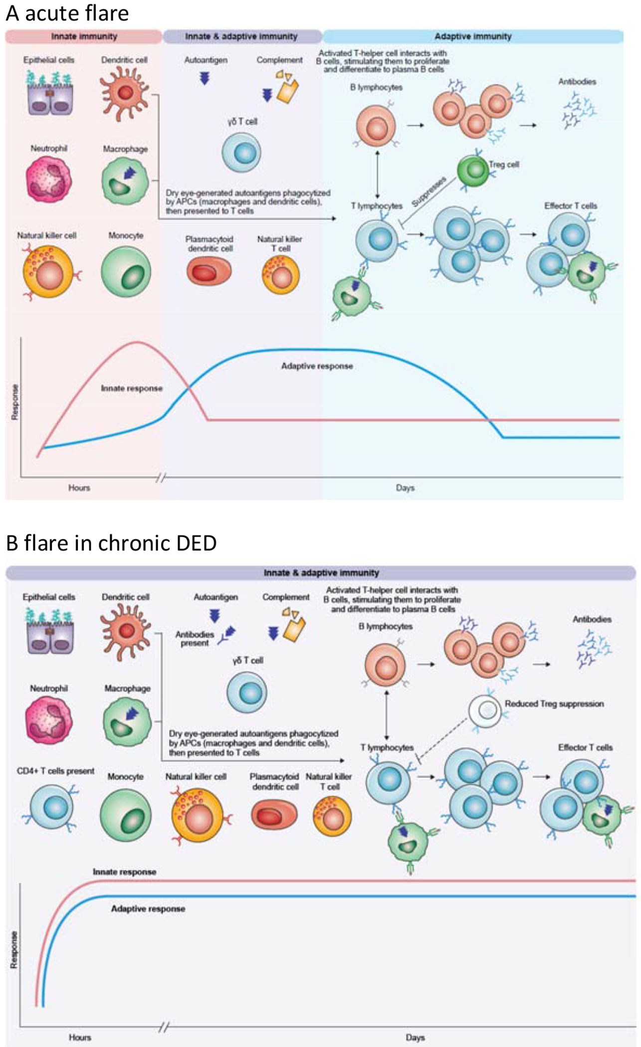
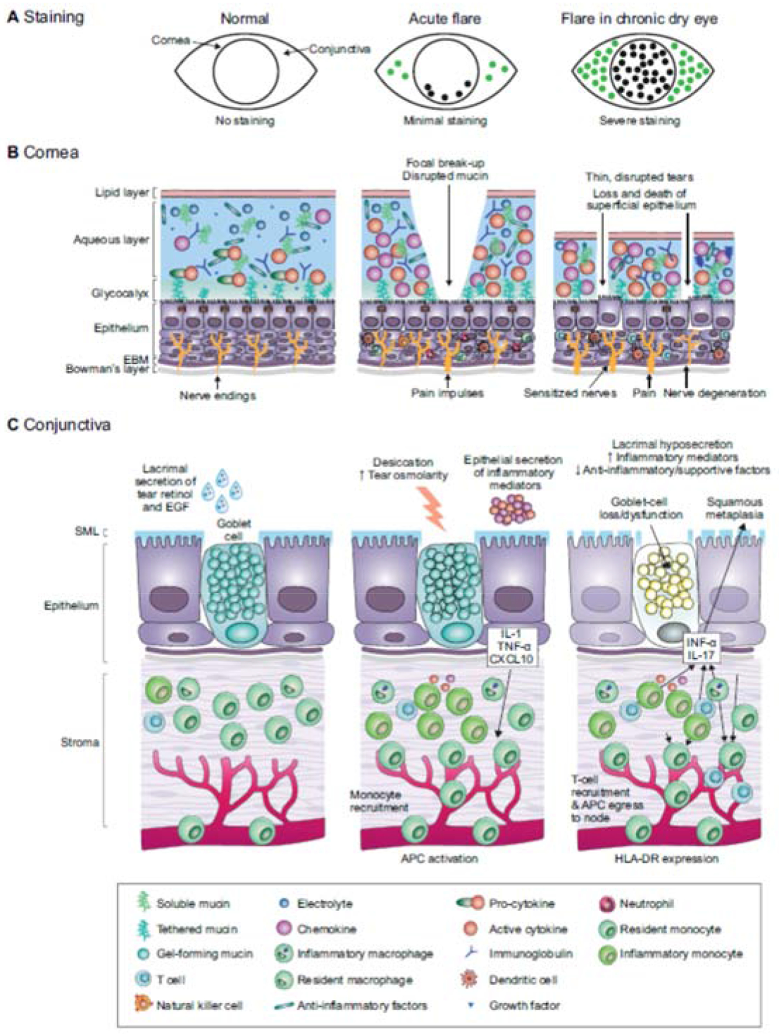
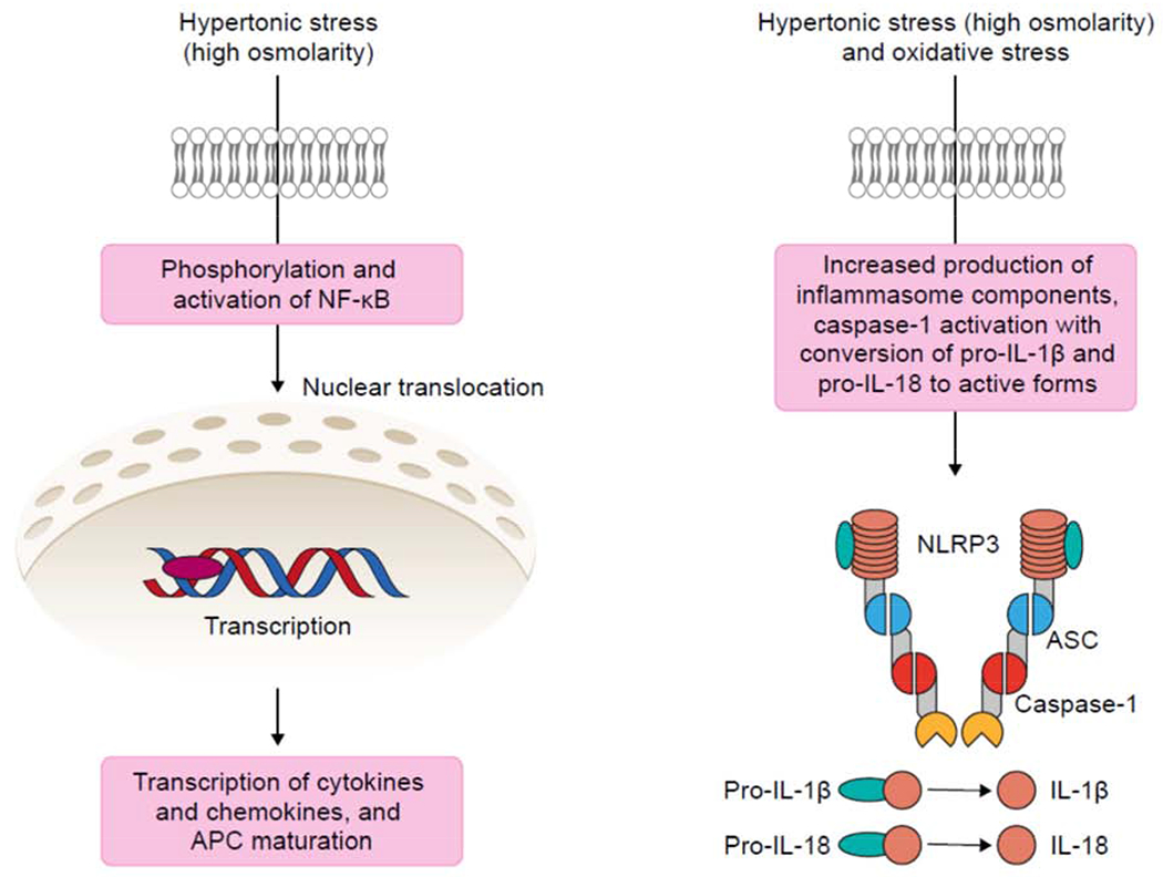
Most patients with chronic dry eye disease (DED) have episodic flares, which can be triggered by a variety of activities and environmental stresses. These flares are typically associated with rapid exacerbation of discomfort symptoms, followed by prolonged elevation of inflammation. In an acute flare, ocular surface inflammation begins with a nonspecific innate immune response, in some cases followed by a slower but more specific adaptive immune response. At the ocular surface, epithelial cells are central to the innate immune response, and we discuss their role in DED flares alongside the other core components. Epithelial cells and other cells of the innate response (neutrophils, monocytes, macrophages and dendritic cells) trigger flares in response to increased osmolarity, detected via pattern receptors on their cell surface. Ultimately, downstream signaling pathways activate innate and adaptive immune responses, with consequent inflammation and symptoms. In chronic DED, pathogenic T cells have infiltrated the ocular surface tissues. The established adaptive immune response is likely to lead to flare-ups at lower thresholds of stress, with inflammation maintained over a longer period. Increased understanding of the inflammatory cascades activated during a flare may guide management and improve outcomes.

Keywords: Adaptive immunity; Conjunctiva; Cornea; Dry eye syndromes; Flare; Innate immunity; Pathology.


Conflict of interest statement

Declaration of competing interest

V.L.P., S.C.P., and M.E.S. are consultants for Kala Pharmaceuticals, Inc. The authors received no compensation related to the development of the manuscript.

Figures

Fig. 1.
Fig. 1.. Proposed concepts and key components of the innate and immune responses over the time-course of a flare.
A. In an acute flare, the innate immune system generates a rapid response, which can activate the adaptive response. B. In chronic DED, components of the adaptive immune response are already activated and there are pathogenic T cells in the ocular surface, that leads to rapid increase in inflammation at lower thresholds. See text for details. APC, antigen-presenting cell
Fig. 2.
Fig. 2.. Normal tissue and disruptions in acute flare or chronic dry eye.
A. Patients with acute flares typically have minimal cornea and conjunctival dye staining, whereas there may be moderate-to-severe superficial punctate epitheliopathy and dye staining in chronic DED. B. Focal tear breakup and osmotic stress activate corneal epithelium and immune cells to produce innate inflammatory mediators (center). In chronic DED, cornea barrier disruption and epithelial cell loss is accompanied by infiltration of myeloid and T cells. C. In an acute flare, inflammatory mediators are produced by stressed conjunctival epithelial cells, including goblet cells that recruit inflammatory cells (center). In chronic DED, goblet cells are lost or dysfunctional, leading to reduced gel-forming mucin secretion that destabilizes the tear film and reduced production of anti-inflammatory factors by the goblet cells (right). APC, antigen-presenting cell; EBM, epithelial basement membrane; EGF, epidermal growth factor; IL, interleukin; INF-α, interferon alpha; SML, secretory mucus layer; TNF-α, tumor necrosis factor alpha
Fig. 3.
Fig. 3.. Signal pathways priming inflammatory cascades.
Innate immune cells (macrophages, dendritic cells, and neutrophils) and epithelial cells of the ocular surface detect hyperosmolarity via pattern receptors. Activation of signal transduction pathways, such as NFκB lead to production of proinflammatory cytokines and trigger innate and adaptive immune responses. Activation of the NLRP3 inflammasome results in IL-1B and IL-18 activation. See text for details. APC, antigen-presenting cell; ASC, apoptosis speck-like protein; NLRP3, NOD-, LRR- and pyrin domain-containing protein 3.

References

    1. Alam J, de Paiva CS, Pflugfelder SC, 2020. Immune–Goblet cell interaction in the conjunctiva. Ocul. Surf 18, 326–334. - PMC - PubMed
    1. Aragona P, Aguennouz M, Rania L, Postorino E, Sommario MS, Roszkowska AM, De Pasquale MG, Pisani A, Puzzolo D, 2015. Matrix metalloproteinase 9 and transglutaminase 2 expression at the ocular surface in patients with different forms of dry eye disease. Ophthalmology 122, 62–71. - PubMed
    1. Benitez del Castillo JM, Wasfy MA, Fernandez C, Garcia-Sanchez J, 2004. An in vivo confocal masked study on corneal epithelium and subbasal nerves in patients with dry eye. Invest. Ophthalmol. Vis. Sci 45, 3030–3035. - PubMed
    1. Bian F, Pelegrino FS, Henriksson JT, Pflugfelder SC, Volpe EA, Li DQ, de Paiva CS, 2016. Differential effects of dexamethasone and doxycycline on inflammation and MMP production in murine alkali-burned corneas associated with dry eye. Ocul. Surf 14, 242–254. - PMC - PubMed
    1. Bradley JL, Ozer Stillman I, Pivneva I, Guerin A, Evans AM, Dana R, 2019. Dry eye disease ranking among common reasons for seeking eye care in a large US claims database. Clin. Ophthalmol 13, 225–232. - PMC - PubMed

Publication types