

Tornwaldt nasopharyngeal cyst: Case series and literature review
- PMID: 33039780
- PMCID: PMC7550824
- DOI: 10.1016/j.ijscr.2020.09.105
Tornwaldt nasopharyngeal cyst: Case series and literature review
Abstract
Background: Tornwaldt cyst (TC) is a relatively rare but benign disease. Although these lesions are asymptomatic and found incidentally during routine ENT examination, the can present with unexplained sinonasal symptoms, such as nasal obstruction, post nasal drip and occipital headache.
Aim: To study the clinical presentations and outcome of Tornwaldt nasopharyngeal cyst cases diagnosed and managed in our center.
Methods: Patients with symptomatic TC who were operated and followed up at our center were selected for this study.
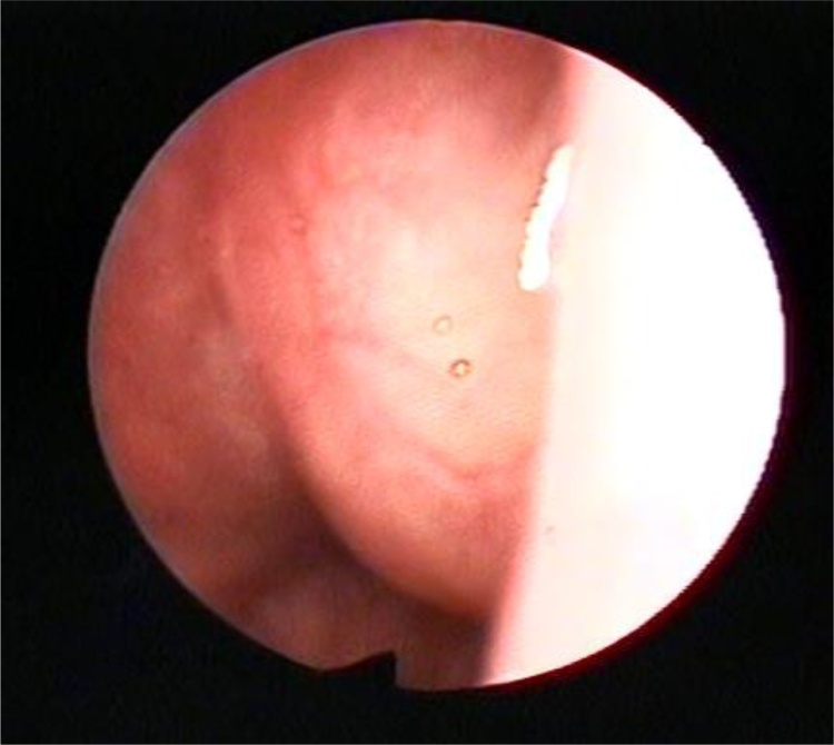
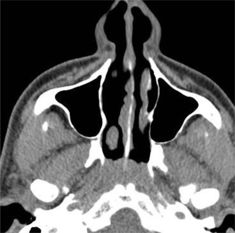
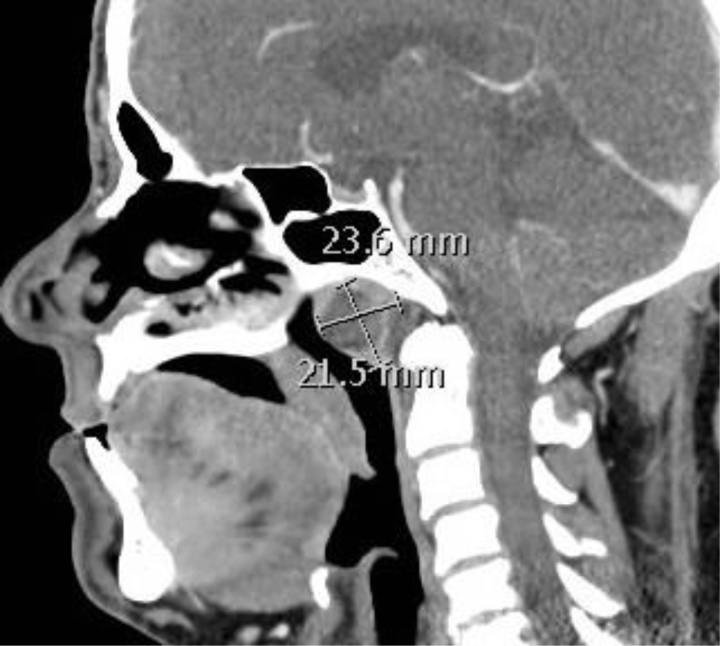
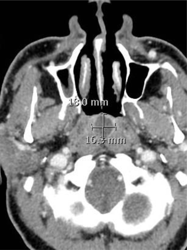
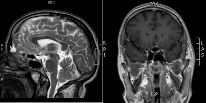
Results: 3 patients with a diagnosis of TC by naso-endoscopy, CT and MRI were included in this study. All patients were males, with age ranging from 54 to 86 years. A trans-nasal endoscopic surgical removal of the cyst was done for all patients with no intra or post-operative complications. All patients were free of symptoms and disease reassurance at follow up.
Conclusion: Although relatively rare, TC should be suspected in any patient complaining of unexplained sinonasal symptoms. Endoscopic surgical excision is a safe and effective maneuver with no cyst recurrence.
Keywords: Nasoendoscopy; Nasopharyngeal cyst; Thornwaldt’s cyst.
Copyright © 2020. Published by Elsevier Ltd.
Figures
References
-
- Moody M.W. Tornwaldt’s cyst: incidence and case report. Ear Nose Throat J. 2007;86:45–47. - PubMed
-
- Rodgers G.K., Chan Khl, Dahl R.E. Antral choanal polyp presenting as obstructive sleep apnea syndrome. Arch. Otolaryngol. Head Neck Surg. 1991;117:914–916. - PubMed
-
- Salib R.J., Sadek S.A., Dutt S.N., Pearman K. Antrochoanal polyp presenting with obstructive sleep apnea and cachexia. Int. J. Pediatric Otorhnolaryngol. 2000;54:163–166. - PubMed
-
- Eloy P., Watelet J.B., Hartert A.S., Bertrand B. Thornwaldt’s cyst and surgery with powered instrumentation. B-ENT. 2006;2:135–139. - PubMed
LinkOut - more resources
Full Text Sources
Research Materials
Miscellaneous
