. 2020 Dec 11;295(50):17200-17214.
doi: 10.1074/jbc.RA120.015025. Epub 2020 Oct 9.

Iron-mediated degradation of ribosomes under oxidative stress is attenuated by manganese

Affiliations

Iron-mediated degradation of ribosomes under oxidative stress is attenuated by manganese

Daniel G J Smethurst et al. J Biol Chem. .

Abstract

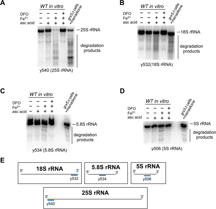
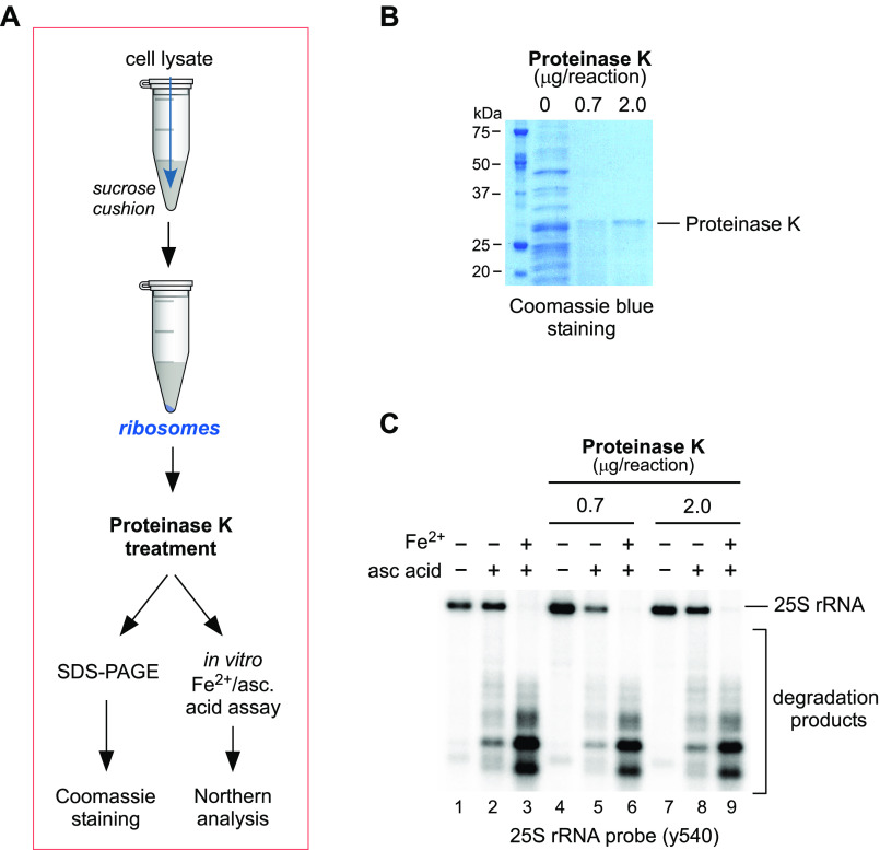
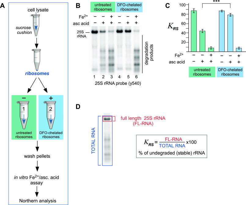
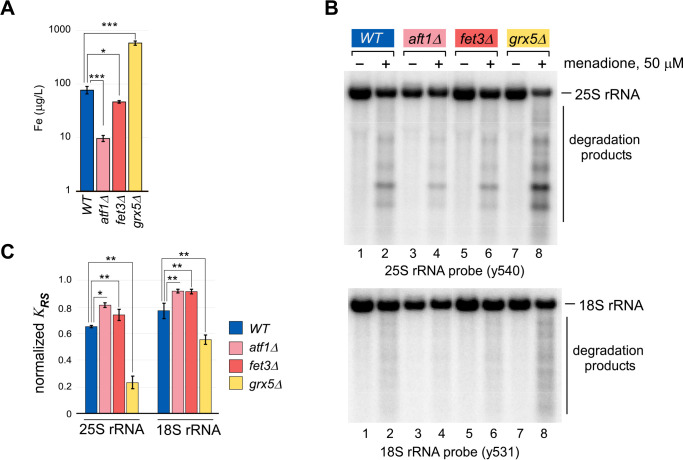
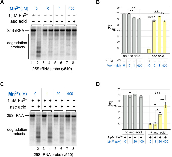
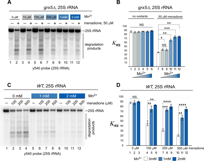
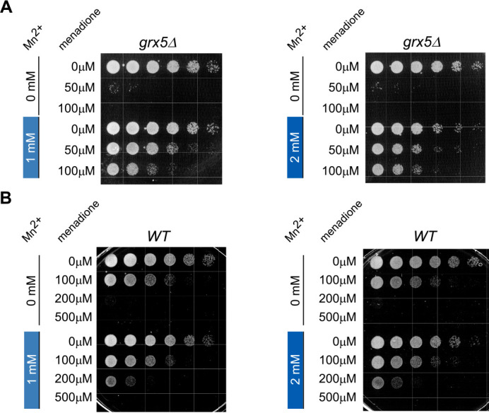
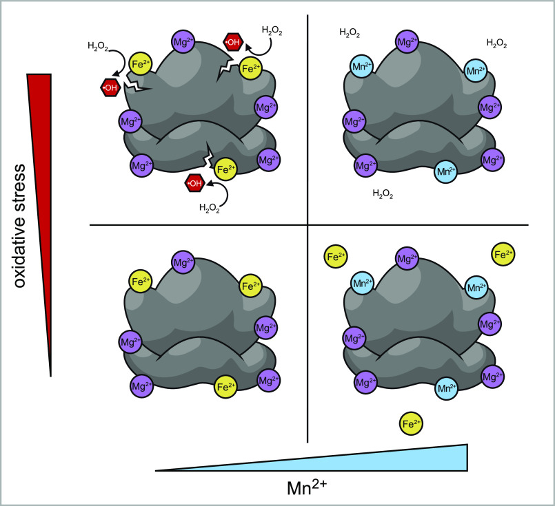
Protein biosynthesis is fundamental to cellular life and requires the efficient functioning of the translational machinery. At the center of this machinery is the ribosome, a ribonucleoprotein complex that depends heavily on Mg2+ for structure. Recent work has indicated that other metal cations can substitute for Mg2+, raising questions about the role different metals may play in the maintenance of the ribosome under oxidative stress conditions. Here, we assess ribosomal integrity following oxidative stress both in vitro and in cells to elucidate details of the interactions between Fe2+ and the ribosome and identify Mn2+ as a factor capable of attenuating oxidant-induced Fe2+-mediated degradation of rRNA. We report that Fe2+ promotes degradation of all rRNA species of the yeast ribosome and that it is bound directly to RNA molecules. Furthermore, we demonstrate that Mn2+ competes with Fe2+ for rRNA-binding sites and that protection of ribosomes from Fe2+-mediated rRNA hydrolysis correlates with the restoration of cell viability. Our data, therefore, suggest a relationship between these two transition metals in controlling ribosome stability under oxidative stress.

Keywords: RNA folding; cell viability; degradation of ribosomes; iron; iron metabolism; manganese; metals; oxidants; oxidative stress; ribosomal ribonucleic acid (rRNA); ribosome; ribosome structure; yeast.


Conflict of interest statement

Conflict of interest—The authors declare that they have no conflicts of interest with the contents of this article.

Figures

Figure 1.
Figure 1.
Ribosomal RNAs from 40S and 60S subunits are degraded in the presence of Fe2+ and ROS both in vitro and in cells. A–D, Northern blotting analysis of in vitro cleavage of rRNA purified from WT cells treated with 0.5 mm ascorbic acid in the presence or absence of 1 μm Fe(NH4)2(SO4)2, using probes for 25S (y540, A), 18S (y532, B), 5.8S (y534, C), and 5S (y506, D) rRNAs. Where indicated, the reactions were pretreated with iron chelator DFO (0.5 mm). In vitro reaction products were resolved in seven repeats on 1.2% agarose–formaldehyde gels (two repeats are shown in A and B; five repeats are shown on Fig. S2A) or in two repeats on 8% polyacrylamide, 8 m urea-containing gels (C and D). Adjacent to these is total RNA from grx5Δ cells that were treated with 50 μm menadione for 2 h at 30 °C. The sequences of all probes used in this study are listed in Table S1. Representative hybridizations are shown. E, schematic representation of the annealing sites of the probes used. See Table S1 for probe sequences.
Figure 2.
Figure 2.
rRNA hydrolysis in vitro occurs independently of ribosome-associated proteins. A, schematic representation of the workflow to test the effect of removing the protein from ribosomes on rRNA cleavage in the presence of Fe2+ and ascorbic acid. Ribosomes were purified from WT cells by centrifugation through a 50% sucrose cushion, and ribosomal pellets containing 10 µg of RNA were treated with the indicated amounts of proteinase K for 5 min on ice or remained untreated. B, samples of purified ribosomes treated with proteinase K at the indicated concentrations were analyzed by SDS-PAGE and stained for total protein content with Coomassie Blue. C, samples of purified ribosomes treated with proteinase K at the indicated concentrations were treated with ascorbic acid and Fe(NH4)2(SO4)2, as in Fig. 1, and analyzed by Northern hybridization using the 25S rRNA probe y540. A representative hybridization is shown.
Figure 3.
Figure 3.
Treatment of purified ribosomes with the iron chelator DFO abolishes rRNA fragmentation. A, schematic representation of the double-cushion workflow for the chelation of ribosome-bound iron, followed by the assessment of this metal's presence within ribosomal structures via an in vitro ascorbate reaction. B, ribosomes purified from WT yeast cells were treated on ice with 0.5 mm DFO for 20 min or remained untreated and pelleted through a second 50% sucrose cushion to remove DFO excess. Equal ribosome amounts, equivalent to 3.5 µg of RNA, were incubated with 0.5 mm ascorbic acid and 1 μm Fe(NH4)2(SO4)2. The reaction products were analyzed by Northern hybridization with probe y540; a representative hybridization is shown. C, assessment of rRNA integrity by calculating the coefficient of rRNA stability (KRS) values. Three replicates of the reactions shown in B were used for the calculation. The error bars represent S.D. ***, P < 0.001 (two-tailed two-sample unequal variance t test). D, calculation of the coefficient of rRNA stability (KRS) exemplified here for 25S rRNA. Radioactive signal of the probe hybridized to undegraded, full-length 25S rRNA (FL-RNA, area shown in red) is divided by the total hybridization signal in the same lane of the blot (TOTAL RNA, blue) and multiplied by 100.
Figure 4.
Figure 4.
Increased cellular iron content correlates with lowered 25S and 18S rRNA stability during oxidative stress. A, whole-cell lysates prepared from equal cell numbers of the indicated yeast strains were analyzed by ICP-MS for levels of iron. Three replicates of each sample from independent isolates were analyzed. ***, P < 0.001; *, P < 0.05 (two-tailed two-sample unequal variance t test). B, indicated yeast cultures were grown overnight in YPDA at 30 °C, diluted with fresh medium to A600 of ∼0.3, grown for an additional 4 h, and treated with 50 μm menadione for 2 h (+) or left untreated (−). RNA was isolated and analyzed by Northern hybridization with the indicated probes. A representative blot from three independent experimental repeats is shown. C, quantification of rRNAs stability in the experiment shown in B. KRS determined as described in Fig. 3D for menadione-treated samples was normalized by the corresponding values in untreated samples for each strain **, P < 0.01; *, P < 0.05, (two-tailed two-sample unequal variance t test).
Figure 5.
Figure 5.
Mn2+ ions compete with Fe2+ in the in vitro cleavage of 25S rRNA. A and B, ribosomes isolated from WT cells were simultaneously treated with both 0.5 mm ascorbic acid (asc acid) and either 1 μm Fe(NH4)2(SO4)2 or the indicated concentrations of MnCl21. Reaction products were analyzed by Northern hybridization with the 25S rRNA probe y540. The KRS values obtained for each reaction represent the mean from three experimental repeats; the error bars represent S.D. The differences between MnCl2-free samples and samples that were treated with 0, 1, and 400 μm MnCl2 were significant. ****, P < 0.0001; **, P < 0.01 (two-tailed two-sample unequal variance t test). A representative blot is shown. C and D, same as in A and B, except that 1 μm Fe(NH4)2(SO4)2 was added into every reaction. Different concentrations of MnCl2 (0, 1, 20, and 400 μm) were also added as indicated. The differences between 25S rRNA stability were nonsignificant in the absence of ascorbic acid in the reaction (D, gray bars) and significant in the presence of ascorbic acid/Fe(NH4)2(SO4)2. ***, P < 0.001; **, P < 0.01; *, P < 0.05 (two-tailed two-sample unequal variance t test).
Figure 6.
Figure 6.
Exogenously supplied manganese protects ribosomes from menadione-induced 25S rRNA degradation in cells. A, mid-log grx5Δ cultures grown in YPDA were shifted to medium supplemented with the indicated concentrations of MnCl2 and grown for 4 h at 30 °C. The cultures were adjusted to the same A600 of ∼0.6 and grown for an additional 2 h in the presence (+) or absence (−) of 50 μm menadione. RNA was isolated and analyzed by Northern hybridization with the 25S rRNA probe y540. A representative blot from three biological replicates is shown. B, quantification of the Northern hybridization data from A. The KRS values for 25S rRNA were determined for every culture treated or untreated with menadione and/or MnCl2. The data show mean values of three independent experiments; error bars represent S.D. The differences between KRS values determined for samples treated with menadione (right columns) grown in MnCl2-free medium and those grown in 1–10 μm, 0.5 mm, 1 mm, and 2 mm MnCl2-containing medium were significant. ***, P < 0.001; **, P <0.01; *, P < 0.05; NS, not significant. Two-tailed two-sample unequal variance t test was used for statistical analysis. C and D, the experiment was performed and the data were quantified as in A and B, except that WT cells were used, MnCl2 ranged from 0 to 2 mm, and menadione concentrations were 0, 100, 200, and 500 μm as indicated. Representative blots from three biological replicates are shown for all experiments. ****, P < 0.0001; ***, P < 0.001; **, P < 0.01; *, P < 0.05; NS, not significant (two-tailed two-sample unequal variance t test).
Figure 7.
Figure 7.
Exogenously supplied manganese restores viability of menadione-treated cells. A and B, overnight grx5Δ (A) and WT (B) cultures grown in YPDA were diluted with fresh YPDA medium to A600 of ∼0.2 and grown for 4 h in the presence of 1 or 2 mm MnCl2 or in the absence of MnCl2. The cultures were next treated with the indicated concentrations of menadione for 2 h at 30 °C with shaking. Thereafter, the cells were collected, washed twice, adjusted to the same cell counts (2 × 10⁶ cells/ml), serially diluted (1:5), and plated on YPDA agar plates. The plates were incubated at 30 °C for ∼2–3 days (WT) or ∼3–5 days (grx5Δ). Viability assays were repeated three times, and representative images are shown.
Figure 8.
Figure 8.
Model for the roles of Fe2+ and Mn2+ in oxidant-induced ribosome damage. Under normal metal homeostasis conditions, rRNA in unstressed cells is bound by divalent metal cations, predominantly Mg2+, as well as some amount of Fe2+, with both cations occupying sites capable of coordinating other metals including Mn2+ (bottom left panel). An increase in the ratio of available Mn2+ to Fe2+ results in the displacement of Fe2+ from these rRNAs sites (bottom right panel). Under oxidative stress, ribosome-bound Fe2+ generates hydroxyl radicals in the Fenton reaction, causing rRNA strand breaks in the vicinity of bound Fe2+ ions (top left panel). Increased Mn2+ concentrations have the opposite effect: by displacing bound Fe2+, Mn2+ prevents rRNA strand breaks produced through Fenton chemistry and makes ribosomes more resistant to oxidative stress conditions (top right panel).

References

    1. Zoroddu, M. A., Aaseth, J., Crisponi, G., Medici, S., Peana, M., and Nurchi, V. M. (2019) The essential metals for humans: a brief overview. J. Inorg. Biochem. 195, 120–129 10.1016/j.jinorgbio.2019.03.013 - DOI - PubMed
    1. Andreini, C., Bertini, I., Cavallaro, G., Holliday, G. L., and Thornton, J. M. (2008) Metal ions in biological catalysis: from enzyme databases to general principles. J. Biol. Inorg. Chem. 13, 1205–1218 10.1007/s00775-008-0404-5 - DOI - PubMed
    1. McCall, K. A., Huang, C., and Fierke, C. A. (2000) Function and mechanism of zinc metalloenzymes. J. Nutr. 130, 1437S–1446S 10.1093/jn/130.5.1437S - DOI - PubMed
    1. Weston, J. (2009) Biochemistry of magnesium. In PATAI'S Chemistry of Functional Groups (Rappoport, Z., ed) John Wiley & Sons, Chichester, UK
    1. Vashishtha, A. K., Wang, J., and Konigsberg, W. H. (2016) Different divalent cations alter the kinetics and fidelity of DNA polymerases. J. Biol. Chem. 291, 20869–20875 10.1074/jbc.R116.742494 - DOI - PMC - PubMed

Publication types