Review
. 2020 Nov;36(11):1285-1298.
doi: 10.1007/s12264-020-00586-4. Epub 2020 Oct 11.

Purinergic Modulation of Activity in the Developing Auditory Pathway

Affiliations
Review

Purinergic Modulation of Activity in the Developing Auditory Pathway

Sasa Jovanovic et al. Neurosci Bull. 2020 Nov.

Abstract

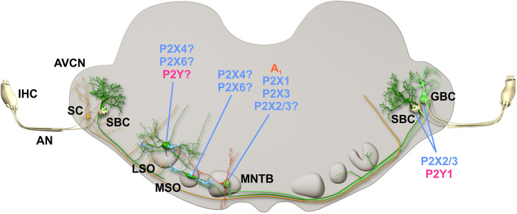
Purinergic P2 receptors, activated by endogenous ATP, are prominently expressed on neuronal and non-neuronal cells during development of the auditory periphery and central auditory neurons. In the mature cochlea, extracellular ATP contributes to ion homeostasis, and has a protective function against noise exposure. Here, we focus on the modulation of activity by extracellular ATP during early postnatal development of the lower auditory pathway. In mammals, spontaneous patterned activity is conveyed along afferent auditory pathways before the onset of acoustically evoked signal processing. During this critical developmental period, inner hair cells fire bursts of action potentials that are believed to provide a developmental code for synaptic maturation and refinement of auditory circuits, thereby establishing a precise tonotopic organization. Endogenous ATP-release triggers such patterned activity by raising the extracellular K+ concentration and contributes to firing by increasing the excitability of auditory nerve fibers, spiral ganglion neurons, and specific neuron types within the auditory brainstem, through the activation of diverse P2 receptors. We review recent studies that provide new models on the contribution of purinergic signaling to early development of the afferent auditory pathway. Further, we discuss potential future directions of purinergic research in the auditory system.

Keywords: Auditory brainstem; Auditory system; Cochlea; Development; Purinergic signaling; Spiral ganglion.


Figures

Fig. 1
Fig. 1
Schematic of the organ of Corti as in prehearing mice, shown in cross section. Two types of sensory cells - inner hair cells (IHCs) and outer hair cells (OHCs) are tightly packed between the immature tectorial membrane (TM) and several types of supporting cells that control the homeostasis of extracellular fluids. P2 receptors are differentially expressed in sensory and supporting cells. The inset depicts the position of the organ of Corti within the temporal bone relative to other middle and inner ear structures. KO, Kölliker’s organ; ISC, inner supporting cell; OSC, outer supporting cell; Type I and II AF, spiral ganglion neuron afferent fibers; PC, pillar cell; BM, basilar membrane; DC, Deiters’ cell.
Fig. 2
Fig. 2
Schematic of the proposed mechanism controlling the inner hair cell membrane potential. ATP, spontaneously released through connexin Cx 26 and Cx 30 hemichannels (1), binds to the auto- and heteroreceptors P2Y1, P2Y2, and P2Y4 (2) on inner supporting cells (ISCs). G-protein coupled P2YRs activate the PLC-IP3 signaling pathway that elevates the intracellular Ca2+ concentration. Connexin gap junctions (3) allow diffusion of IP3 and Ca2+ to neighboring ISCs, thus building an intercellular Ca2+ wave that reaches the vicinity of IHCs. Elevated [Ca2+]i activates the TMEM16A channel (4), leading to Cl efflux that is balanced by K+ efflux through the leak K+ channels (5). The rise in extracellular K+ concentration depolarizes the IHC, resulting in a series of Ca2+-dependent APs shaped mainly by the KV delayed rectifier K+ channel (6) and the CaV1.3 Ca2+ channel (7). Further sculpting of the IHC firing pattern is achieved via SK2 K+ channel (8) that is activated by the Ca2+ influx through the surrounding α9α10 nicotinic ACh receptors (9) and P2X3 receptors (10). Additional Ca2+ entry and IHC depolarization might be achieved through P2X2 receptors (11) located in the proximity of stereocilia. AF, spiral ganglion neuron afferent fiber; EF, medial olivocochlear efferent fiber.
Fig. 3
Fig. 3
Mammalian auditory brainstem with schematic wiring. Acoustic information is transduced into graded receptor potential by the inner hair cells (IHCs). This analog information is encoded into action potentials, conveyed by auditory nerve (AN) fibers. In the cochlear nucleus, the obligatory first central station along the afferent pathway, the information is segregated into streams processing different sound features. In the anteroventral part of the cochlear nucleus (AVCN), the major targets of AN fibers are stellate cells (multipolar cells, SCs), spherical bushy cells (SBCs), and globular bushy cells (GBCs). The medial superior olive (MSO) is the first stage of processing of interaural time differences through the computation of bilateral excitatory inputs from the SBC plus an inhibitory projection from the medial nucleus of the trapezoid body (MNTB). Note that MNTB neurons are activated by the contralateral GBCs. Interaural level differences are first calculated in the lateral superior olive (LSO), where excitatory input from the ipsilateral SBC and inhibitory input from the ipsilateral MNTB converge. Excitatory neurons are shown in green and yellow, inhibitory neurons in red.

References

    1. Burnstock G. Purinergic nerves. Pharmacol Rev. 1972;24:509–581. - PubMed
    1. Burnstock G. Introduction to purinergic signalling in the brain. Adv Exp Med Biol. 2020;1202:1–12. - PubMed
    1. Abbracchio MP, Burnstock G, Verkhratsky A, Zimmermann H. Purinergic signalling in the nervous system: an overview. Trends in Neurosciences. 2009;32:19–29. - PubMed
    1. Bowser DN, Khakh BS. Vesicular ATP is the predominant cause of intercellular calcium waves in astrocytes. J Gen Physiol. 2007;129:485–491. - PMC - PubMed
    1. Ho T, Jobling AI, Greferath U, Chuang T, Ramesh A, Fletcher EL, Vessey KA. Vesicular expression and release of ATP from dopaminergic neurons of the mouse retina and midbrain. Front Cell Neurosci. 2015;9:389. - PMC - PubMed

Substances

LinkOut - more resources