. 2020 Dec 4;127(12):1552-1565.
doi: 10.1161/CIRCRESAHA.120.317415. Epub 2020 Oct 12.

Genetic Regulation of Atherosclerosis-Relevant Phenotypes in Human Vascular Smooth Muscle Cells

Affiliations

Genetic Regulation of Atherosclerosis-Relevant Phenotypes in Human Vascular Smooth Muscle Cells

Redouane Aherrahrou et al. Circ Res. .

Abstract

Rationale: Coronary artery disease (CAD) is a major cause of morbidity and mortality worldwide. Recent genome-wide association studies revealed 163 loci associated with CAD. However, the precise molecular mechanisms by which the majority of these loci increase CAD risk are not known. Vascular smooth muscle cells (VSMCs) are critical in the development of CAD. They can play either beneficial or detrimental roles in lesion pathogenesis, depending on the nature of their phenotypic changes.

Objective: To identify genetic variants associated with atherosclerosis-relevant phenotypes in VSMCs.

Methods and results: We quantified 12 atherosclerosis-relevant phenotypes related to calcification, proliferation, and migration in VSMCs isolated from 151 multiethnic heart transplant donors. After genotyping and imputation, we performed association mapping using 6.3 million genetic variants. We demonstrated significant variations in calcification, proliferation, and migration. These phenotypes were not correlated with each other. We performed genome-wide association studies for 12 atherosclerosis-relevant phenotypes and identified 4 genome-wide significant loci associated with at least one VSMC phenotype. We overlapped the previously identified CAD loci with our data set and found nominally significant associations at 79 loci. One of them was the chromosome 1q41 locus, which harbors MIA3. The G allele of the lead risk single nucleotide polymorphism (SNP) rs67180937 was associated with lower VSMC MIA3 expression and lower proliferation. Lentivirus-mediated silencing of MIA3 (melanoma inhibitory activity protein 3) in VSMCs resulted in lower proliferation, consistent with human genetics findings. Furthermore, we observed a significant reduction of MIA3 protein in VSMCs in thin fibrous caps of late-stage atherosclerotic plaques compared to early fibroatheroma with thick and protective fibrous caps in mice and humans.

Conclusions: Our data demonstrate that genetic variants have significant influences on VSMC function relevant to the development of atherosclerosis. Furthermore, high MIA3 expression may promote atheroprotective VSMC phenotypic transitions, including increased proliferation, which is essential in the formation or maintenance of a protective fibrous cap.

Keywords: cell proliferation; coronary artery disease; genome-wide association study; human genetics.


Figures

Figure 1:
Figure 1:. Schematic representation of the overall study design and major results.
Illustration of phenotypic and genotypic characterization of VSMC. The risk variant in the 1q41 CAD locus affects atherosclerosis by reducing MIA3 expression, which, in turn, negatively regulates the proliferation of VSMCs, which is important for the formation of a protective fibrous cap.
Figure 2:
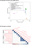
Figure 2:. Genotypic and phenotypic characterization of VSCM donors.
A) Principal component analysis of the genotypes of the 151 donors in our population and 1000 Genomes populations. The colors indicate different 1000 Genomes reference samples. Donors in our study are represented with “+” and black color. B) Heatmap of pairwise correlations between 12 VSMC cellular phenotypes. The size of the circles and different colors represent the correlation range (r) from –1 to +1. Orange indicates a perfect negative correlation, whereas blue indicates a perfect positive correlation. The color key of the correlations is shown at the bottom. “light color” indicates a low correlation, and “dark color” indicates a high correlation. Statistically significant (FDR<5%) correlations are shown with colored circles.
Figure 3:
Figure 3:. Regional association and genotype-phenotype plots of the four genome-wide significant loci associated with VSMC phenotypes.
Associations with genetic variants around the lead SNP for each significant locus are shown using LocusZoom for (A–C) calcification under the osteogenic stimulus and (D) relative proliferation in IL-1β-containing media. The left y-axis represents the −log10 (P-value) of the SNP associations. The right y-axis shows the recombination rate. Linkage disequilibrium (r2) of each SNP with the lead SNP is color-coded. (E–H) Box and whisker plots of the associated VSMC phenotypes in relation to the genotype of the four lead variants. P-values were determined using the linear mixed-model regression in performing GWAS.
Figure 4:
Figure 4:. Heatmap of effect sizes for significant associations between CAD variants and VSMC phenotypes.
79 of the 158 CAD GWAS loci genotyped in our population showed a nominal association (P-value<0.05) with at least one VSMC phenotype. Rows show 12 VSMC phenotypes, and columns show the index variants in the CAD loci. The color key of the correlations is shown on the left. The colors refer to the SNP weight (beta) direction and magnitude, ranging from −3 (blue) to 3.5 (red). Only statistically significant associations (P-value<0.05) are indicated with a colored box. Negative effect sizes (blue) indicate that risk allele was associated with lower VSMC phenotype, whereas positive effect sizes (red) indicate that risk allele was associated with a higher VSMC phenotype.
Figure 5:
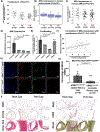
Figure 5:. Association of MIA3 with VSMC proliferation.
The risk allele (G) of SNP rs67180937 is associated with A) lower proliferation, B) lower MIA3 expression in human arteries aorta, and C) VSMCs. P-values in A and C were determined using the linear mixed-model regression, whereas the P-value in B was obtained from the GTEx database. VSMCs from two heterozygous carriers of the risk allele were transduced with three distinct shRNAs against MIA3 showed significant D) MIA3 downregulation and E) lower proliferation. F) Correlation of MIA3 expression with proliferation. G) Representative immunofluorescence images of brachiocephalic artery (BCA) lesions from VSMCOct4-WT/WT ApoE−/− and VSMCOct4-Δ/Δ ApoE−/− mice, which were fed 18 weeks of hypercholesterolemic Western diet, were stained for MIA3, ACTA2, and eYFP. H) Quantification of the frequency of eYFP+ MIA3+ ACTA+ cells as a percent of total eYFP+ VSMC in advanced BCA lesions from VSMCOct4-WT/WT ApoE−/− and VSMCOct4-Δ/Δ ApoE−/− mice. Colocalization of MIA3 with ACTA2 and eYFP is shown in Supplementary Figure 14. Single-channel images with isotype controls are shown in Online Figure XV. I) Results of MIA3, ACTA2, CD68, Pico-Sirius Red (Bright Field), and Movat staining in human coronary artery fibrous cap atheroma. IgG control images are shown in Online Figures XVI and XVII. The representative images in G and I were chosen since they captured the critical lesion features, including the three anatomical layers, cell composition, and evident staining. P-values presented in panels E and F are from one-way ANOVA with Tukey’s multiple comparison test, while in H are from comparisons using the T-test.

References

    1. Frismantiene A, Philippova M, Erne P, Resink TJ. Smooth muscle cell-driven vascular diseases and molecular mechanisms of VSMC plasticity. Cellular Signalling. 2018;52:48–64. - PubMed
    1. Bennett MR, Sinha S, Owens GK. Vascular Smooth Muscle Cells in Atherosclerosis. Circ Res. 2016;118:692–702. - PMC - PubMed
    1. Shankman LS, Gomez D, Cherepanova OA, et al. KLF4-dependent phenotypic modulation of smooth muscle cells has a key role in atherosclerotic plaque pathogenesis. Nat Med. 2015;21:628–37. - PMC - PubMed
    1. Benjamin EJ, Virani SS, Callaway CW, et al. Heart Disease and Stroke Statistics—2018 Update: A Report From the American Heart Association. Circulation. 2018;137:e67–e492. - PubMed
    1. McPherson R, Tybjaerg-Hansen A. Genetics of Coronary Artery Disease. Circ Res. 2016;118:564–78. - PubMed

Publication types

MeSH terms

Substances